
Taeniastrotos pleuronichthydis ( Yamaguti, 1939 )
|
publication ID |
https://doi.org/ 10.5281/zenodo.170310 |
|
DOI |
https://doi.org/10.5281/zenodo.5669935 |
|
persistent identifier |
https://treatment.plazi.org/id/6005083B-FFEA-1D01-3546-9113FC51954C |
|
treatment provided by |
Plazi |
|
scientific name |
Taeniastrotos pleuronichthydis ( Yamaguti, 1939 ) |
| status |
|
Taeniastrotos pleuronichthydis ( Yamaguti, 1939)
( Figs. 1–5 View FIGURE 1 View FIGURE 2 View FIGURE 3 View FIGURE 4 View FIGURE 5 )
Anchistrotos pleuronichthydis Yamaguti, 1939: 410 ; Izawa, 1986: 82
Taeniastrotos pleuronichthydis ( Yamaguti, 1939) : Dojiri & Cressey, 1987: 230
Material examined. Four adult Ψ from Pseudorhombus cinnamoneus (Temminck & Schlegel) captured on 17 April, 1971, in Tanabe Bay, Wakayama Prefecture (33 41'N, 135 20'E), coll. K. Izawa; 11 adult Ψ and 5 adult ɗ from Kareius bicoloratus (Basilewsky) and Pseudopleuronectes yokohamae (Günther) collected 2 October, 1971, Ise Bay, Mie Prefecture (34 45'N, 136 35'E), coll. K. Izawa; 3 adult Ψ and 2 adult ɗ from K. bicoloratus and P. yokohamae captured 15 February, 1979, Ise Bay, Mie Prefecture (34 36'N, 136 35'E), coll. K. Izawa. Voucher specimens (7 Ψ and 3 ɗ) are deposited in the Australian Museum (AM P.68816 and P.68817; P.70245–P.70248).
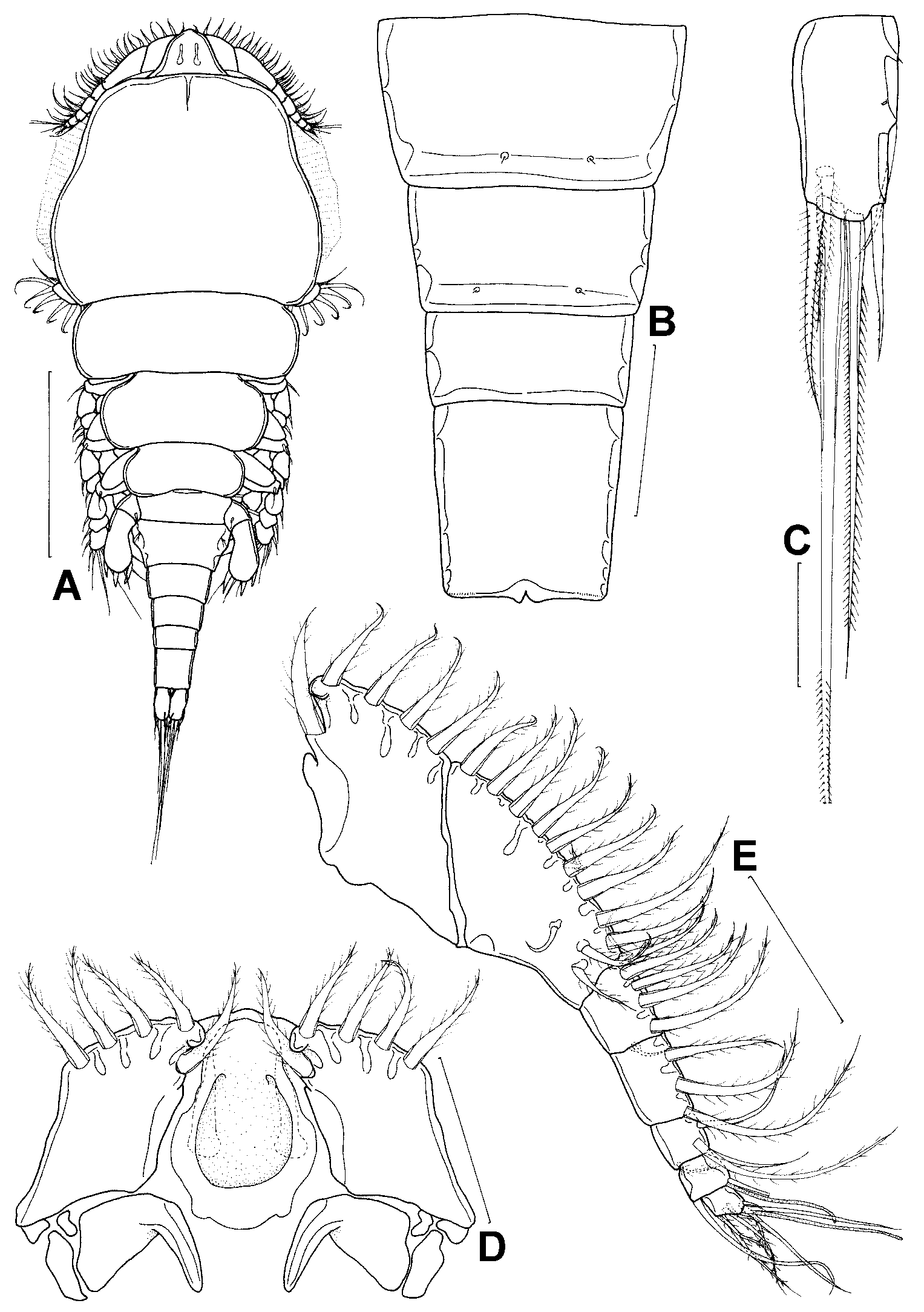
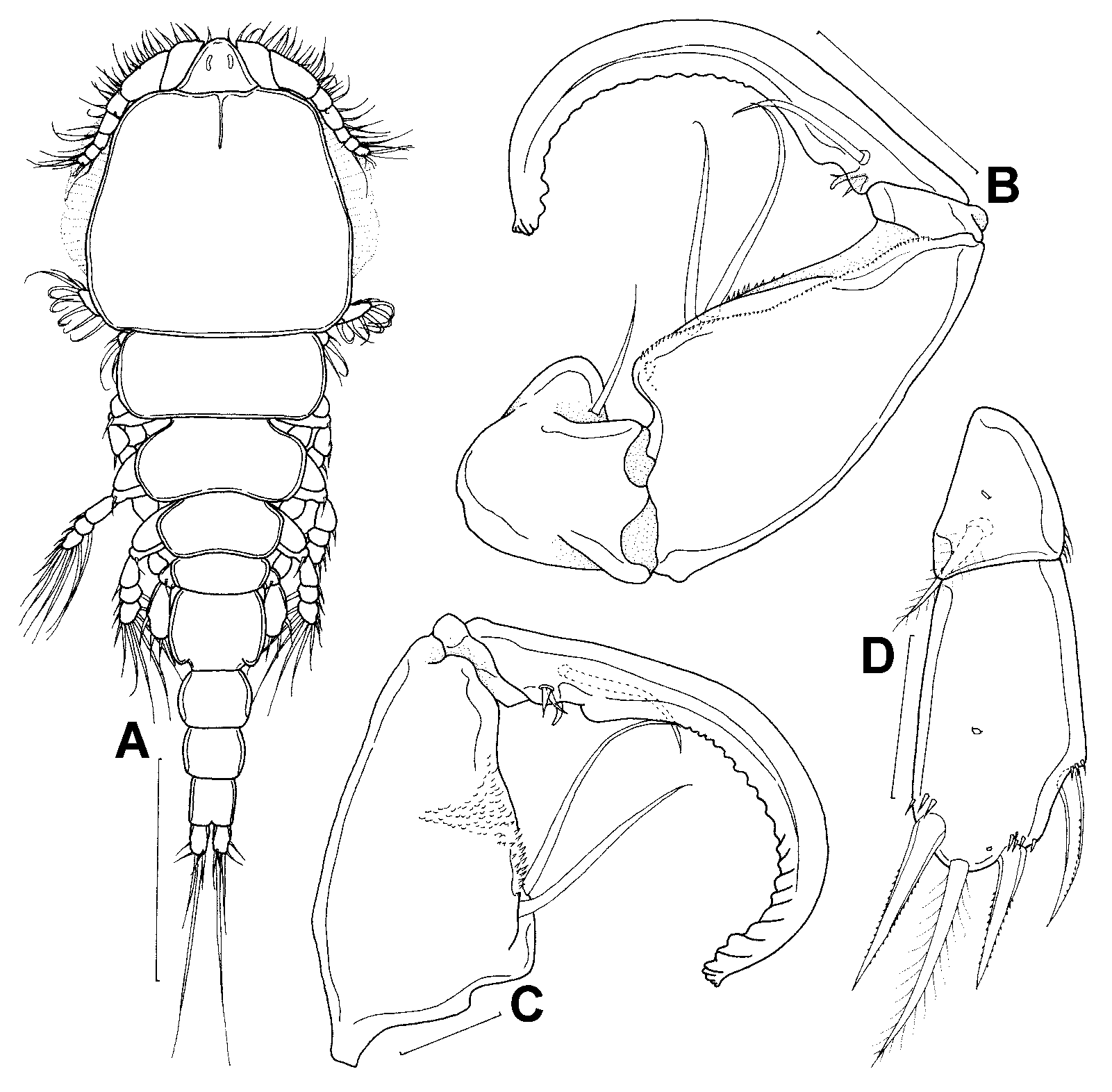
Description. Female ( Fig. 1 View FIGURE 1 A): Total body length (excluding setae on caudal rami) 1.49 ± 0.23 mm. Prosome 0.98 ± 0.15 mm long and 0.57 ± 0.09 mm wide, representing 66% of total body length; composed of cephalothorax (first pedigerous somite fused with cephalosome) and 3 free pedigerous somites. Lateral margins of cephalothorax ornamented with marginal membranes and narrower anteriorly. Second pedigerous somite 0.47 ± 0.08 mm wide; pedigerous somites 3 and 4 decreasing in width posteriorly. Urosome comprised of pedigerous somite 5, genital somite and 4 free abdominal somites. Genital somite wider (0.21 ± 0.03 mm) than long (0.08 ± 0.02 mm). Abdomen ( Fig. 1 View FIGURE 1 B) 0.30 ± 0.05 mm long and 0.13 ± 0.03 mm wide; first 2 abdominal somites bearing a pair of sensillae on posteroventral margin; ventral surface of anal somite with transverse row of minute spinules posterolaterally. Caudal ramus ( Fig. 1 View FIGURE 1 C) longer (64 ± 8 µm) than wide (33 ± 3 µm), bearing 1 minute proximolateral element and 1 midlateral, 1 dorsal, 2 subterminal and 2 terminal setae. Dorsal seta pinnate; midlateral seta naked; outer subterminal seta with barbules on inner margin; outer terminal and inner subterminal setae ornamented with long, fine spinules along margins; inner terminal seta longest, twice length of outer terminal seta, and ornamented with short, fine spinules along margins.
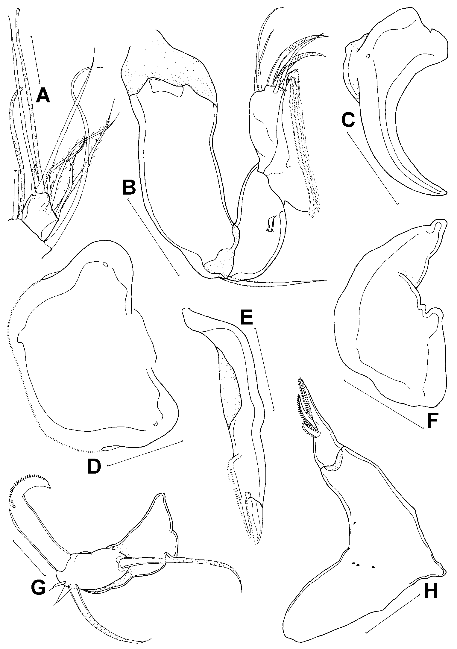
Rostral area ( Figs 1 View FIGURE 1 A, D) protuberant, with 2 internal, chitinous rods and a ventral, horseshoeshaped, sclerotised structure; area between horseshoe structure concave. Antennule ( Fig. 1 View FIGURE 1 E) 7segmented; armature formula: 5, 15, 5, 3, 4, 2 + 1 aesthetasc and 7 + 1 aesthetasc; first segment bearing rounded projection anteromedially; anterodistal seta on last segment share common base with aesthetasc ( Fig. 2 View FIGURE 2 A). Strong, uncinate process situated posterior to each antennule base ( Fig. 1 View FIGURE 1 D). Antenna ( Fig. 2 View FIGURE 2 B) composed of coxobasis and 2 endopodal segments; coxobasis longer than length of endopodal segments combined, and bearing long, bristled seta distally; first endopodal segment with 1 inner seta; second endopodal segment bearing 2 unequal, pectinate processes, 3 clawlike spines and 4 unequal setae; large, pectinate process with 1 seta and several rows of spinules; short, pectinate process with minute, blunt seta and single row of spinules. Postantennal process ( Fig. 2 View FIGURE 2 C) with a wide base and curved tine.
Labrum ( Fig. 2 View FIGURE 2 D) spinulated along posterior margin. Mandible ( Fig. 2 View FIGURE 2 E) with 1 terminal and 1 subterminal blade; terminal blade spinulated along posterior margin; articulation of subterminal blade with gnathobase indistinct, with 2 proximal rows of spinules and 1 distal row of spinules. Paragnath ( Fig. 2 View FIGURE 2 F) unornamented. Maxillule ( Fig. 2 View FIGURE 2 G) lobate bearing 2 long, naked setae, 2 short, naked setae, 1 long, broad seta armed with a distal row of spinules, and anterior knoblike process. Maxilla ( Fig. 2 View FIGURE 2 H) 2segmented; syncoxa unarmed; basis armed with 1 spinulated, terminal process and 2 spinulated setae.
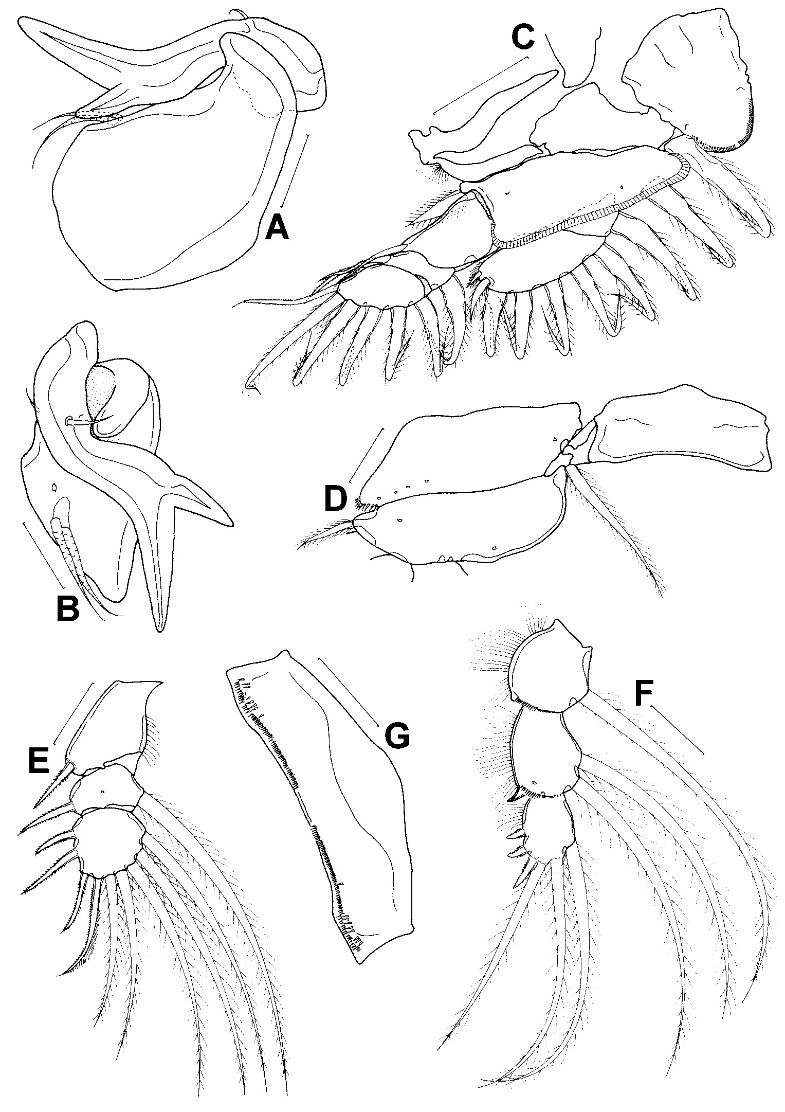
Maxilliped ( Figs 3 View FIGURE 3 A–B) 3segmented; syncoxa (not illustrated) ringlike, bearing 1 naked seta; basis armed with 2 proximal, naked setae and mediodistal protrusion; terminal claw sigmoidshaped, bearing 2 naked setae and accessory claw.
Legs 1–4 biramous ( Figs 3 View FIGURE 3 C–4F) with 3segmented rami, except leg 1 endopod 2 segmented. Armature on rami of legs 1 to 4 as follows (Roman numerals = spines; Arabic numerals = setae; int. = intermediate spine):
Coxa Basis Exopod Endopod
Leg 1 01 11 10; 11; 7 01; 7
Leg 2 01 10 I0; I1; III, I, 5 01; 02; II, I, 3
Leg 3 01 10 I0; I1; II, I, 5 01; 02; II, I, 2
Leg 4 00 10 I0; I1; II, I, 5 0int.; 0int.; I, 3 int.
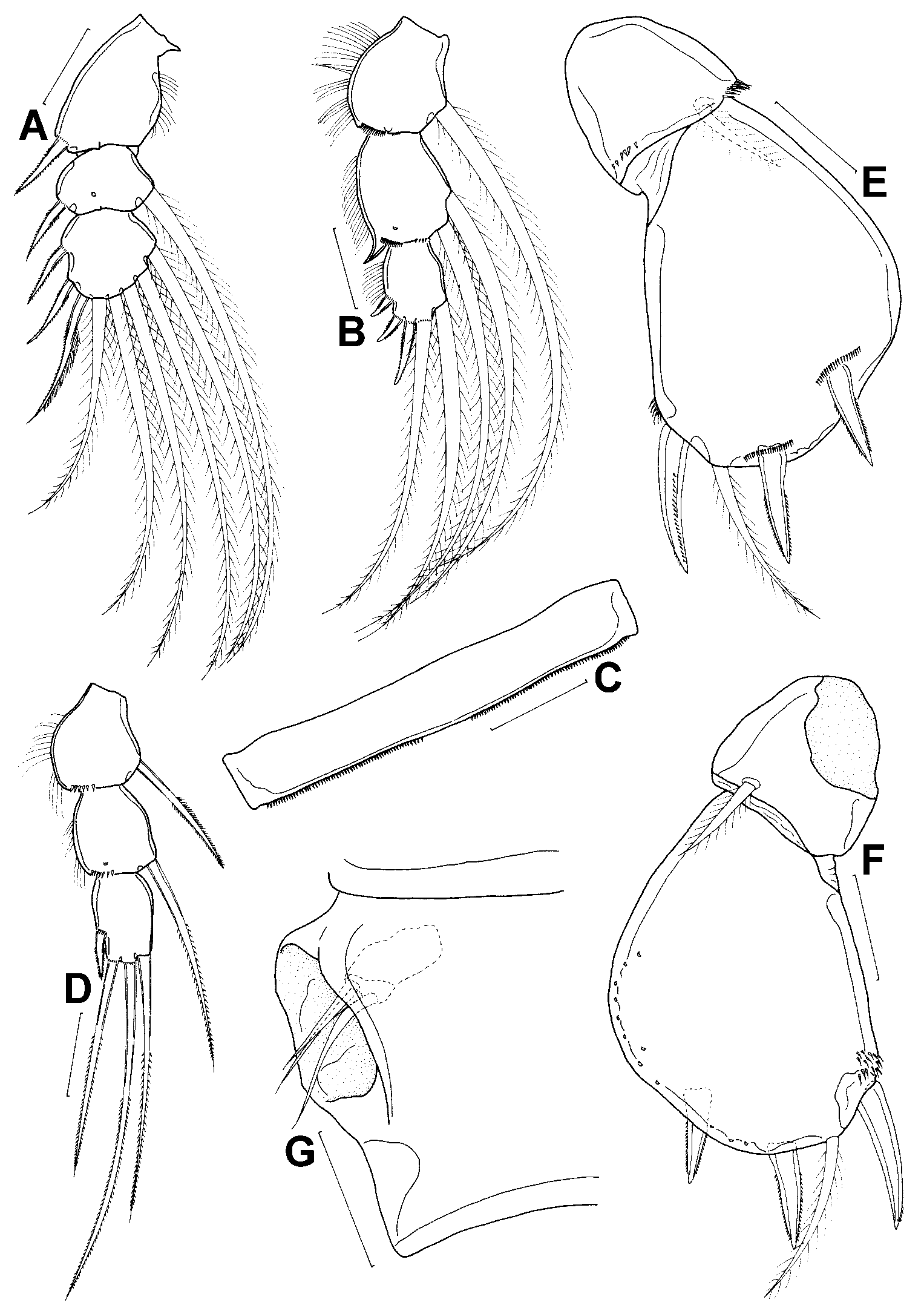
Leg 1 ( Fig. 3 View FIGURE 3 C) protopod and rami flattened and expanded. Intercoxal sclerite subtriangular; posterior margin rounded, armed with fine spinules. Coxa with patch of setules on outer border; basis ornamented with marginal membrane posteriorly. First 4 outer exopodal setae bristled along margins; 2 outermost setae on terminal endopodal segment shorter than 5 inner setae; outer margin armed with patch of setules. Leg 2 intercoxal sclerite and basis ( Fig. 3 View FIGURE 3 D) unornamented; coxa bearing large spinules on distolateral margin. Leg 2 exopodal spines ( Fig. 3 View FIGURE 3 E) weakly sclerotized; first 5 spines spinulated along margins and apical spine on terminal segment finely spinulated on outer edge and armed with pinnules on inner edge; single row of minute spinules at base of most spines; row of setules present on inner margin of first exopodal segment. Leg 2 endopodal segments ( Fig. 3 View FIGURE 3 F) ornamented with row of setules on outer borders and spinules on posterior margins; second endopodal segment with distolateral spiniform process; spines on terminal segment finely spinulated on margins. Leg 3 intercoxal sclerite ( Fig. 3 View FIGURE 3 G) spinulated along posterior margin; coxa and basis similar to that in leg 2. Ornamentation of leg 3 exopod ( Fig. 4 View FIGURE 4 A) and endopod ( Fig. 4 View FIGURE 4 B) similar to that in leg 2; spines on terminal endopodal segment ( Fig. 4 View FIGURE 4 B) slightly longer than those in leg 2. Leg 4 intercoxal sclerite ( Fig. 4 View FIGURE 4 C) wide and short; coxa and basis similar to that in leg 2 except inner coxal seta absent; exopod similar to that in leg 3. Leg 4 endopodal segments ( Fig. 4 View FIGURE 4 D) with larger spinules on posterior margins than on legs 2 and 3; second segment lacking posterolateral process; terminal segment without outer row of setules. Leg 5 ( Figs 4 View FIGURE 4 E–F) uniramous, 2 segmented. Protopodal segment armed with 1 dorsolateral, pinnate seta, a row of long spinules and row of short spinules; exopodal segment concave on medial surface and armed with 3 spinulated spines and 1 pinnate seta; spinules present at base of each spine. Leg 6 ( Fig. 4 View FIGURE 4 G) vestigial, represented by 3 unequal, naked setae at egg sac attachment area.
Male ( Fig. 5 View FIGURE 5 A): Total body length (excluding setae on caudal rami) 0.90 ± 0.20 mm. Prosome 0.59 ± 0.11 mm long and 0.33 ± 0.04 mm wide. Second pedigerous somite 0.26 ± 0.03 mm wide; remaining pedigerous somites decrease in width posteriorly. Genital somite wider (0.12 ± 0.02 mm) than long (0.08 ± 0.03 mm). Abdomen 0.15 ± 0.06 mm long and 0.09 ± 0.01 mm wide, composed of 3 free somites; ventral surface of abdominal somites ornamented as in female. Caudal ramus longer (37 ± 3 µm) than wide (22 ± 3µm), bearing similar elements as in female.
Maxilliped ( Figs 5 View FIGURE 5 B–C) 4segmented; first segment compact, bearing 1 naked seta; second segment elongate, armed with 2 long, naked setae, patch of minute spinules and single row of spinules on posterior surface, and large patch of spinules and small, truncate denticles on anterior surface; third segment small, unornamented; terminal segment a strongly curved claw, bearing 1 posterior seta, 2 anterior setae, and 1 hyaline process and single row of large, platelike denticles on inner margin. Leg 5 ( Fig. 5 View FIGURE 5 D) with dorsolateral pinnate seta and a row of long spinules on protopodal segment; free exopodal segment slimmer than in female, lacking medial concavity; spines on terminal segment slimmer than in female; spinules at base of each spine fewer in number than in female.
Remarks. The specimens in the present study conform to the descriptions and illustrations provided by Yamaguti (1939) and Izawa (1986). However, there are several discrepancies between our observations and those of Yamaguti (1939) and Izawa (1986) in terms of the ornamentation of the anal somite, segmentation and armature of the antennule, armature of the maxillule, maxilla and maxilliped, segmentation of leg 1 exopod, ornamentation of legs 2–4, armature of leg 4 endopod and ornamentation of leg 5 of the adult male and female ( Table 1). These differences are minor, and are most likely attributed to variations in the interpretation of fine morphological details by each investigator rather than to intraspecific variation.
Taeniastrotos pleuronichthydis is characterised by the following combination of apomorphies: (1) a horseshoeshaped sclerotised structure on the ventral surface of the rostrum; (2) an anteromedial projection on the first antennule segment; (3) a robust, uncinate process posterior to each antennule base; (4) pair of postantennal processes; (5) a sigmoidshaped maxilliped claw bearing an accessory process; (6) an inner coxal seta on legs 2 and 3; and (7) an inner intermediate spine on the first and second endopodal segments of leg 4. Although the female of T. pleuronichthydis possesses a broad, spinulated seta on the maxillule, a sigmoidshaped maxilliped claw and a body shape similar to T. californiensis and T. tragus , the absence of a ventral, corrugated, shieldlike rostral area excludes it from the genus Taeniastrotos . The species T. pleuronichthydis is also not affiliated with Anchistrotos , the genus to which it was originally assigned, as it has five setae on the maxillule rather than six, lacks two long, whiplike setae on the maxilliped claw and the second endopodal segment of leg 1 is armed with seven setae rather than six. Species of Irodes Wilson, 1911 , Phagus Wilson, 1911 , Pseudotaeniacanthus Yamaguti and Yamasu, 1959 and Scolecicara Ho, 1969 possess the plesiomorphic inner coxal seta on legs 2 and 3 similar to T. pleuronichthydis . Nonetheless, T. pleuronichthydis cannot be placed in any of these genera due to noticeable differences in body tagmosis, the cephalothoracic appendages and armature of leg 4 ( Table 2 View TABLE 2 ). This species represents a new genus, which is diagnosed below.
No known copyright restrictions apply. See Agosti, D., Egloff, W., 2009. Taxonomic information exchange and copyright: the Plazi approach. BMC Research Notes 2009, 2:53 for further explanation.
|
Kingdom |
|
|
Phylum |
|
|
Class |
|
|
Order |
|
|
Family |
|
|
Genus |
Taeniastrotos pleuronichthydis ( Yamaguti, 1939 )
| Tang, Danny & Izawa, Kunihiko 2005 |
Taeniastrotos pleuronichthydis (
| Dojiri 1987: 230 |
Anchistrotos pleuronichthydis
| Izawa 1986: 82 |
| Yamaguti 1939: 410 |