
Bostrycapulus, OLSSON & HARBISON, 1953
|
publication ID |
https://doi.org/10.1111/j.1096-3642.2005.00162.x |
|
DOI |
https://doi.org/10.5281/zenodo.10545350 |
|
persistent identifier |
https://treatment.plazi.org/id/603AD32D-FFA3-FFE9-D879-F9EE08F4CB83 |
|
treatment provided by |
Diego |
|
scientific name |
Bostrycapulus |
| status |
|
BOSTRYCAPULUS OLSSON & HARBISON, 1953 View in CoL
Type species: Bostrycapulus aculeatus (Gmelin) by original designation.
Original description
‘Shell widely slipper-shaped, with a strongly eccentric apex, closely appressed and spirally coiled towards the left side (viewed dorsally). Surface with strong, radial riblets or threads, the primary ones often becoming scabrous or spiniform. Diaphragm as in Crepidula s. s., its edge nearly straight, the muscle scar below small but distinct’.
Morphological description
Shell: externally, the shell is relatively flattened and more coiled, but generally similar to that of Crepidula species. The internal septum extends about half the length of the shell, and the anterior margin is indented medially and notched on the animal’s left side. A distinct but small medial ridge or crease extends from the medial indentation to the posterior shell margin near the apex. The small lunar muscle scar on the animal’s right side anterior to the shelf is often more deeply indented than in Crepidula species. The shell is distinctly coiled with about one single whorl after the protoconch–teleoconch boundary. The apex is appressed, usually occurring slightly above the posterior shell margin on the right; it is not excavated. External shell sculpture ranges from widely spaced large scale-like plicate spines to tightly packed pointed granular bumps along fine spiral ribs. Shell colour ranges from overall cream with scattered brown markings to solid chocolate brown, sometimes with a pale streak and occasionally solid tan. The markings are sometimes speckled and often streaky. No teleoconch characters have been found to unambiguously diagnose species in the genus.
Protoconch: the size of the protoconch varies between species depending on the mode of development but is less than two whorls and is often eroded in adult specimens. Hatchlings and embryos show a linear pattern of fine, widely spaced granules on the protoconch. Protoconch characters can be used to diagnose several species.
Pigmentation: the head, neck, foot and mantle are cream, but there is a matt black marbled area along the edge of the foot. Large yellow or orange splotches are scattered along the neck lappets and concentrated on the lips and tentacles. Black pigment also occurs on the dorsal side of the head and neck. The intensity of all pigmentation varies, with some animals showing almost no black pigment. The black pigment is retained in preserved or fixed material, although the yellow and orange markings are lost. There are no diagnostic differences in pigmentation among the species described here.
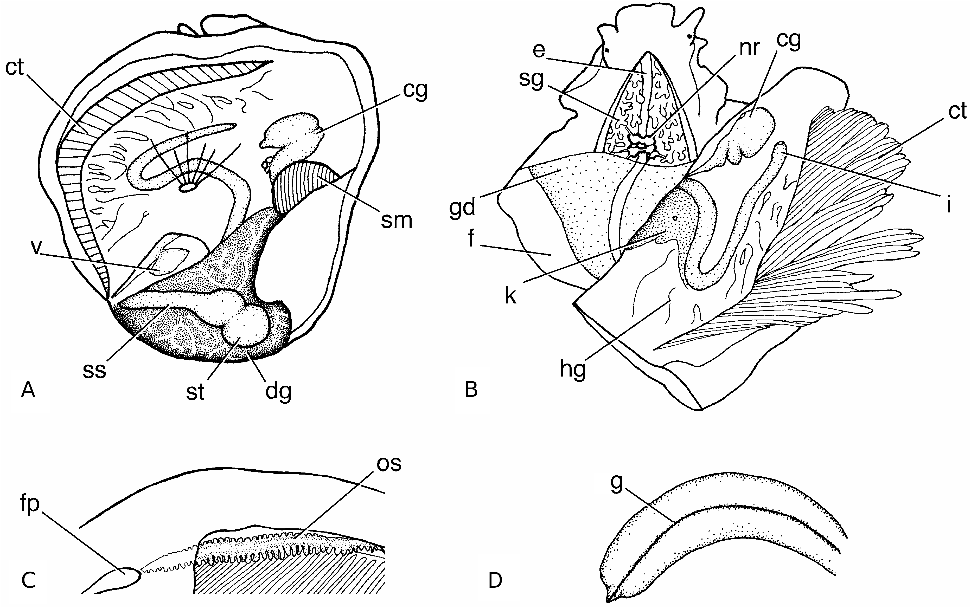
Anatomy: the overall anatomy of Bostrycapulus spp. is similar to that of other calyptraeids ( Kleinsteuber, 1913; Werner & Grell, 1950; B. aculeatus s.l. described by Simone (2002) ( Fig. 9 View Figure 9 )). The foot is round with a rectangular propodium and extends slightly more than half the length of the shell. There are no mesopodial flaps. The corners of the propodium are not extended laterally and cannot extend free of the rest of the foot. The neck is dorsoventrally flattened with lappets along each side and with a narrow food groove travelling forward to the tentacle on the right side. Tentacles are stubby, with a simple black eye on the lateral side about a third of the way to the distal end. The lips are equal in size with small, thin jaws embedded in the dorsal side. Tentacles narrow suddenly, immediately distal to the eye. The food pouch at the anterior medial edge of the mantle is surrounded by thick flaps. The tissue connection between the mantle margin and the foot extends anterior to the foot and to the shelf on the animal’s left side. The osphradium is a dark, tightly packed strip of bipectinate filaments at the base of the gill filaments. The anterior filaments are smaller than the posterior filaments. The osphradium extends from the food pouch to slightly within the mantle cavity. The long, narrow gill filaments are somewhat thickened at their base. The salivary glands are huge, filling the entire neck and extending from the buccal mass, externally past the nerve ring, to the anterior margin of the visceral mass. They are intricately branched along their entire length.
When removed from the shell the distal third of the viscera curves to the animal’s right. The tapered mantle cavity and gills extend about two thirds of the way to the tip of the viscera on the dorsal left side. The crescent-shaped shell muscle extends dorsally from the foot to the shell roof on the right side. A small, dorsal attachment muscle runs from within the dorsal mantle tissue above the intestine to the medial shell roof just anterior to the shelf.
The stomach is visible dorsally to the right of the posterior end of the mantle cavity. The oesophagus runs ventrally in the viscera and enters the stomach posteroventrally. The short style sac runs laterally from the stomach to the left margin of the visceral mass in the dorsal viscera posterior to the mantle cavity. The distal end of the style sac narrows to connect with the intestine, which runs directly to the right side in the ventral visceral mass. The distal loop of the intestine is visible in the dorsal wall of the mantle cavity. This arrangement of the digestive system with respect to the mantle cavity is distinct from the arrangement in Crepidula , where the mantle cavity extends to the end of the visceral mass and the style sac is ventral to the mantle cavity. The brown digestive gland surrounds the stomach and extends to the end of the visceral mass. In fresh and ethanol-preserved material a network of thick white vessels running through the digestive gland is clearly visible. These vessels are not visible in formalin-fixed material.
The heart and kidney are similar to Crepidula species. The heart and pericardial cavity are visible in the dorsal side of the viscera. The pericardial cavity is at an angle to the anterio-posterior axis and extends along the posterior margin of the mantle cavity. In Crepidula species the pericardial cavity is orientated anterior-posteriorly. The hollow kidney is located in the roof of the mantle cavity anterior to the pericardial cavity and posterior to the distal loop of the intestine. The nephrostome opens into the mantle cavity midway between the pericardial cavity and the distal loop of the intestine.
The cream or yellow gonad is somewhat external to the digestive gland and covers almost the entire ventral side of the visceral mass in females and the anterior ventral side in males. The seminal vesicle is a convoluted, narrow tube in the right anterior dorsal margin of the viscera below the mantle cavity and opens into the open-grooved vas deferens. The vas deferens runs to the base of the penis where an open sperm groove runs medially on the ventral side to its distal end. The thick flattened penis ends bluntly with a very small papilla. The penis is usually considerably longer than the tentacles and often exceeds the animal’s body length in small males. In females the visceral oviduct and gonopericardial duct join at the right anterior dorsal margin of the visceral mass where the albumen gland extends up into the roof of the mantle cavity. Several seminal receptacles connect to the albumen gland. Distal to the seminal receptacles, the two lobes of the capsule gland converge and open directly into the mantle cavity through the genital pore. The female genital papilla is absent. All species described here show evidence of protandry.
The nerve ring is located at the posterior margin of the neck just anterior to the visceral mass and completely embedded in the salivary glands. The nerve ring is the same as in C. fornicata ( Werner & Grell, 1950) . A pair of buccal ganglia are located against the dorsal medial margin of the buccal mass.
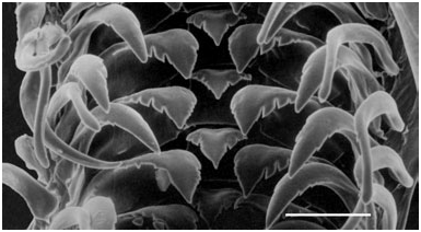
Radula : the taenoioglossate radula ( Fig. 10 View Figure 10 ) is similar to that of other calyptraeids. In Crepidula the major cusps are straight-sided (e.g. Collin, 2000a), producing a dagger-shaped or triangular cusps. In Bostrycapulus the sides of the major cusps on the rachidian and lateral teeth are sinuous. The minor cusps on all teeth are more appressed to the body of the tooth than in other species. The number of denticles on each tooth varies significantly among rows within an individual and among individuals ( Table 3).
Development: the transparent, thin-walled egg capsules of Bostrycapulus species are typical of all calyptraeids. The stalks are wide, flattened ribbons and not thread-like as in some species. The female broods the capsules between the neck and substrate and propodium until hatching. Differences in development are diagnostic among species.
There are currently eight recognized species in Bostrycapulus (see Table 4 for summary).
No known copyright restrictions apply. See Agosti, D., Egloff, W., 2009. Taxonomic information exchange and copyright: the Plazi approach. BMC Research Notes 2009, 2:53 for further explanation.