
Dinosauria Owen, 1842
|
publication ID |
https://doi.org/ 10.11606/1807-0205/2021.61.75 |
|
persistent identifier |
https://treatment.plazi.org/id/61008826-892B-6F12-FE9C-FA08FA8BFC05 |
|
treatment provided by |
Felipe |
|
scientific name |
Dinosauria Owen, 1842 |
| status |
|
Systematic Paleontology Dinosauria Owen, 1842
Saurischia Seeley, 1888 Sauropoda Marsh, 1878 Titanosauriformes Salgado, Coria & Calvo, 1997 Somphospondyli Wilson & Sereno, 1998 Titanosauria Bonaparte & Coria, 1993 Lithostrotia Upchurch, Barrett, & Dodson, 2004 Lithostrotia indet. ( Fig. 3 View Figure 3 )
Description
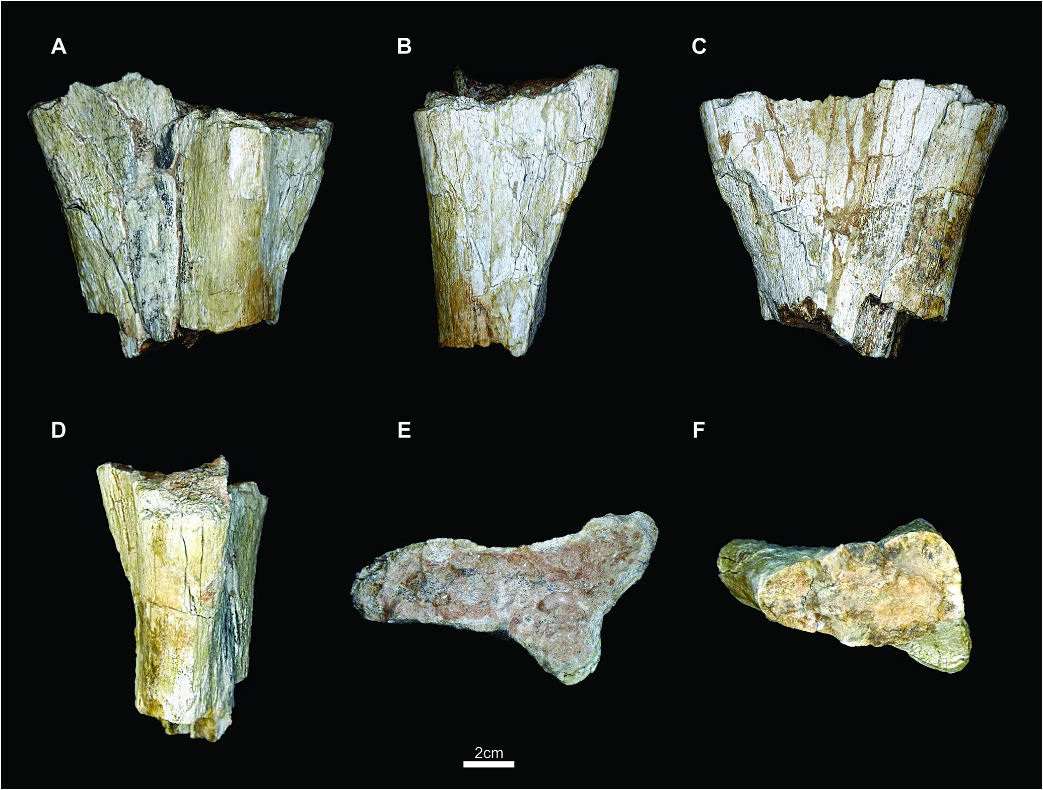
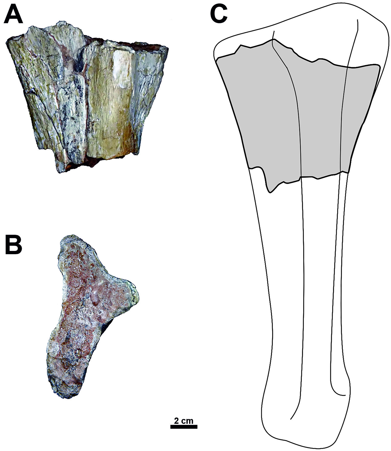
Paleo-UFG/V-0039 is recognized as a proximal fragment of the right ulna, based on its morphology. The bone is thin in its central portion and becomes robust proximally. The distalmost part of the fossil is slightly craniocaudally expanded. In a proximal view, the ulna is triradiate and ‘V’-shaped, a feature observed in other titanosauriforms (Upchurch et al., 2015). The irregular fractures in the most distal portion indicate a post-dia- genetic feature, a taphonomic signature less common for the Marilia Formation titanosaur bones (e.g., Bandeira et al., 2018).
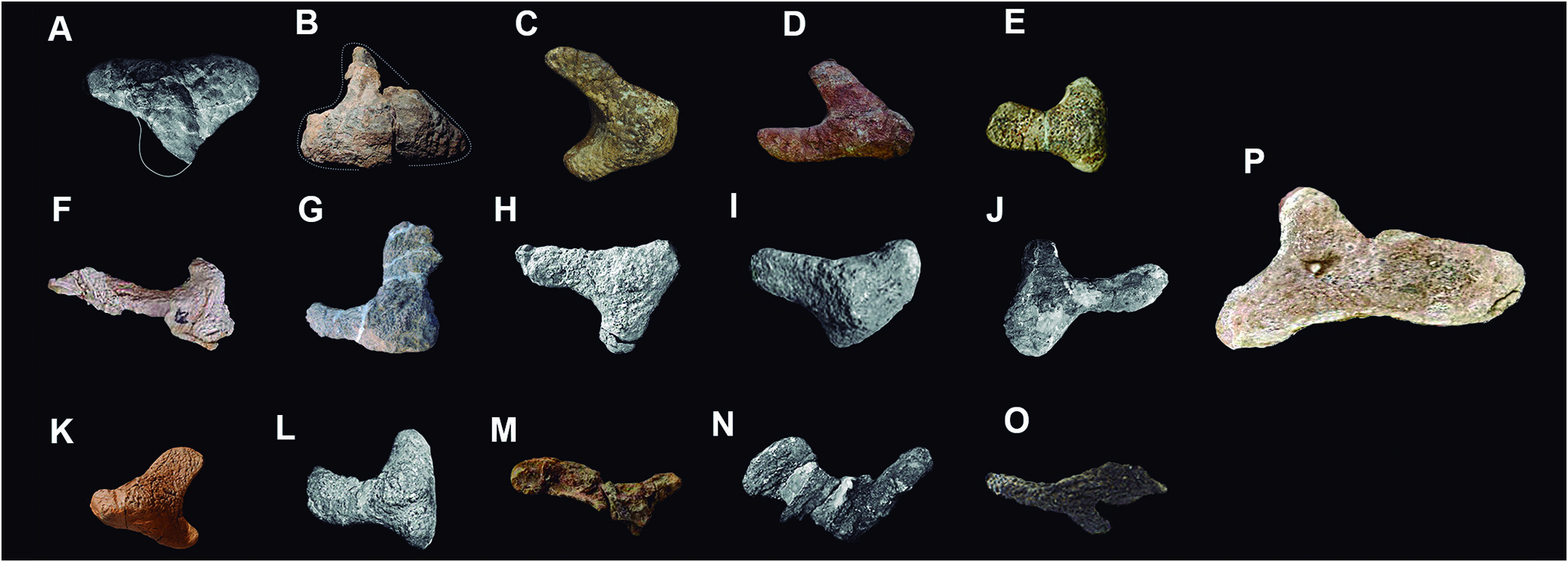
Despite incomplete, this morphology indicates that the bone was genuinely wider at the proximal end and narrowed towards its distal end. The craniomedial and craniolateral processes are prominent ( Fig. 4 View Figure 4 ); but, as in other sauropods, the caudal process is much more developed and larger than the former (e.g., Poropat et al., 2015; Upchurch et al., 2015; Ullmann & Lacovara, 2016; González-Riga et al., 2019).
Although this area is not completely preserved, the craniomedial process also is marked by the concave articular surface of the humerus ( Fig. 5 View Figure 5 ), while the craniolateral process inclines ventrally, distancing itself from the olecranon, in lateral view ( Fig. 3 View Figure 3 ). The caudomedial face exhibits a shallow medial concavity (for insertion of the flexor carpi ulnaris muscle). The ulna shaft is not preserved, but the cross section of the broken distal part of the bone shows that the shaft was subtriangular in this region, due to a caudodistally descending longitudinal crest of the olecranon. The proximal portion of the ascending crest of the olecranon curves slightly medially. In addition, a longitudinal relief ridge defines the medial border of the radial articular face in cranial view.
No known copyright restrictions apply. See Agosti, D., Egloff, W., 2009. Taxonomic information exchange and copyright: the Plazi approach. BMC Research Notes 2009, 2:53 for further explanation.