
Microtendipes umbrosus Freeman, 1955
|
publication ID |
https://doi.org/ 10.11646/zootaxa.4320.3.8 |
|
publication LSID |
lsid:zoobank.org:pub:5C046843-3E95-4D75-A891-50559A12C05E |
|
DOI |
https://doi.org/10.5281/zenodo.6028279 |
|
persistent identifier |
https://treatment.plazi.org/id/6107879D-C440-FD38-E6F7-FB1A763A9185 |
|
treatment provided by |
Plazi |
|
scientific name |
Microtendipes umbrosus Freeman |
| status |
|
Microtendipes umbrosus Freeman View in CoL
( Figures 1 View FIGURE 1 , 5A View FIGURE 5 )
Microtendipes umbrosus Freeman, 1955:32 View in CoL ; Freeman 1961: 720. Microtendipes tamaogouti Sasa, 1983: 7 View in CoL . Syn. nov.
Microtendipes shounagasaki Sasa, 1989a: 30 . Syn. nov.
Microtendipes kamoprimus Sasa, 1989b: 62 . Syn. nov.
Microtendipes amamihosoides Sasa, 1990: 116 View in CoL . Syn. nov.
Microtendipes hibaraquintus Sasa, 1993: 75 . Syn. nov.
Microtendipes tokarafegeus Sasa & Suzuki, 1995: 263 View in CoL . Syn. nov. Microtendipes simantofegeus Sasa, Suzuki & Sakai, 1998: 53 View in CoL . Syn. nov. Microtendipes simantogeheus Sasa, Suzuki & Sakai, 1998: 54 View in CoL . Syn. nov. Microtendipes tusimabeceus Sasa & Suzuki, 1999: 4 View in CoL . Syn. nov. Microtendipes tusimacedeus Sasa & Suzuki, 1999: 5 View in CoL . Syn. nov. Microtendipes sakhalinensis Zorina, 2001: 35 View in CoL . Syn. nov.
Material examined. Syntypes of Microtendipes tamaogouti , 10 M, 8 F (NSMT-I-Dip 56 69–5680), labelled, “ No. 67: 51–62”, respectively, JAPAN: Tokyo, Okutama, Tama River , 12.vi.1981 . Holotype of Microtendipes shounagasaki , M (NSMT-I-Dip 4639), labelled, “ No. 152: 47”, JAPAN: Toyama, Shou River , 25.viii.1988 . Holotype of Microtendipes kamoprimus , M (NSMT-I-Dip 4 660), labelled, “ No. 163: 1”, JAPAN: Kyoto, Kamo River , 12.x.1988 . Holotype of Microtendipes amamihosoides , M (NSMT-I-Dip 4686), labelled, “ No. 178: 96”, JAPAN: Kagoshima, Amami Island, Yakkachi River , 18.iii.1989 (emerged 10.iv.1989) . Holotype of Microtendipes hibaraquintus , M (NSMT-I-Dip 4843), labelled, “ No. 223: 36”, JAPAN: Fukushima, Kitashiobara, Lake Hibara , 6.viii.1991 . Holotype of Microtendipes tokarafegeus , M (NSMT-I-Dip 5011), labelled, “ No. 290: 15”, JAPAN: Kagoshima, Nakanoshima Island , 20.v.1994 . Holotype of Microtendipes simantofegeus , M (NSMT-I-Dip 5199), labelled, “ No. 358: 47”, JAPAN : Kochi, Nakamura, Shimanto River , 26.iv.1998 . Holotype of Microtendipes simantogeheus , M (NSMT-I-Dip 5202), labelled, “ No. 358: 53”, JAPAN : Kochi, Nakamura, Shimanto River , 26.iv.1998 . Holotype of Microtendipes tusimabeceus , M (NSMT-I-Dip 5140), labelled, “ No. 353: 69”, JAPAN: Nagasaki, Tsushima Island, Uchiyama , Izuhara , 24.iii.1998 ; Holotype of Microtendipes tusimacedeus , M (NSMT- I-Dip 5139), labelled, “ No. 353: 68”, JAPAN: Nagasaki, Tsushima Island, Uchiyama , Izuhara , 24.iii.1998 . Paratype of Microtendipes ginzanefeus , M ( NSMT), labelled, “ No. 403: 46”, JAPAN: Hokkaido, Mt. Ginzan , 2.ix.2000 . Non-types. Le / Pe /M ( SUM), JAPAN: Fukushima, Hirono, Asami River , 15.viii.2001 (emerged 30.viii.2001) ; M ( SUM), Fukushima, Iwaki, Yaguki , 15.vii.2012 (emerged 30.vii.2012) ; M, L ( SUM), Tochigi, Ichikai, Miage , 1.ix.1989 (emerged 10.ix.1989) ; Pe /M ( SUM), Kanagawa, Kiyokawa, Miyagase , 23.ii.1994 (emerged 27.iii.1994) ; Le/Pe/M (SUM), as previous except 26.v.1996 (emerged 26.vi.1996); 2 M ( SUM), Shizuoka, Sunto, Shimizu-cho, Kakita River , 3.iii.1985 ; 2 Le / Pe /F, 2 L ( SUM), Shizuoka, Shimizu, Yanbara River , 3.iii.1985 (emerged 10.v.1985) ; Pe/M (SUM), as previous except 3.iv.1985 (emerged 8.iv.1985); 3 M, Le/Pe/M, 3 F, 3 Le/F, 13 Pe, 4Le, 13L (SUM), as previous except 16.vi.1985 (emerged 19–30.vi.1985); Pe /M ( SUM), Shizuoka, Shimizu, Ihara River , 12.ix.1988 (emerged 20.ix.1988) ; Pe /M ( SUM), Shizuoka, Kujiragaike , 19.xi.1987 (emerged 25.xi.1987) ; 2 L ( SUM), Shizuoka, Kakegawa, Osuka-cho , 14.i.1989 ; 2 L (SUM), as previous except 27.i.1989; Pe/M, Pe/F (SUM), as previous except 11.iii.1989 (emerged 16.iii.1989); 4 Le / Pe /M ( EJNU), CHINA: Guangdong, Guangzhou, Bage villa, 20.iii.2015 (emerged 1.v.2015) ; 4 M (EJNU), as previous except 29.xi.2015; Pe /M ( EJNU), Hainan, Wuzhi Mt. , 3.xii.2011 ; 3 M, 4 Pe ( EJNU), Yunnan, Honghe, Jinping County, Maandi Town , 8.vi.2017 ; 2 M, 4 Pe ( EJNU), Yunnan, Pu’er , Ximeng County, 20.i.2015 ; 2 M ( EJNU), Fujian, Mt. Wuyi , 9.viii.2014 ; Pe, M ( EJNU), Zhejiang, Xiangshan County, 15.vi.2017 .
Description. Male (n = 15). Total length 3.7–5.6, 4.6 mm.
Coloration. Thorax brown with 3 scutal vittae, anepisternum II, preepisternum and postnotum darkened. Abdomen green with dark segments VII–IX. Wing ( Figure 1A View FIGURE 1 ) with faint cloud around RM and FCu or more extensively, occasionally on apical half. Foreleg yellow with dark markings; femur dark brown on middle and apex; tibia variable in extent of brown areas, darkened only on both ends or along its entire length; occasionally ta1 broadly darkened basally. Mid and hind legs yellow with darkened knees.
Head. Temporals 14–24, 20 (13), uniserial, partially biserial. Frontal tubercles absent. AR 1.7–2.1, 2.0. Clypeus trapezoid with 27–42, 32 setae. Lengths of palpomeres 1–5 (µm): 60–75, 67 (13); 55–70, 64 (13); 260– 325, 291 (13); 275–335, 298 (13); 330–460, 388 (13), respectively. Pm4/Pm3 0.97–1.1, 1.0 (13); Pm5/Pm4 1.1–1.4, 1.3 (13). Pm3 apically with 4–6, 5 sensilla clavata, longest 18–23, 20 µm long.
Thorax. Lateral antepronotals 3–8, 5; acrostichals 5–12, 8, concentrated at apex of scutum; dorsocentrals 12– 18, 15, uniserial, occasionally biserial anteriorly; prealars 3–8, 4, uniserial; scutellars 24–35, 28.
Wing. Length 2.5–3.6, 3.0 (11) mm. VR 1.1–1.2, 1.1 (11). Vein R2+3 ending close to apex of R1. R, R1 and R4+5 with 22–35, 26 (12); 18–28, 23 (12); 35–53, 42 (12) setae, respectively. Squama with 11–20, 15 setae.
Legs. Forefemur with 2 rows of proximally directed setae on outer side. Foretibia apically truncate, unarmed. Mid and hind tibiae each with 2 combs and 1 recurved spur. Mid ta1 with 5–10, 7 (13) sensilla chaetica, distalmost located 0.47–0.54, 0.51 (13) from base. Lengths and proportions of leg segments as in Table 1.
Hypopygium ( Figure 1B View FIGURE 1 ). Anal tergite with anterior bands; median setae 1–5, 2, arising from pale pits on each end of tergal bands; anal point parallel-sided, with truncate apex. Superior volsella sickle-shaped, rounded apically, with one basal and 3–7, 5 dorsal setae; occasionally with sparse microtrichia basally. Median volsella poorly developed, consisting of small tubercles with 1–4, 2 setae, occasionally absent. Gonostylus 118–165, 138 (11) µm long, 3.2–3.6, 3.5 (11) times as long as broad at middle, apically with short and stout setae.
Female (n = 9). Total length 2.5–4.2, 3.3 mm.
Coloration. Similar to male.
Head. Temporals 18–24, 21. Antenna with 5 flagellomeres; terminal flagellomere 160–200, 177 (8) µm long, shorter than preceding 2 flagellomeres together; AR 0.34–0.41, 0.37 (8). Clypeus with 32–45, 40 setae. Lengths of palpomeres 1–5 (µm): 50–75, 57; 60–70, 62; 280–310, 293; 290–340, 308; 350–460, 390, respectively. Pm4/Pm3 1.0–1.1, 1.0; Pm5/Pm4 1.2–1.4, 1.3. Pm3 with 5–6, 5 sensilla clavata, longest 15–23, 19 µm long.
Thorax. Lateral antepronotals 1–5, 3; acrostichals 6–12, 9; dorsocentrals 17–25, 21; prealars 3–6, 4; scutellars 25–37, 31.
Wing. Length 2.3–3.5, 2.7 mm. VR 1.2–1.3, 1.2 (8). Veins R, R1 and R4+5 with 22–37, 29; 23–41, 30; and 55– 89, 71 setae, respectively. Squama with 11–22, 15 setae.
fe ti ta1 ta2 ta3 ta4 ta5 LR BR Male P1 1167–1523 1167–1599 1548–2005 711–990 660–914 558–736 279–406 1.1–1.4 2.1–2.6
1322 1343 1753 825 753 632 311 1.3 2.4 P2 1269–1700 1091–1523 711–990 355–533 279–406 178–254 102–152 0.62–0.67 3.5–4.2
1453 1267 831 431 324 209 123 0.66 3.9 P3 1421–1878 1218–1650 939–1294 558–787 406–584 228–355 127–178 0.75–0.79 3.5–5.0
1626 1400 1081 652 484 290 142 0.77 4.5
Female P1 1244–1523 1167–1472 1675–2030 787–964 711–888 584–736 279–330 1.3–1.4
1376 1300 1839 854 792 669 305 1.4
P2 1269–1599 1142–1447 685–888 381–508 279–381 178–228 102–152 0.60–0.62
1435 1300 789 427 319 206 121 0.61
P3 1421–1777 1244–1624 939–1218 584–736 431–558 254–330 127–152 0.74–0.79
1616 1416 1085 (8) 650 (8) 492 (8) 282 (8) 133 (8) 0.76 (8)
Legs. Mid ta1 with 16–26, 21 sensilla chaetica, distalmost located 0.52–0.60, 0.55 from base. Lengths and proportions of leg segments as in Table 1.
Genitalia ( Figure 1C View FIGURE 1 ). Sternite VIII with 16–26, 20 (6) setae on each side. Gonocoxapodeme strong. Gonapophysis VIII broad, rounded caudally. Gonocoxite IX with 1–3, 2 (7) setae. Lateral plate of segment X triangular without setae. Postgenital plate triangular. Notum 150–165, 158 (4) µm long, 2.0–2.6, 2.3 (4) times as long as ramus. Labium with microtrichia. Seminal capsule oval, 65–75, 72 µm long, 1.1 times as long as broad, and 0.45–0.48, 0.46 (4) times as long as notum, with cylindrical neck.
Pupa (n = 23). Total length 4.7–6.7, 5.4 mm.
Coloration. Exuviae pale brown with somewhat infuscated thorax and abdomen.
Cephalothorax. Cephalic tubercles ( Figure 1D View FIGURE 1 ) conical, 100–160, 127 (22) µm long, 1.3–1.5, 1.4 (22) times as long as basal width in mounted exuviae. Frontal setae absent. Dorsum of thorax strongly pebbled along median suture.
Abdomen ( Figure 1E View FIGURE 1 ). Tergites I and VII without spinulation; II–V each with more or less extensive, triangular spinule patch; VI with posterior transverse spinule band, usually interrupted medially; VIII with central spinule patch and posterior transverse spinule band; IX with somewhat strong central spinule patch, and occasionally very weak anterolateral spinule patches. Tergites II–VI each with anterior transverse band of spines. Tergite II with row of 45–89, 62 (21) caudal hooklets; its row 0.40–0.51, 0.45 (21) times as long as tergal width. Conjunctives III/IV and IV/V each with spinule band. Segment IV with vortex. Segment V with 3 Lt-setae on each side; VI–VII each with 4 Lt-setae, occasionally VI with 3 Lt-setae; VIII with 5 Lt-setae. Anal comb ( Figure 1F View FIGURE 1 ) on segment VIII with 2–4, 3 (21) teeth becoming smaller anteriorly. Anal lobe 275–400, 326 (21) µm long, 1.5–2.0, 1.7 (21) times as long as broad, with 47–77, 56 (22) lateral taeniae; with dorsal seta simple, located 0.19–0.29, 0.24 (20) from apex. Male genital sac 0.96–1.1, 1.1 (12) times as long as anal lobe.
Fourth instar larva (n = 31). Body length 7.2–10.6, 8.7 (7) mm.
Coloration. Head generally yellowish, with dark brown postoccipital margin, in alcoholic specimen.
Head. Length 434–545, 480 (12) µm long; cephalic index 0.72–0.76, 0.73 (11). Antenna ( Figure 1G View FIGURE 1 ) 0.33– 0.39, 0.36 (11) times as long as head capsule, with 6 segments. Lengths of first to sixth segments (µm): 80–110, 93 (19); 20–28, 23 (19); 23–33, 26 (19); 14–28, 20 (19); 10–18, 15 (19); 6–10, 8 (19). AR 0.9–1.0, 1.0 (19). First segment with ring organ located 0.24–0.31, 0.28 (19) from base; blade 105–155, 121 (10) µm long, extending far beyond apex of terminal segment; accessary blade very small, 8–10, 9 (4) µm long. Each of second and third segments laterally with Lauterborn organ 18–25, 23 (18) µm long. Third segment laterally with style 8–13, 10 (17) µm long. Labral lamella with 11–18, 15 (23) teeth. Premandible ( Figure 1H View FIGURE 1 ) 88–130, 102 (27) µm long, with 3 teeth. Pecten epipharyngis with 3 equal-sized teeth ( Figure 1I View FIGURE 1 ). Mandible ( Figure 1J View FIGURE 1 ) 155–215, 175 (19) µm long with seta subdentalis 40–65, 50 (16) µm long, curved apically, reaching distalmost inner tooth; seta interna 4- branched. Mentum ( Figure 1K View FIGURE 1 ) 143–190, 157 (19) µm wide; median tooth bifid, pale, 30–45, 35 (19) µm wide, with very small central tooth. Ventromental plate 70–100, 82 (18) µm long, 120–160, 137 (18) µm wide, with 28– 35, 30 (20) striae; distance between both plates 0.47–0.51, 0.50 (19) times as broad as width of mentum. Postmentum 175–233, 193 (28) µm long.
Body. With 8 anal setae.
Remarks. Microtendipes umbrosus Freeman is distributed in Africa and Australia ( Freeman & Cranston 1980, Cranston & Martin 1989). In Australian populations, the pupa was drawn with three Lt-setae on abdominal segment VI ( Cranston 2000) but across a series of specimens this number varies (3 or 4) including between one side and the other (P.S. Cranston, Canberra, Australia, pers. comm.). Of 23 Japanese specimens examined here, three specimens (13 %) have three Lt-setae on one side of the segment VI and four Lt-setae on the other, 20 (87 %) possessing four Lt-setae on each side of the segment. The features of the Japanese specimens are consistent with Freeman (1955, 1958, 1961) for the males and females and with those of Australian pupal and larval exuviae associated with their adults (P.S. Cranston, Canberra, Australia, pers. comm.) for the pupae and larvae.
After re-examinations of the syntype males and females of M. tamaogouti Sasa and the holotype males of M. shounagasaki Sasa , M. kamoprimus Sasa , M. amamihosoides Sasa , M. hibaraquintus Sasa , M. tokarafegeus Sasa & Suzuki , M. simantofegeus Sasa, Suzuki & Sakai , M. simantogeheus Sasa, Suzuki & Sakai , M. tusimabeceus Sasa & Suzuki and M. tusimacedeus Sasa & Suzuki , it was evident that all features of M. umbrosus are common to these taxa with little difference between them, and thus, we regard these as junior synonyms of M. umbrosus .
Judging from the original morphological description of the male, the Russian species M. sakhalinensis Zorina, 2001 also may be conspecific with M. umbrosus .
The male of M. umbrosus resembles that of Palaearctic M. pedellus ( De Geer, 1776) in the coloration of thorax and legs, and also the general appearance of the hypopygium, but differs in the wing with a faint cloud (at least around the vein FCu) and the poorly developed median volsella, bearing 0–4 setae on small tubercles. In M. pedellus , the male is characterized by the wing without any marking, and the median volsella with a bundle of setae on a well-developed tubercle ( Langton & Pinder 2007: 110, fig. 219 C).
Microtendipes umbrosus View in CoL is most common in Japanese Microtendipes View in CoL . However, not only wide variation in the leg and wing markings but also the inadequate justifications for differentiation made by Sasa and his co-workers have led to much confusion. In the description of M. tamaogouti, Sasa (1983: 7) View in CoL wrote, “Dorsal appendage with a slightly expanded base bearing 2 or 3 long inner setae, and a finger-like process with rounded apex and bearing 4 setae in the middle on the dorsal surface.” He failed to distinguish the setae of the median volsella from those of the superior volsella.
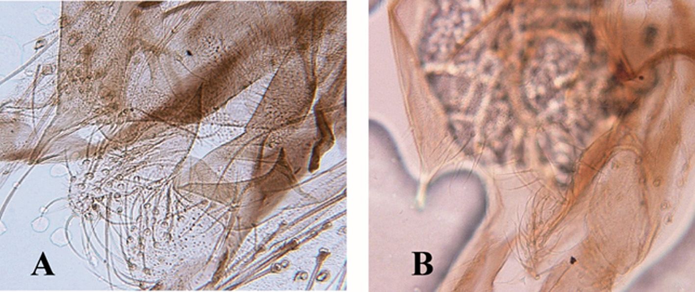
In describing M. tusimacedeus, Sasa & Suzuki (1999: 5) View in CoL wrote, ”Dorsal appendage wide and sickle-shaped”, and drew a curiously short superior volsella (p. 43, fig. 3g). Re-examination of the holotype proved that they overlooked the apical portion of the volsella folded by the mounting procedure ( Figure 5A View FIGURE 5 ). Further, in the description of M. simantofegeus, Sasa et al. (1998: 53) View in CoL even miscalculated the value of male antennal ratio. The correct value is 1.6, not 0.97.
Microtendipes ginzanefeus Sasa & Suzuki, 2001 View in CoL was erected on the basis of two male specimens collected from Hokkaido, northern Japan. Re-examination of the type series showed that the original description is not based on the holotype, but on the paratype, which is a male of M. umbrosus View in CoL .
Two species groups, the M. pedellus View in CoL group and the M. rydalensis View in CoL group, based on the pupal and larval morphology are recognized currently in this genus ( Pinder & Reiss 1983: 324 for the larva, 1986: 334 for the pupa). Microtendipes umbrosus View in CoL belongs to the M. pedellus View in CoL group, because the pupa has long transverse bands of anterior spines on the abdominal tergites II–VI, a central spinule patch on the anal tergite, and five Lt-setae on the abdominal segment VIII, and the larva possesses a bifid median tooth in the mentum, three equal-sized teeth in the pecten epipharyngis, and three teeth in the premandible.
Recently the first author collected many specimens of M. umbrosus from Zhejiang, Fujian, Guangdong, Hainan and Yunnan Provinces in Oriental China. Collections from Thailand by several collectors from Chiang Mai and Kasertsart University, deposited in the Australian National Insect Collection (ANIC, Canberra, Australia), show that specimens of Microtendipes , predominantly larvae but including pupae and adults, are common in standing and flowing waters. These can be allocated to M. umbrosus in our current understanding, with distribution extending from19°26’N in Chiang Rai Province to 9°18’ N in Phang Nga Province, including provinces of Chiang Mai, Kampaeng Phet, Lamphung, Loei, Pechabun, Prachuap Kiri Khan, Ranong, Sakorn Nakorn and Sra Kaew, at elevations ranging from sea level (‘post-tsunami’ ponds) to 600 m above sea level on Doi Inthanon (Chiang Mai) (P.S. Cranston, pers. comm). In Australia, the species is restricted to the state of Queensland, between 17°01’S to 27°06’S, including the tropical lakes Barrine and Eacham ( Cranston & Dimitriadis 2004). The larvae were very abundant early colonizers of an experimental artificial stream channel fed by water derived from a eutrophic dam in South East Queensland (specimens deposited in ANIC, P.S. Cranston, pers. comm.).
For a morphologically defined but somewhat variable species with such a wide range, we can assume that molecular data will show geographically discrete populations or cryptic species, as with Polypedilum nubifer ( Cranston et al. 2016) . However, sampling across such a wide range is time consuming, impractical and well beyond the scope of this study. Furthermore, the species in the range of its type locality (Africa, Kenya, Nyanza) would need to be sampled, as would specimens from throughout the range including species described as endemic to China but potentially synonyms of other named species.

No known copyright restrictions apply. See Agosti, D., Egloff, W., 2009. Taxonomic information exchange and copyright: the Plazi approach. BMC Research Notes 2009, 2:53 for further explanation.
|
Kingdom |
|
|
Phylum |
|
|
Class |
|
|
Order |
|
|
Family |
|
|
Genus |
Microtendipes umbrosus Freeman
| Niitsuma, Hiromi 2017 |
Microtendipes tokarafegeus
| Zorina 2001: 35 |
| Sasa 1999: 4 |
| Sasa 1999: 5 |
| Sasa 1998: 53 |
| Sasa 1998: 54 |
| Sasa 1995: 263 |
Microtendipes hibaraquintus
| Sasa 1993: 75 |
Microtendipes amamihosoides
| Sasa 1990: 116 |
Microtendipes shounagasaki
| Sasa 1989: 30 |
Microtendipes kamoprimus
| Sasa 1989: 62 |
Microtendipes umbrosus
| Sasa 1983: 7 |
| Freeman 1961: 720 |
| Freeman 1955: 32 |