
Capnogryllacris xujuni Zhang & Bian, 2022
|
publication ID |
https://doi.org/ 10.11646/zootaxa.5190.3.6 |
|
publication LSID |
lsid:zoobank.org:pub:720FA2BB-4C63-40D9-9289-5D284F4106E4 |
|
DOI |
https://doi.org/10.5281/zenodo.7138344 |
|
persistent identifier |
https://treatment.plazi.org/id/61228783-2721-FF8D-BB9A-9866041710EC |
|
treatment provided by |
Plazi |
|
scientific name |
Capnogryllacris xujuni Zhang & Bian |
| status |
sp. nov. |
Capnogryllacris xujuni Zhang & Bian sp. nov.
Oiv烟ëȃ
Figures 1–5 View FIGURE 1 View FIGURE 2 View FIGURE 3 View FIGURE 4 View FIGURE 5
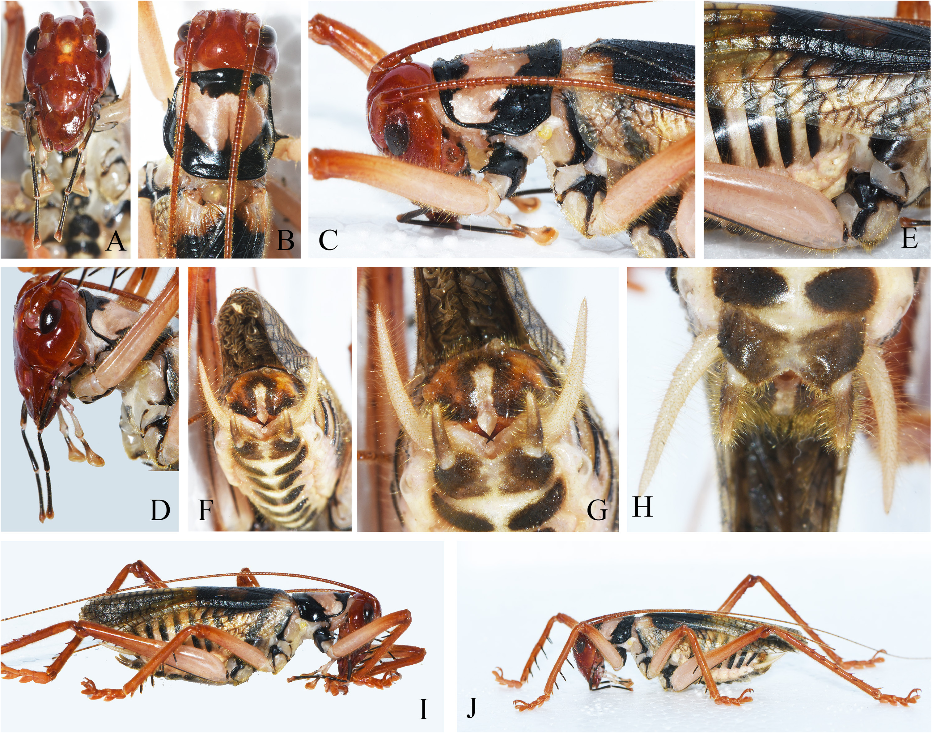
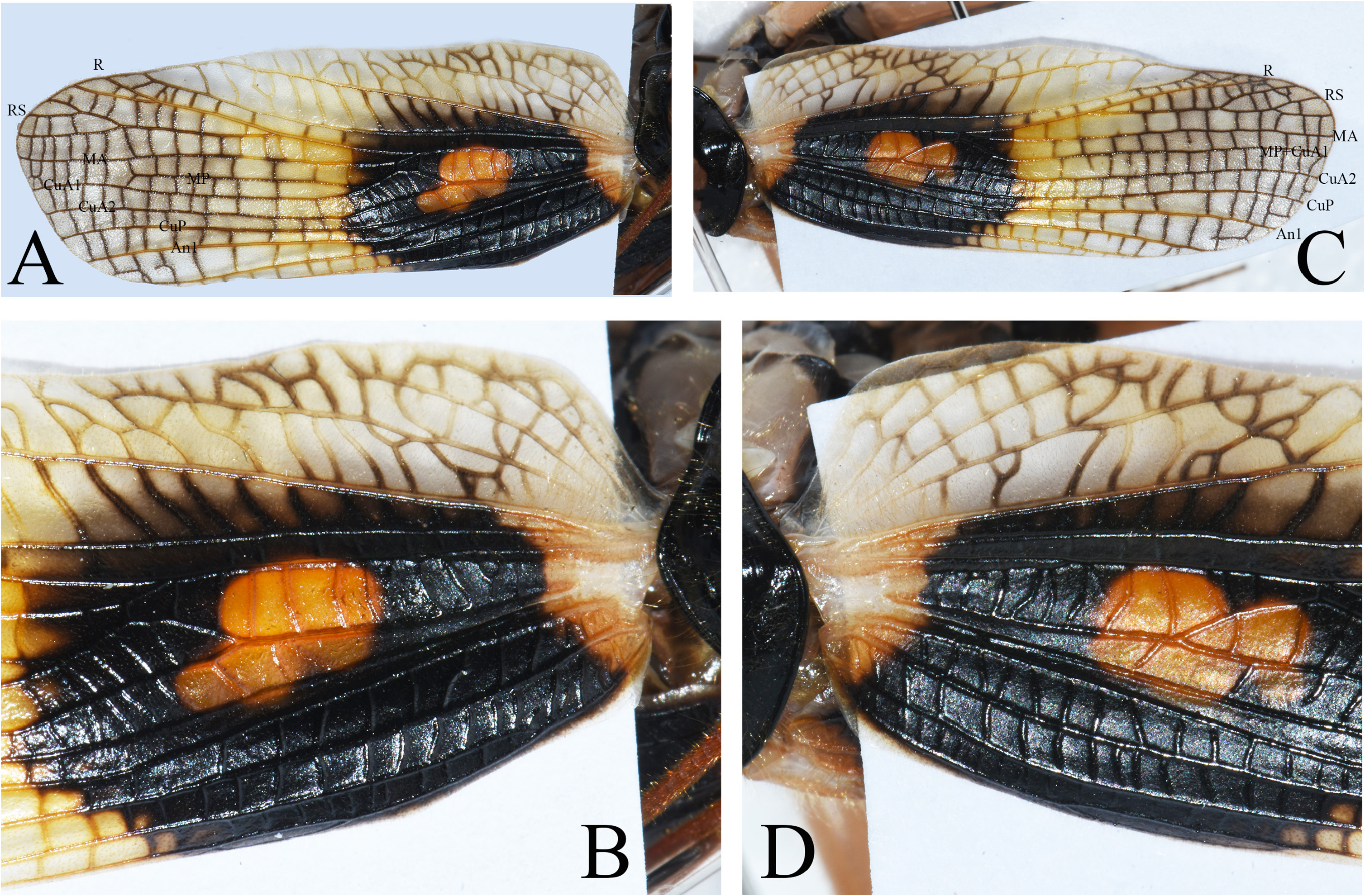
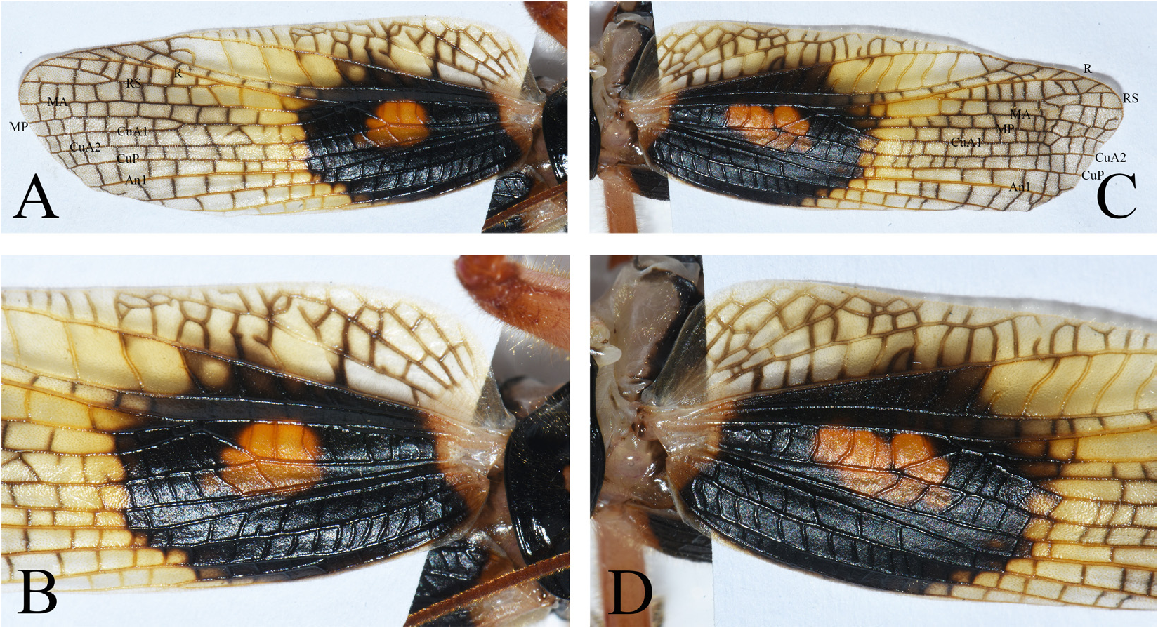
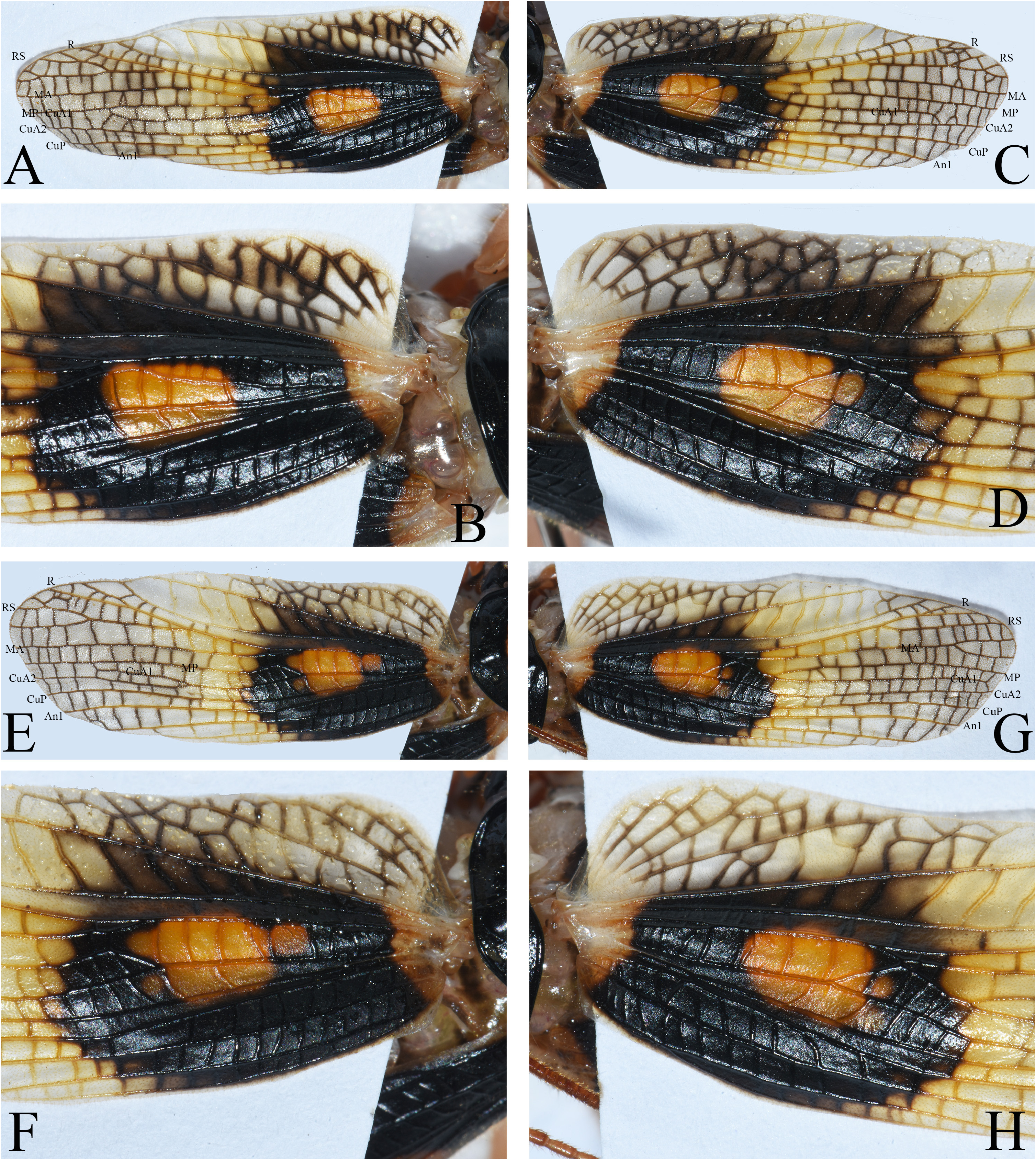
Diagnosis. The new species differs from other species of Capnogryllacris in: head red ( Figs. 1A View FIGURE 1 , 3A View FIGURE 3 ), pronotum with black margins which widened at anterior and posterior margins ( Figs. 1B View FIGURE 1 , 3B View FIGURE 3 ); basal area of tegmina with large black spots surrounding an orange spot in the middle ( Figs. 2 View FIGURE 2 , 4 View FIGURE 4 , 5 View FIGURE 5 ); spines of male ninth abdominal tergite only crossed at subapical area, subgenital plate with a triangular concavity at posterior margin ( Fig. 1H View FIGURE 1 ); female subgenital plate with medial grooves narrower separated from each other at basal area, posterior margin bilobate ( Fig. 3E–G View FIGURE 3 ).
Description. Male. Large species. Face ovoid with sparse punctures ( Fig. 1A View FIGURE 1 ). Fastigium verticis wider than scape. Apical segments of maxillary palpi slightly longer than subapical ones, apices faintly swollen ( Fig. 1A View FIGURE 1 ). Apical segments of labial palpi obviously swollen. Ocelli distinct. Anterior margin of pronotum slightly projected, posterior margin arched; anterior angle of lateral lobes rounded, posterior angle slightly extending downwards ( Fig. 1B View FIGURE 1 ). Humeral sinus indistinct. Fore coxae with 1 spine ( Fig. 1C View FIGURE 1 ). Fore and middle femora unarmed on ventral surfaces; hind femora ventrally with 8–11 internal and 7–8 external spines. Fore and middle tibiae with 5 pairs of ventral spurs, including 1 pair of apical ones; hind tibiae with 5–6 pairs of spines on dorsal surface, apices with 1 pair of dorsal spurs and 2 pairs of ventral spurs. Second and third abdominal tergites with few largely reduced, indistinct stridulatory pegs or not ( Fig. 1E View FIGURE 1 ). Eighth abdominal tergite prolonged. Ninth abdominal tergite globular, with a shallowly furrow in midline, basal area swollen on each side of furrow, at apex with an acute spine, its subapical area crossing each other and directing downward and forward ( Fig. 1F–G View FIGURE 1 ). Subgenital plate wider than long, posterior margin with a triangular concavity ( Fig. 1H View FIGURE 1 ); styli slightly longer than subgenital plate, inserted at apico-lateral angles ( Fig. 1H View FIGURE 1 ).
Female. Subgenital plate nearly trapezoidal; middle area of basal margin with 2 small black grooves ( Fig. 3G View FIGURE 3 ); the lateral margins of basal areas convex, then narrowing, posterior margin bilobate ( Fig. 3E–F View FIGURE 3 ). Ovipositor moderately upcurved, dorsal and ventral margins smooth, apices subacute ( Fig. 3H View FIGURE 3 ).
Wings. Tegmina justly reaching or slightly surpassing the apex of abdomen, but not reaching the apices of hind femora ( Figs. 1I–J View FIGURE 1 , 3I View FIGURE 3 ); hind wings slightly longer than tegmina. Tegminal veins ( Figs. 2 View FIGURE 2 , 4 View FIGURE 4 , 5 View FIGURE 5 ): Radius with RS branching before apical third area of tegmen, both forked near tip; media anterior free from base, then it fuses with radius, after both veins separated again; cubitus anterior at base single branched, after divides into two branches before basal third, the anterior branch makes a curvature and receives a short connection branch from MA, shortly after it divides into MP and CuA1 (sometimes they do not reach the tip of tegmen ( Figs. 2A View FIGURE 2 , 4A View FIGURE 4 , 5C, E View FIGURE 5 )), or not divided instead runs as fused MP+CuA1 till tip ( Figs. 2C View FIGURE 2 , 5A View FIGURE 5 ), the posterior branch (CuA2) undivided. Cubitus posterior undivided, free throughout; with 4 anal veins, the last two with common stem.
Coloration. General color light brown ( Figs. 1I–J View FIGURE 1 , 3I View FIGURE 3 ). Head red, eyes black, ocelli pale yellow ( Fig. 1A View FIGURE 1 , 3A View FIGURE 3 ). Margins of pronotum black which extended at anterior and posterior margins, the posterior black band nearly V-shaped ( Fig. 1B View FIGURE 1 , 3B View FIGURE 3 ). Coxae with black spots, apical areas of femora and tibiae of all legs rufous ( Figs. 1I–J View FIGURE 1 , 3I View FIGURE 3 ). Tegmina yellowish brown, towards anterior and posterior margins lighter; basal area with large black spots surrounding an orange spot in middle; veins brown ( Figs. 2 View FIGURE 2 , 4 View FIGURE 4 , 5 View FIGURE 5 ). Each abdominal tergite with black band on posterior margin; each abdominal sternite with 1 pair of black spots. Apical half of female subgenital plate and ovipositor black ( Fig. 3H View FIGURE 3 ).
Material examined. Holotype: male, Jinuoshan, Jinghong, Yunnan, August , 2022, coll. by Xujun Wang . Paratypes: 1 male and 1 female, the other information as holotype; 2 males and 2 females, Jinuoshan, Jinghong , Yunnan, August 16, 2022, coll. by Bu .
Measurements (mm). Male: BL 32.0–34.5, PL 7.2–7.5, HFL 19.7–21.0, TL 22.5–23.5; Female: BL 32.5–33.6, PL 7.3–8.0, HFL 20.0–20.5, TL 21.5–26.5, OvL 25.5–26.0.
Distribution. Yunnan (Jinghong).
Etymology. Named in honour of Mr. Xujun Wang (ƗOiv) who send us some of the specimens.

No known copyright restrictions apply. See Agosti, D., Egloff, W., 2009. Taxonomic information exchange and copyright: the Plazi approach. BMC Research Notes 2009, 2:53 for further explanation.