
Lodosocoris santhae Salini
|
publication ID |
https://doi.org/ 10.11646/zootaxa.5072.1.6 |
|
publication LSID |
lsid:zoobank.org:pub:1A99BDDE-A379-4137-A4CA-50A8C2DE42C6 |
|
DOI |
https://doi.org/10.5281/zenodo.5729908 |
|
persistent identifier |
https://treatment.plazi.org/id/612CF105-FFD9-C87C-FF12-FD76FE35F766 |
|
treatment provided by |
Plazi |
|
scientific name |
Lodosocoris santhae Salini |
| status |
sp. nov. |
Lodosocoris santhae Salini sp. nov.
LSID: urn:lsid:zoobank.org:act:18EE4C38-0A6C-405E-BDEC-3ACD056A09BA
( Figs. 1–20 View FIGURES 1–2 View FIGURES 3–7 View FIGURES 8–12 View FIGURES 13–17 View FIGURES 18–20 )
Type locality. India: Pasighat , Arunachal Pradesh [28.0619° N, 95.3260° E] GoogleMaps .
Type material. Holotype: ♂ ( NIM), ‘ INDIA: Arunachal Pradesh, Pasighat, Sirukki Waterfall , 04.xi.2017, Amala.U // HOLOTYPUS, LODOSOCORIS SANTHAE sp.nov., det. Salini. S., 2021[p, red label]’. Holotype is pinned, the dissected male genitalia placed in a plastic microvial with glycerol attached to the same pin, all parts intact.
Paratypes (1 ♂ 2 ♀): India: Arunachal Pradesh: 1 ♀, Pasighat, Sirukki Waterfall, 04.xi.2017, Amala. U. ( NIM), dissected female genitalia placed in a plastic microvial with glycerol attached to the same pin and the dissected abdominal terga and sterna glued on card ; 1 ♂ 1 ♀, Pasighat, 19.iii.2017, ex Mango, Mahendiran. G. ( NIM), left hind leg of female specimen glued on card. Each bearing the following identification label : ‘ PARATYPUS / LODOSOCORIS SANTHAE / sp. nov. / det. Salini. S., 2021 [p, yellow label] ’
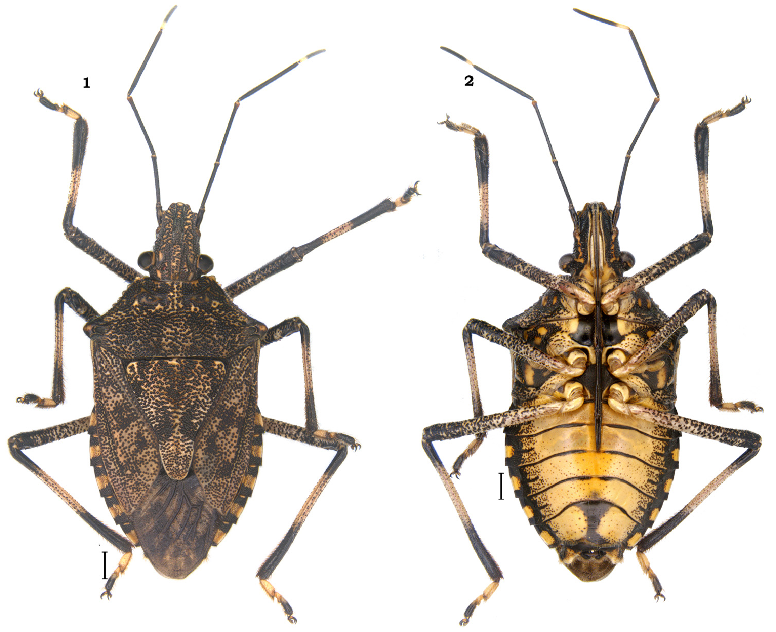
Description. Colouration. Dorsum ( Fig. 1 View FIGURES 1–2 ) including head dark brown to black, mottled with irregular, yellowish markings; lateral margins of head, pronotum (except lateral margins of humeri) and hemelytra, pale yellow. Antennae, black except ventral half of antennal segment I and base of antennal segment V, yellow. Connexivum bright yellow except anterior and posterior 1/3 rd of connexival segments including the short spinous process on posterolateral margins of each connexival segments, black; small, narrow, transverse stripe joining with anterior margin of each connexival segment pale white, sometimes with reddish tinge. Membrane, smoky brown with base, venation and a few markings on the membrane, black.
Ventral surface ( Fig. 2 View FIGURES 1–2 ) medially bright yellow with the head, thoracic sterna and abdominal sterna laterally black except the following: base of head, narrow lateral stripe anteriorly connecting base of antennae, rim of bucculae including the denticle-like process on anterior apex, three moderate sized irregular spots each on pro and mesopleura: two adjacent to respective coxal cavities and one towards lateral margin, outer margins of ostiolar plate, peritreme, a longitudinal spot on evaporatorium, a median large round spot on lateral margins of each abdominal sternites III–VII, bright yellow. Mesosternum medially with a large shining black spot and a moderate sized black spot present medially on metasternum. Labium with segment I yellow, segments II–III black medially and yellow laterally, segment IV, black. Spiracular outline,lateral muscle scars of abdominal sternites III–VII and a small nearly round spot posterolaterad of each spiracle and a median, longitudinal spot on abdominal sternite VII, black. Legs ochraceous (sometimes with elongate reddish streak especially on dorsal surface of femora and tibiae of all legs) except anterior 1/4 th and posterior 1/3 rd of all tibiae, black; tarsi ochraceous except tarsal segments III, black.
Integument and vestiture. Dorsum including head, pronotum, scutellum and hemelytra covered with dense, round punctures, black. Connexivum with dense, fine concolourous punctures. Ventral side of head and thoracic sterna with coarse, dense punctures, black. Abdomen on ventral side with punctures fine, denser towards sublateral margin. Legs with dense, coarse punctures, black.
Body not completely glabrous: lateral margin of pronotum including margin basal to humeri with medium sized silvery white setae. Antennae with short, pale, semi erect setae sparsely distributed. On ventral side, lateral margins of propleura and ventral side of humeri with short, thin silvery white setae. Abdomen on ventral side with short pubescence. Labium and legs with semi erect, thin pale yellow setae. Male genitalia (caudal 1/3rd including the dorsal and ventral rim, the finger-like processes laterally on infoldings of ventral rim) possess moderately elongate, golden setae. Female genitalia (valvifers VIII, IX, abdominal segment X, laterotergite VIII and IX) with moderately elongate, golden setae.
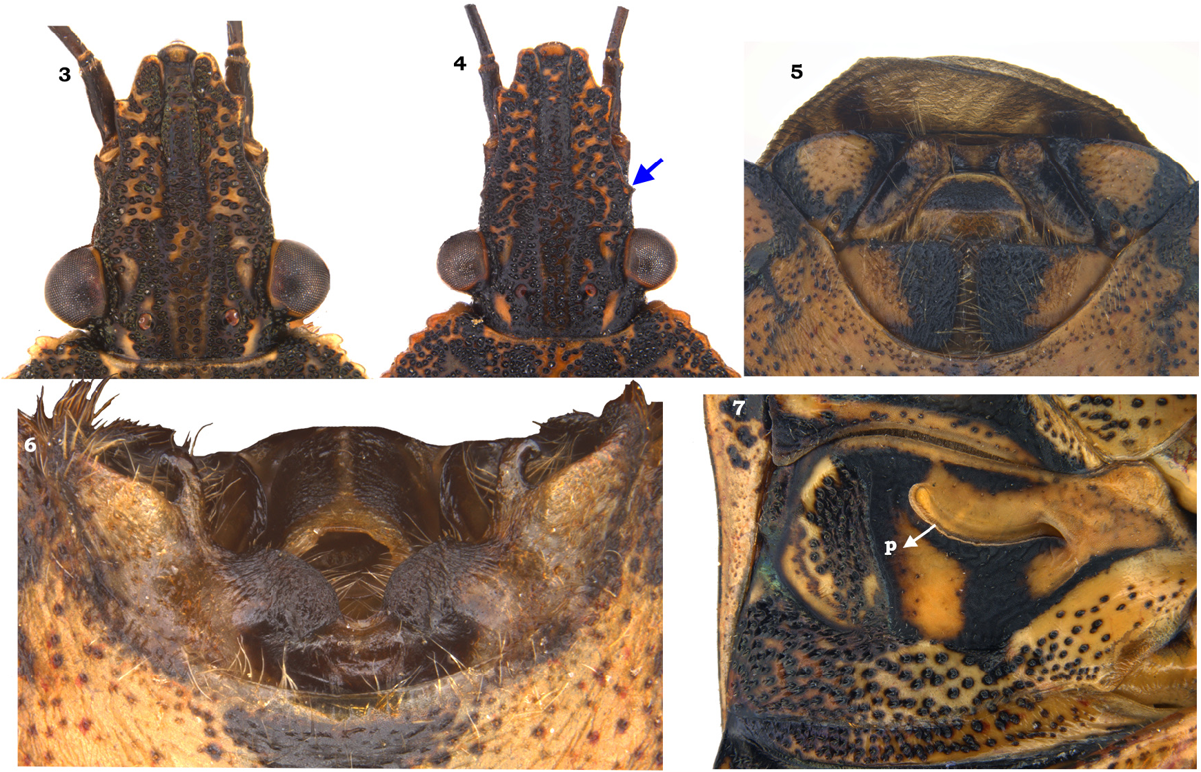
Structure. Head ( Figs. 3–4 View FIGURES 3–7 ) with mandibular plates as long as or shorter than clypeus; lateral margins of mandibular plates with minute, triangular tooth in front of compound eyes ( Fig. 4 View FIGURES 3–7 ), sometimes absent ( Fig. 3 View FIGURES 3–7 ). Antennomeres from shortest to longest: I<IIa<IIb≤IV<III, scape (I) shortest and stoutest; remaining segments cylindrical and slender. Bucculae shorter than labiomere I, anterior apex with short, acute denticle. Labium reaching middle of ventrite IV, sometimes reaching posterior margin of ventrite IV ( Fig. 2 View FIGURES 1–2 ).
Pronotum. Anterolateral angles lacking denticle, rather rectangulate; posterolateral angles indistinct, posterolateral margins of pronotum oblique and posterior margin straight. Disc of pronotum moreorless flat with a narrow transverse impression on anterior half of pronotal disc.
Scutellum. Longer than wide at base, apex rounded. Disc of scutellum more or less flat, central 1/3 rd of scutellar disc laterally with oblique impression.
Hemelytra. Membrane translucent, widely rounded apically, bearing 6–7 prominent and simple veins.
Thoracic pleura and sterna. Evaporatorium developed as subtrapezoidal patch on metapleuron, and a narrow transverse stripe anteriad of metathoracic spiracle on mesopleuron.
Legs. Tarsi with II tarsomere shortest, tarsomere I and III subequal.
Pregenital abdomen nearly as broad as pronotum across humeri. Posterolateral angles of ventite III–VII with short, stout and acute spines.
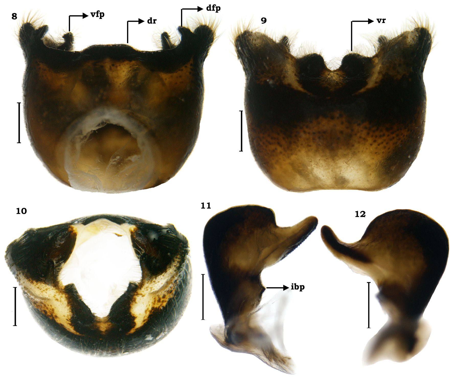
Male genitalia ( Figs. 6 View FIGURES 3–7 , 8–17 View FIGURES 8–12 View FIGURES 13–17 ). Genital capsule ( Figs. 8–10 View FIGURES 8–12 ) quadrangular with posterolateral lobes (=caudal lobes) well developed. Dorsal rim (dr) shallow, widely excavated concave with narrow, median emargination, the anterior margin of median emargination slightly concave; dorsal rim laterally ends in short, roughly subtriangular finger-like projection (dfp) placed at oblique angle to posterolateral lobes, apex of finger-like projection rounded ( Fig. 8 View FIGURES 8–12 ); infoldings of dorsal rim well developed, deeply impressed laterad of dorsal sinus; dorsal sinus semiovate. Ventral rim (vr) shallowly concave with 1+1 tumescence at the centre separated by a U-shaped margin; infoldings of ventral rim laterally with short, stout finger-like process (vfp), placed inner and basal to posterolateral lobes. Paramere ( Figs. 11–12 View FIGURES 8–12 ) with crown broad, semicircular and plate-like. A beak-like, upwardly directed projection, inner laterad to crown. Outer margin of parameral crown arcuately rounded, forming U-shaped concavity with beaklike projection; narrow, elongate sclerotized region with small, scale-like structures, extending from base to apex of beak-like projection. Base of the crown with another short, blunt projection on inner margin (ibp); stem indistinct; apodeme plate-like. Articulatory apparatus (arp) ( Fig. 13 View FIGURES 13–17 ). Basal plate and support bridge complex, fused to form a cup-like structure; capitate processes (cp) modified into small, cone-like structures. Phallus ( Figs. 14–17 View FIGURES 13–17 ). Phallotheca broad and constricted proximally; dorsal walls lightly convex in lateral view; ventral wall convex anterior 2/3 rd and slightly concave towards posterior end (=proximal end), proximal end of phallotheca ventromedially with short, stout tubercle (vt); fully inflated phallus with single pair of conjunctival processes, large, membranous and subdivided into mainly two parts: a basal part (bp) encircling the processes of aedeagus and the apical part (ap) possessing one pair of elongate arm-like lateral processes; a pair of short finger-like processes(flp) at centre of apical conjunctival processes, bend towards ventral side and 1+1 narrow, rod-like, short and apically acuminate processes (aap) originating from basal portion of conjunctival processes in dorsolateral position, resting over phallotheca, extending up to anterior 1/4 th of phallotheca; basal part of conjunctival process on ventral side modified into two subtriangular lobes (sl), joined proximally and separated distally. Processes of aedeagus (pa) well sclerotized, large rod-like paired structures transversely positioned, fused proximally, but separated throughout its length and resting over the shallow inner groove of the subtriangular lobes; aedeagus (ad) elongate tube-like slightly S-shaped with apex directed towards ventral side; phallotreme oblique.
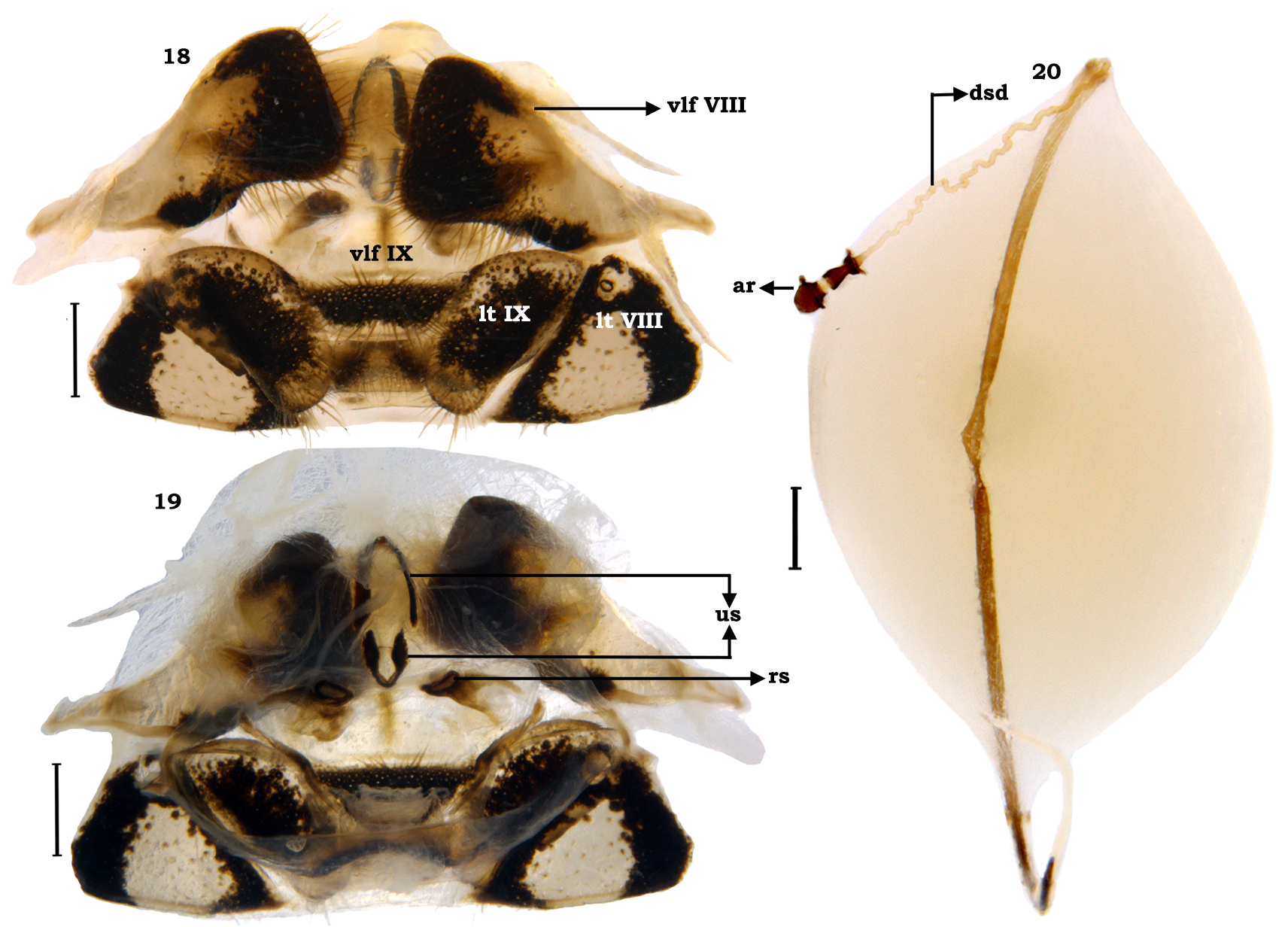
Female genitalia ( Figs. 18–20 View FIGURES 18–20 ). Terminalia. Valvifers VIII (vlf VIII) transverse, broad and roughly subquadrangular, with mesial margins distinctly straight; inner posterolateral angles rounded; posterior margin slightly concave, sublaterally slightly extended into flap-like; valvifers IX (vlf IX) single, transverse, large sclerite, anterior half less sclerotized and fused with ring sclerites ventrally, posterior half roughly trapezoidal with nearly straight posterior margin; laterotergites IX (lt IX) oblique, elongate, roughly oval, most of the central region impressed; laterotergite VIII (lt VIII) subtriangular fused centrally with a narrow, straight region. Gynatrium. Spermathecal opening is guarded by two U-shaped sclerites (us) facing each other, anterior one slightly large and dorsal one small; a pair of ring sclerites (rs) visible as small round structures ( Fig. 19 View FIGURES 18–20 ). Spermatheca ( Fig. 20 View FIGURES 18–20 ). Spermathecal dilation long, regularly, obliquely fluted externally; distal spermathecal duct (dsd) narrow, helical and longer than proximal one; proximal spermathecal duct narrow and tubular; proximal flange shorter than distal flange; apical receptacle (ar) hemispherical, without any ductules ( Fig. 20 View FIGURES 18–20 ).
Measurements (mm).Males(n=2);median(minimum–maximum).Bodylength15.66(15.57–15.74);head:length 3.30 (3.25–3.35), width (including eyes) 2.92 (2.90–2.93), interocular width 1.65 (1.58–1.72); lengths of antennal segments:I—0.98(0.96–0.99),IIa—2.00(1.92–2.08),IIb—2.49(2.42–2.55),III—3.01(2.69–3.32),IV—2.61(2.43–2.78); lengths of labial segments: I—1.64 (1.60–1.67), II—3.03 (2.98–3.08), III—2.46 (2.43–2.49), IV—1.50 (1.41–1.58); pronotum: length 3.19 (3.17–3.2), width (including humeri) 7.24 (7.17–7.3); scutellum: length 4.99 (4.90–5.08), width (at basal angles) 4.41 (4.35–4.46).
Females (n = 2); median (minimum–maximum). Body length 18.66 (18.38–18.94); head: length 3.90 (3.80– 4.00), width (including eyes) 3.14 (3.07–3.21), interocular width 1.79 (1.70–1.88); lengths of antennal segments: I –0.96 (0.94–0.97), IIa—2.04 (1.96–2.12), IIb—2.43 (2.29–2.57), III—3.03 (2.98–3.08), IV –2.55 (2.47–2.62); length of labial segments: I –1.70 (1.68–1.71), II—3.33 (3.31–3.35), III—2.82 (2.74–2.89), IV—1.59 (1.49–1.68); pronotum: length 3.74 (3.61–3.86), width (including humeri) 8.29 (7.89–8.69); scutellum: length 6.19 (6.17–6.20), width (at basal angles) 5.01 (4.90–5.12).
Differential diagnosis. This species can be distinguished from the only known congener, L. azhari by the male and female genitalia characters. The distinguishing characters are as given in the table 1 and diagnostic key.
Etymology. This species is dedicated to first author’s mother, late Mrs. Santhamma. The specific epithet is a noun in genitive case formed by adding -ae to the stem of the personal name, Santh as per the article 31.1.2. of ICZN.
Bionomy. Unknown. Specimens were collected from the bark of an unknown tree as well as from Mangifera indica L. (F. Anacardiaceae )
Distribution in India. Arunachal Pradesh: Pasighat, Sirukki waterfall. As of now its distribution is restricted to Arunachal Pradesh, north-eastern state of India.
General distribution. India.
Remarks. This is the second species described for the genus Lodosocoris and the first species recorded from India. One of the diagnostic characters for this species: The body is not completely glabrous with the lateral margins of pronotum including margin basal to humeri, lateral margins of propleura and ventral side of humeri possessing medium sized silvery white setae. Another peculiar character found in this species, the narrow, helical, distal spermathecal duct of females, which though visible in the images provided in the original description of L. azhari by Ahmad & Afzal (1986), it is not mentioned by them. This species is distinguished from its only congener by the characters given in Table 1 View TABLE 1 .

| NIM |
Museum d'histoire naturelle de Nîmes |
No known copyright restrictions apply. See Agosti, D., Egloff, W., 2009. Taxonomic information exchange and copyright: the Plazi approach. BMC Research Notes 2009, 2:53 for further explanation.
|
Kingdom |
|
|
Phylum |
|
|
Class |
|
|
Order |
|
|
Family |
|
|
Genus |