
Gnathia maculosa, Ota, Yuzo & Hirose, Euichi, 2009
|
publication ID |
https://doi.org/ 10.5281/zenodo.187941 |
|
DOI |
https://doi.org/10.5281/zenodo.6214527 |
|
persistent identifier |
https://treatment.plazi.org/id/6216B324-8C70-FFAE-58B9-FA97FC0491BA |
|
treatment provided by |
Plazi |
|
scientific name |
Gnathia maculosa |
| status |
sp. nov. |
Gnathia maculosa View in CoL sp. nov.
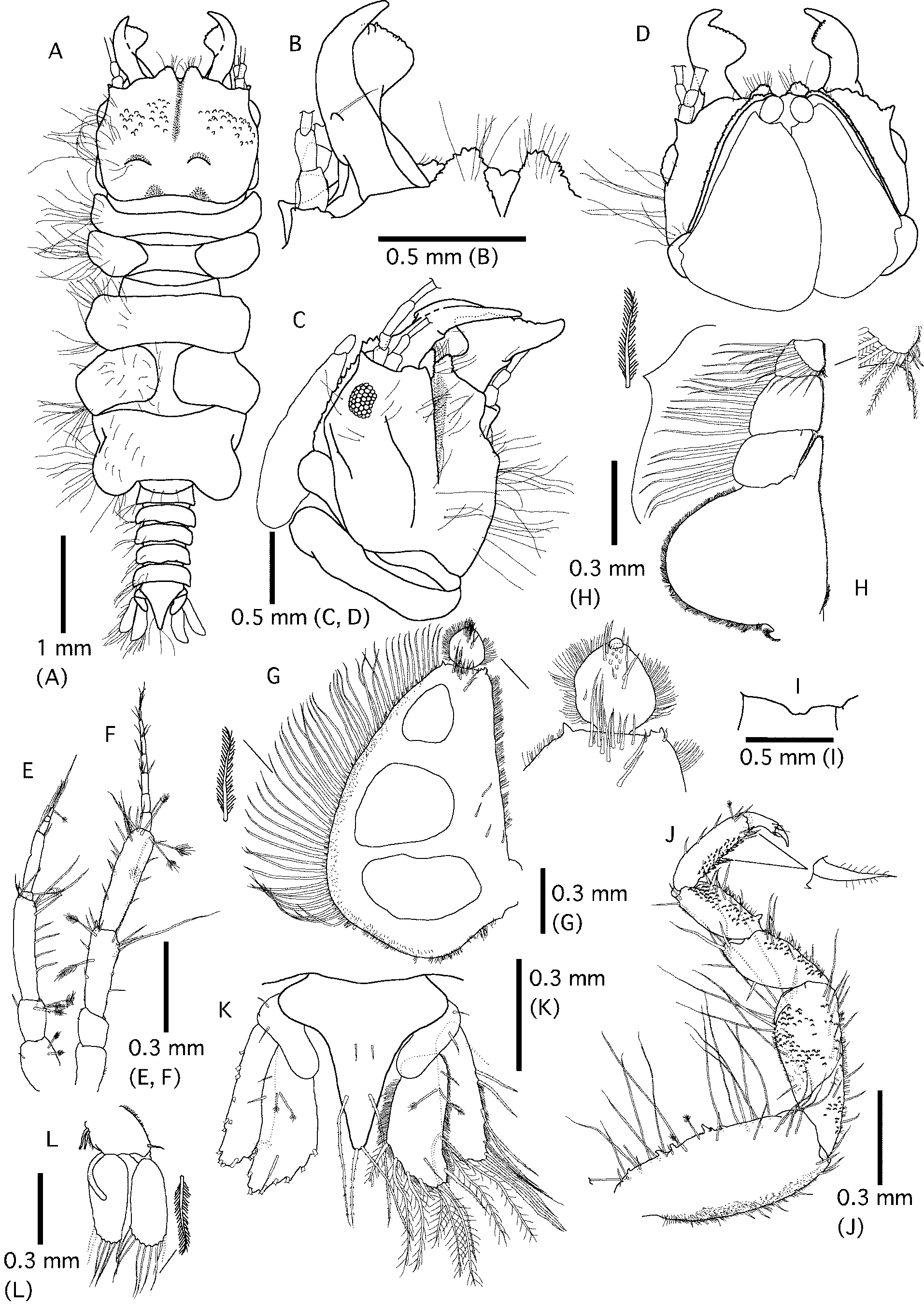
( Figs 1–3 View FIGURE 1 View FIGURE 2 View FIGURE 3 , 5 View FIGURE 5 A)
Material examined. Holotype. Male, 5.7 mm (NSMT-Cr 20425), from gill chambers of blotched fantail ray Taeniura meyeni Müller & Henle, 1841 , gill net, Nakagusuku Bay (26ºN, 127ºE), Okinawa Island, Ryukyu Archipelago, Japan, 25 May 2007, coll. Y. O t a.
Paratypes. 3 males, 3 females and 5 praniza larvae from the same locality and host as the holotype (NSMT-Cr 20426). 9 males, 2 females, and 2 praniza larvae from the same locality and host as the holotype ( NMV J46718 View Materials ). 2 praniza larvae from the same locality and host as the holotype ( KMNH IvR 500, 414). 9 males and 12 females from a Javanese cownose ray Rhinoptera javanica caught by a gill net in Nakagusuku Bay, 16 May 2007, coll. Y. Ota ( KMNH IvR 500, 413).
Description.
Male ( Fig. 1 View FIGURE 1 ). Body 3.9–5.8 mm (mean ± SD; 5.0 ± 0.6 mm, n = 21) long, color of live specimens white. Cephalosome ( Fig. 1 View FIGURE 1 A–D) almost square, with posterior margin convex, about one-fifth of total length. Frontal border ( Fig. 1 View FIGURE 1 B) with several setae, medially notched by 2 frontolateral processes; notch anteriorly opened. Apex of frontolateral process serrate. Eyes well developed, lateral and sessile, about one-fifth length of cephalosome. Supraocular lobe slightly prominent. Paraocular ornamentation present as several tubercles around the eyes. Dorsal sulcus deep and narrow.
Pereonites ( Fig. 1 View FIGURE 1 A) sparsely covered with setae. Pereonites 1–7 about half of total length. Pereonite 1 very short, not fused, concave anteriorly. Pereonites 2, 3, and 4 of same width. Pereonite 2 twice as long as pereonite 1; lateral margins extend anteriorly. Pereonite 3 slightly longer than pereonite 2. Pereonite 4 with constriction anteriorly, twice as long as pereonite 3. Pereonites 5 and 6 together subequal in length to pereonites 2–4 combined. Pereonites 5 and 6 with areae laterales and lobi laterales, respectively. Pereonite 7 short and narrow, overlapping pleonite 1.
Pleonites ( Fig. 1 View FIGURE 1 A) with several setae on lateral margins subequal in length. Pleonites and pleotelson about one-fourth of total length. Pleonal epimera not pronounced.
Pleotelson ( Fig. 1 View FIGURE 1 K) triangular, with middle of outer margins convex; 3 pairs of small setae on center, lateral margin, and distal apex.
Antenna 1 ( Fig. 1 View FIGURE 1 E) composed of 3 peduncular and 5 flagellar articles; 2 and 3 feather-like bristles on external margins of peduncles 1 and 2, respectively. Internal margins of peduncles 1 and 2 with 1 seta. Several setae on external and distal margins of peduncle 3. Length ratio of peduncular articles about 1:1:2. Featherlike bristles on flagellar articles 1 and 5. Aesthetascs on articles 3 and 5. Article 5 terminates in 3 long setae. Length ratio of 5 flagellar articles about 1:12:2:1:2.
Antenna 2 ( Fig. 1 View FIGURE 1 F) slightly longer than antenna 1; 2 and 3 feather-like bristles and several setae on peduncle articles 3 and 4, respectively. Length ratio of peduncular articles about 1:1:2:3. Few setae on distal margins of flagellar articles 1–6. Article 7 terminates in 4 long setae. Length ratio of flagellar articles approximately 2:2:2:2:2:2:1.
Mandibles ( Fig. 1 View FIGURE 1 B–D) three-fourths length of cephalosome. Carina unarmed. Apex curved internally. Dentate blade occupies about two-fifths length of mandible. Basal neck and erisma prominent. One mandibular seta on middle of dorsal surface.
Pylopod ( Fig. 1 View FIGURE 1 D, G) with 3 articles, ventral surfaces of article 1 with 19 setae, article 2 with 18 setae. Article 1 with 3 areolae, large and elliptical, fringed by fine setae on external margin, and 47 plumose setae on internal margin. Article 2 circular and fringed by fine setae. Article 3 minute.
Maxilliped ( Fig. 1 View FIGURE 1 H) with fine setae on lateral margin. Endite reaches palp article 2. Palp articles 1–4 with 6, 8, 5 and 8 plumose setae on external margins, respectively. Article 4 terminates in 4 setae.
Pereopods ( Fig. 1 View FIGURE 1 J) subequal in shape and bearing many setae; longer on outer margin than inner margin. Basis oblong; with 2 feather-like bristles on outer margin. Ischium about three-fourths length of basis, becoming larger distally. Merus about half length of ischium. Carpus elliptical and about four-fifths length of merus. Propodus rectangular, about 1.2 times longer than carpus; with 2 spines on inner-mid and inner-distal margins. Dactylus with few setae. Length of dactylus and unguis combined about half that of propodus. All pereopods similar in shape and size.
Penes ( Fig. 1 View FIGURE 1 I) composed of 2 contiguous papillae; not prominent.
Pleopodal peduncle ( Fig. 1 View FIGURE 1 L) fringed with fine setae, 1 seta on outer distal corner; coupling hook on inner margin. Both pleopodal rami oval-shaped and equal in length. All pleopods subequal in shape. Pleopod 1 exopod with 7 plumose setae, 6 plumose setae on endopod; pleopod 2 exopod with 9 plumose setae, endopod with 7 plumose setae. Appendix masculina extends beyond half of endopod of pleopod 2. Exopod of pleopods 3–5 with 9 setae, endopod with 8 setae.
Uropodal rami ( Fig. 1 View FIGURE 1 K) subequal in length, extending beyond apex of pleotelson. Exopod with 15 setae on margin. Endopod withng 12 setae on margin and 2 coupling setae on dorsal surface.
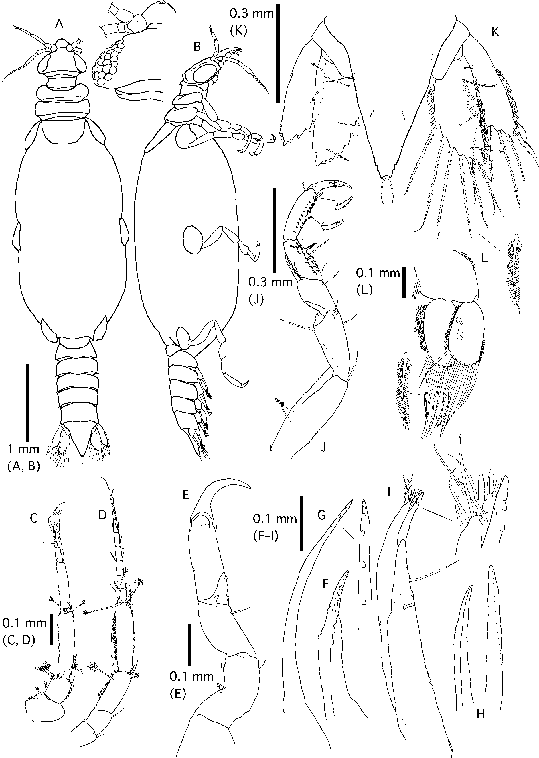
Female ( Fig. 2 View FIGURE 2 ). Body 4.1–5.5 mm (4.5 ± 0.3 mm, n = 17), color of live specimens light orange or yellow.
Cephalosome ( Fig. 2 View FIGURE 2 A) elliptical. Frontal margin slightly convex. Eyes well developed, lateral and sessile.
Pereonites ( Fig. 2 View FIGURE 2 A, B) with several setae on lateral margins and dorsal surface, swollen between pereonites 3 and 6. Pereonite 1 very short, not fused. Pereonites 2 and 3 with lateral margins that extend anteriorly. Pereonites 3–6 oval, with 3 sutures. Lateral shields of pereopods 4–6 visible dorsally. Pereopod 7 visible dorsally, short and narrow.
Pleonites ( Fig. 2 View FIGURE 2 A, B) with several setae subequal in length. Pleotelson ( Fig. 2 View FIGURE 2 G) triangular with an acute apex. 5 pairs of simple setae on dorsal surface and apex.
Antenna 1 ( Fig. 2 View FIGURE 2 C) composed of 3 peduncular and 4 flagellar articles. Pedunclar articles covered with scales and fine setae. Aesthetasc on articles 2–4. Article 4 terminates in 3 setae. Antenna 2 ( Fig. 2 View FIGURE 2 D) composed of 4 peduncular and 7 flagellar articles. Pedunclar articles covered with scales and fine setae.
Pylopod ( Fig. 2 View FIGURE 2 E) with 3 articles; article 2 rectangular; article 3 minute.
Maxilliped ( Fig. 2 View FIGURE 2 F) endite sparsely covered with fine setae, reaching half of article 1 of palp. Basis and articles 1–4 with 2, 7, 9, 8, and 9 plumose setae, respectively.
Pereopods ( Fig. 2 View FIGURE 2 G) bearing fewer setae than those of male, covered with scales and fine setae on ischium, merus, and carpus. Each article more slender in shape than those of male. Pereopods 5 and 6 larger than other pereopods.
Pleopodal peduncle ( Fig. 2 View FIGURE 2 I) has a few setae on lateral margin. Exopods fan-shaped and with 8 (pleopod 3) or 9 (pleopods 1, 2, 4, and 5) plumose setae on distal margins. Endopods subequal in shape and size to exopods; 7 (pleopod 3) or 8 (pleopods 1, 2, 4, and 5) plumose setae on distal margins. All pleopods subequal in shape.
Uropodal peduncle and both rami ( Fig. 2 View FIGURE 2 H) subequal in shape. Uropodal rami extend beyond apex of pleotelson. Exopod bears 12 long setae on margin. Endopod bears 9 setae on margin and 2 pairs and 7 featherlike bristles on dorsal surface.
Praniza larva ( Figs. 3 View FIGURE 3 , 5 View FIGURE 5 A). Body 4.2–5.8 mm (5.1 ± 0.5 mm, n = 7). Thorax of live specimen dark red or black with white speckles or dapples ( Fig. 5 View FIGURE 5 A). Other parts light brown or white.
Cephalosome ( Fig. 3 View FIGURE 3 A) triangular. Eyes well developed, and lateral and sessile.
Pereonite 1 ( Fig. 3 View FIGURE 3 A, B) very short, reaching lateral margin. Anterior margins of pereonites 2 and 3 concave. Lateral shields of pereopods 4–6 visible in dorsal view, elliptical. Pereonite 7 visible in dorsal view; short and narrow, overlapping pleonite 1.
Pleonites 1–4 subequal in length. Epimera not prominent in dorsal view. Pleotelson ( Fig. 3 View FIGURE 3 K) triangular, with dentate margin at apex, bearing 2 pairs of simple setae on posterior lateral margin and apex.
Antenna 1 ( Fig. 3 View FIGURE 3 C) composed of 3 peduncular and 4 flagellar articles. Internal margins of peduncles 2 and 3 fringed by fine setae. An aesthetasc each on articles 2–4. Antenna 2 ( Fig. 3 View FIGURE 3 D) composed of 4 peduncular and 7 flagellar articles; longer than antenna 1. Internal margins of peduncles 3 and 4 fringed by fine setae.
Gnathopod ( Fig. 3 View FIGURE 3 E) with pereopodal shape. Ischium 2 times as long as basis. Merus subequal in length to ischium. Carpus reduced. Propodus with distal bulbous protrusion. Mandible ( Fig. 3 View FIGURE 3 F) with 9 teeth on inner margin. Maxillule ( Fig. 3 View FIGURE 3 G) with slender article; 6 teeth on apex. Paragnath ( Fig. 3 View FIGURE 3 H) slightly curved. Maxilliped ( Fig. 3 View FIGURE 3 I) composed of a basis and two-articled palp. Basis with coupling hook on inner margin and an endite with one seta. Apex of palp 1 has 4 fine setae, 2 stout setae, 2 teeth, and endite; style-like with 3 teeth facing ventrally. 5 setae and 1 spine on distal margin of palp 2.
Pereopods ( Fig. 3 View FIGURE 3 J) more slender shape and more scarce setae than those of male. Pereopods 5 and 6 larger than the other pereopods.
Pleopodal peduncles ( Figs. 3 View FIGURE 3 L) with coupling hook on inner margin. Exopods fan-shaped and 8 (pleopod 1) or 9 (pleopod 2–5) plumose setae on distal margins. Endopods subequal in shape and size to exopods, and eight (pleopods 1–5) plumose setae on distal margins. All pleopods subequal in shape.
Uropodal peduncle and both rami ( Fig. 3 View FIGURE 3 K) subequal in shape to those of male. Uropodal rami fringed with fine setae and do not reach apex of pleotelson.
Etymology. The specific name maculosa is derived from Latin, meaning “the color disposed in broad, irregular blotches” and referring to the larval thorax.
Remarks. Gnathia maculosa is most similar to G. trimaculata . However, G. m a c u l o s a is distinguished from G. trimaculata by deeper and narrower dorsal sulcus on cephalosome, a narrower body, and the shape of the pylopod (slender pylopod in G. trimaculata ) ( Coetzee et al., 2009). The overall proportions and mandibles of G. m a c u l o s a are also similar to G. odontomachus Cohen & Poore, 1994 . However, G. odontomachus is smaller in its total length (2.9 mm) than G. m a c u l o s a and has a mediofrontal process ( Cohen & Poore, 1994). Gnathia capillata , G. grandilaris , and G. pantherina have also recorded from elasmobranchs. These species can be distinguished from the following features. Gnathia capillata has a shallow and wide dorsal sulcus and a notch on article 1 of the pylopod ( Nunomura & Honma, 2004). The total length of G. grandilaris is larger (5.7–8.3 mm), and has a medio-frontal process on frontal boarder and more slender pylopod with two areolae ( Coetzee et al., 2008). Gnathia pantherina has pectinate scales on the pleotelson, fine setae on peduncle articles of the antennae, and no areola on the pylopod (Smit & Basson, 2002).
Gnathia camuripenis View in CoL and G. l i m i c o l a might be found with G. m a c u l o s a, as their locales are near each other. However, G. camuripenis View in CoL and G. limicola View in CoL are smaller (the former is longest 2.8 mm; the latter is longest 2.6 mm) and have prominent penes fused into a laterally compressed blade ( Tanaka, 2004; Ota et al. 2007). Thus, adult males of these species are easily distinguished from those of G. m a c u l o s a.
Only a few detailed descriptions of adult females are available for gnathiids. In the genus Gnathia View in CoL , female mouthparts have been described in detail only for the following six species, i.e. G. c a m u r i p e n i s, G. limicola View in CoL , G. africana View in CoL , G. pantherina View in CoL , G. gurjanovae View in CoL , and G. trimaculata View in CoL (Smit & Basson, 2002; Smit et al., 2002; Tanaka, 2004; Golovan, 2006; Ota et al., 2007; Coetzee et al., 2009). Of these species, the females of G. trimaculata View in CoL are most similar to the females of G. m a c u l o s a. However, G. trimaculata View in CoL is distinguished from G. m a c u l o s a by a wider pleotelson and the simple pappose setae on the frontal boarder ( Coetzee et al., 2009).
The praniza larvae of G. pantherina , G. capillata , G. grandilaris and G. trimaculata may be found with those of G. m a c u l o s a, as they are ectoparasites on elasmobranchs. The apex of the pleotelson does not extend beyond the uropodal rami in these 3 species, except for G. capillata (Smit & Basson, 2002; Nunomura & Honma, 2004; Coetzee et al., 2008, 2009). Gnathia capillata is distinguished from G. m a c u l o s a by a large plumose seta on peduncle 4 of antenna 2 ( Nunomura & Honma, 2004).


| NMV |
Museum Victoria |
No known copyright restrictions apply. See Agosti, D., Egloff, W., 2009. Taxonomic information exchange and copyright: the Plazi approach. BMC Research Notes 2009, 2:53 for further explanation.