
Phanuromyia ricaniae Nam, Lee & Talamas, 2020
|
publication ID |
https://doi.org/ 10.11646/zootaxa.4890.1.6 |
|
publication LSID |
lsid:zoobank.org:pub:2E403C31-D3F6-4113-8872-133749E61EDC |
|
DOI |
https://doi.org/10.5281/zenodo.4323718 |
|
persistent identifier |
https://treatment.plazi.org/id/6278D001-890D-FF8B-09F1-E865FEB9F863 |
|
treatment provided by |
Plazi |
|
scientific name |
Phanuromyia ricaniae Nam, Lee & Talamas |
| status |
sp. nov. |
Phanuromyia ricaniae Nam, Lee & Talamas , sp. n.
urn:lsid:zoobank.org:act:xxxx
https://bioguid.osu.edu/xbiod_concepts/509748
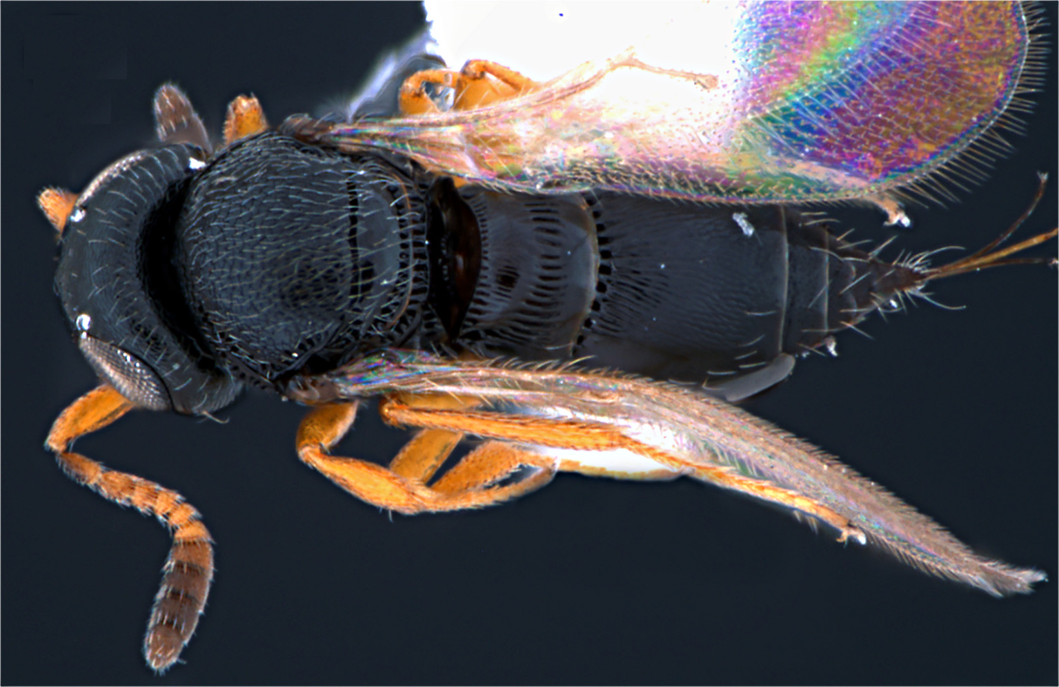
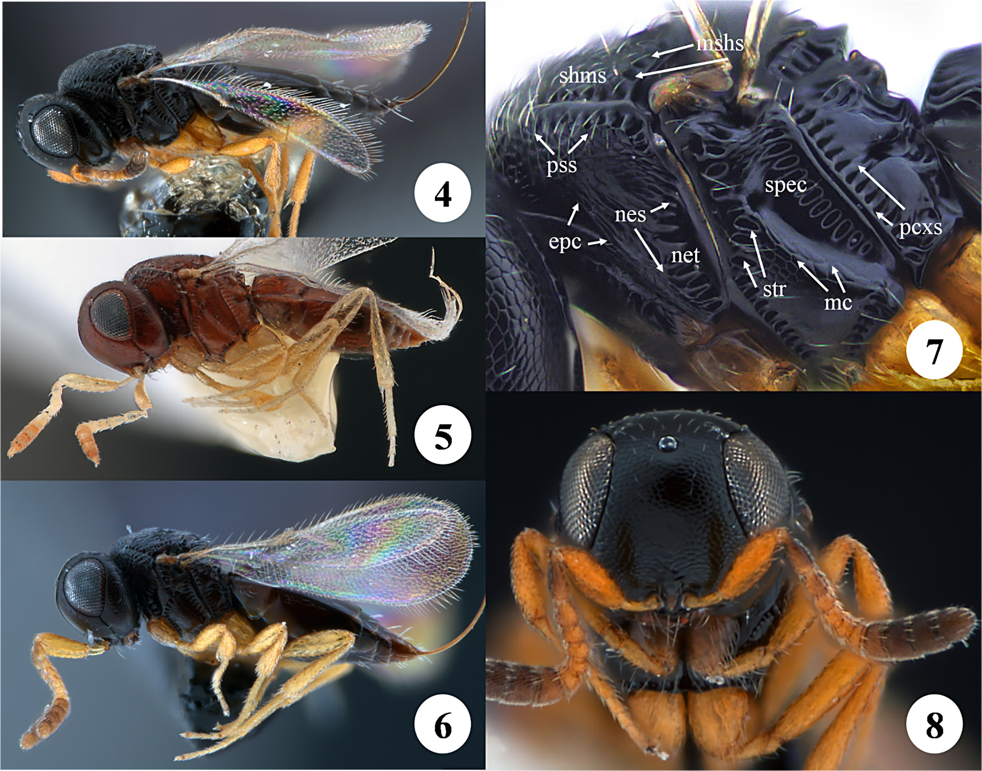
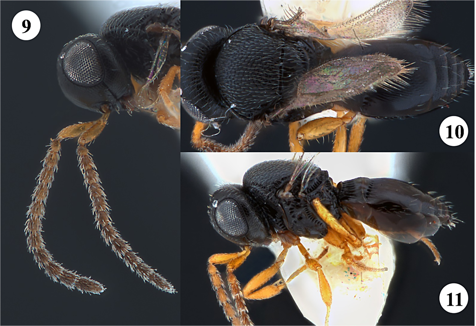
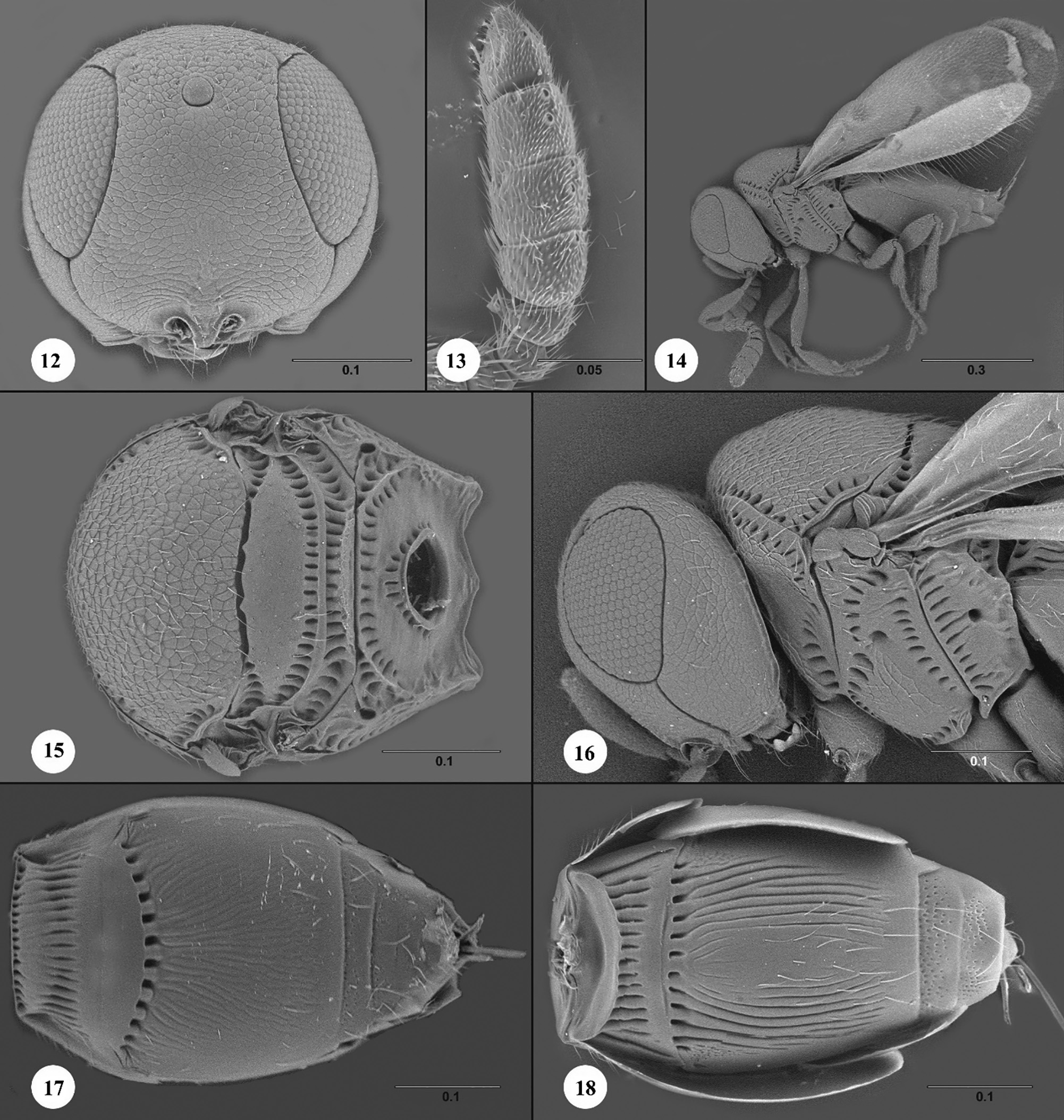
Figures 3–18 View Figure 3 View FIGURE 4–8 View FIGURE 9–11 View FIGURE 12–18
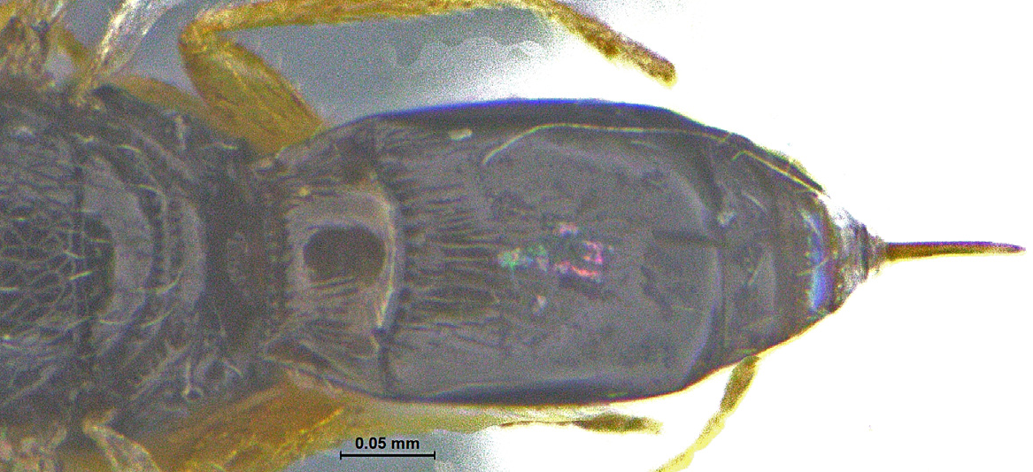
Description. Color of body: light brown to black. Female body length: 0.9–1.09 mm (n=22). Male body length: 0.9–1.06 mm (n=13).
Head. Claval formula: 1-2-2-1. Shape of clypeus: convex, with obtuse point medially. Number of clypeal setae: 6. Central keel: absent. Antennal scrobe: absent, undifferentiated from remainder of frons. Sculpture of frons: reticulate microsculpture. Sculpture of gena: reticulate microsculpture. Setation of compound eye: absent. Position of lateral ocellus: contiguous with compound eye. Shape of vertex: broadly rounded. Anterior margin of occipital carina: weakly crenulate. Hyperoccipital carina: absent.
Mesosoma. Epomial carina: present, short and often obscured by head in lateral view. Pronotal cervical sulcus: present as a smooth furrow. Sculpture of lateral pronotal area: obliquely rugose. Pronotal suprahumeral sulcus: indicated by punctures. Netrion sulcus: foveate, complete dorsally. Prespecular sulcus: foveate, extending from subalar pit to mesopleural pit. Sculpture of speculum: transversely rugulose. Anterior mesepisternal area: present as an elevated, smooth area. Sculpture of femoral depression: smooth. Mesopleural carina: absent. Episternal foveae: present, forming a continuous line with foveae of postacetabular sulcus that extends to mesopleural pit. Postacetabular sulcus: comprised of distinct foveae. Sculpture of anteroventral mesopleuron: with microsculpture directly ventral to femoral depression, otherwise smooth. Mesopleural epicoxal sulcus: foveate. Posterior mesepimeral area: narrow, present as a strip of lesser width than mesepimeral sulcus. Paracoxal sulcus: foveate, present along entire anterior margin of metapleuron. Transverse portion of metapleural sulcus: indicated by furrow extending to the anterior and posterior margins of the metapleuron. Sculpture of ventral metapleural area: smooth. Sculpture of dorsal metapleural area: smooth. Microsculpture of mesoscutum: reticulate microsculpture. Notaulus: absent. Mesoscutal suprahumeral sulcus: present as a distinct line of cells. Mesoscutal humeral sulcus: present as a distinct line of foveae. Sculpture of mesoscutellum: smooth, sometimes with reticulate microsculpture laterally. Metascutellum: line of foveae. Sculpture of metanotal trough: foveate. Length of postmarginal vein: about 3 times as long as stigmal vein. Length of marginal vein: short, about 1/4 length of stigmal vein. Sculpture of lateral propodeal area: foveate along anterior margin of lateral propodeal carina. Sculpture of metasomal depression: with line of fovea along periphery and surrounding propodeal foramen, otherwise smooth.
Metasoma. Sculpture of T1 between basal costae and median band of foveae: striate between basal costae and median band of foveae, striae sometimes incomplete medially. Median band of foveae on T1: present. Sculpture of T1 posterior to median band of foveae: smooth. Setation of laterotergite 1: present as a patch in anterodorsal portion. Sculpture of T2 posterior to basal foveae: longitudinally strigose, reticulate posteriorly. Setation of laterotergite 2: present along dorsal margin. Sculpture of T3–T5: covered in cuticular pores, otherwise smooth. Setation of T3–T7: with sparse, long setae. Sculpture of S2: longitudinally striate. Sculpture of S3–S4: covered in cuticular pores, otherwise smooth. Setation of S3–S6: with long setae laterally.
Etymology. The epithet “ricaniae” is the genitive case form of Ricania , the genus from which this species has been reared.
Material Examined. Holotype, female: SOUTH KOREA: Dong-gu Daejeon, Jungang-ro , 36°19’56.3”N 127°26’02.5”, Daejeon St. 215, 8.VIII.2016, sweeping, S. Nam, SNUP0010003 (deposited in SNU) . Paratypes: (63 females, 12 males) CHINA : 44 females, 10 males, SCAU 3040575–3040583, 3048582–3048583 ( SYSBM) , SCAU 3044143–3044185 ( SYSBM) SOUTH KOREA: 19 females, 2 males, CNC1558300–CNC1558302 ( CNCI) ; SNUP0010001 View Materials – SNUP0010002 View Materials , SNUP0010004 View Materials – SNUP0010019 View Materials ( SNU) .
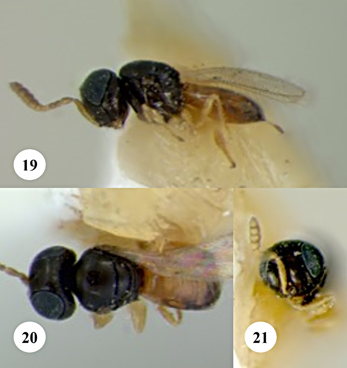
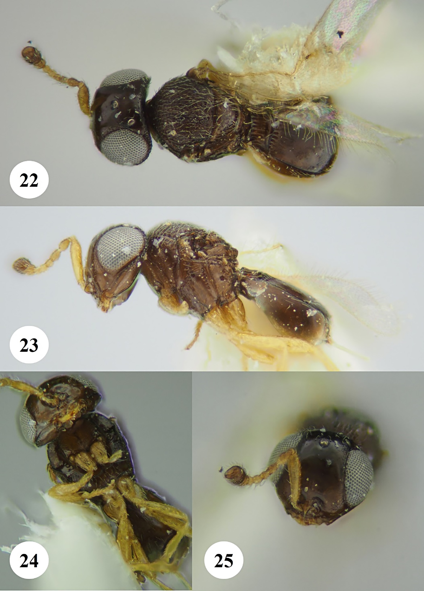
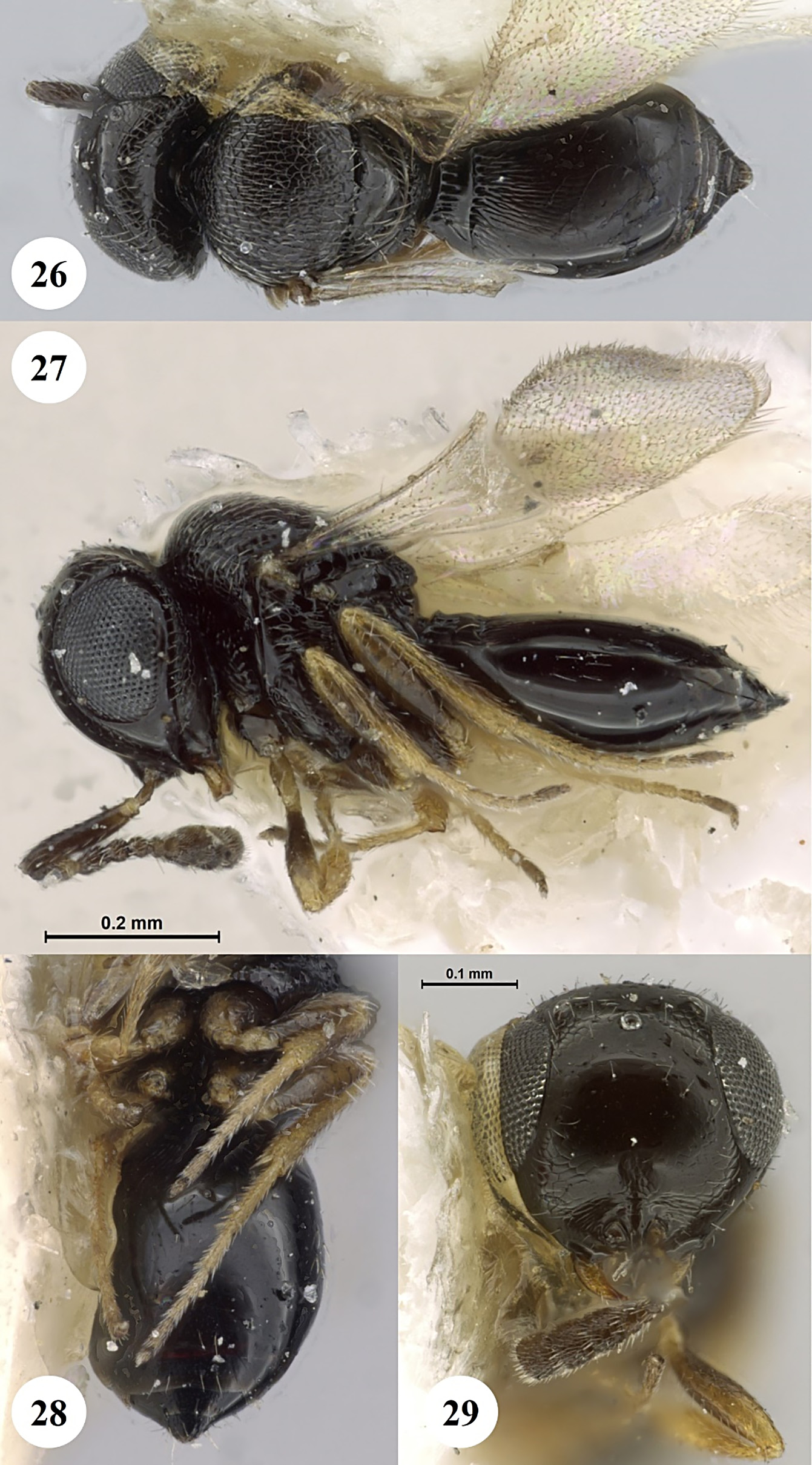
Diagnosis. In the absence of a large-scale revision of Asian Phanuromyia , we compared P. ricaniae to the images of Phanuromyia species presented in Veenakumari & Mohanraj (2014) and Veenakumari & Mohanraj (2019), the description of P. bidentata by Johnson & Musetti (2003), and holotype images of three other Asian species: P. flaviventris (Kozlov & Kononova) ( Figs 19–21 View FIGURE 19–21 ), P. nioba (Kozlov & Kononova) ( Figs 22–25 View FIGURE 22–25 ), and P. marshakovi (Kozlov & Kononova) ( Figs 26–29 View FIGURE 26–29 ). Among these, P. ricaniae is most similar to P. andamanensis and P. nabakovi Veenakumari , which share a similar pattern of mesosomal sulci, coloration, and surface sculpture. Phanuromyia ricaniae and P. andamanensis can easily be separated by the length of striae on S2. These striae extend nearly to the end of the sternite in P. ricaniae and are only half the length of the sternite in P. andamanensis (see Figure 8 View FIGURE 4–8 in Veenakumari & Mohanraj 2014). Separating P. ricaniae from P. nabakovi is less straightforward because the sculpture of S2 is not described or illustrated in Veenakumari & Mohanraj (2014). Figure 29 View FIGURE 26–29 in Veenakumari & Mohanraj (2014) illustrates a paracoxal sulcus comprised of very small foveae that are small and poorly defined, particularly in the ventral half of the metapleuron whereas in P. ricaniae these foveae are robust and tend to be elongate. Metasomal tergite 1 is described as “weakly costate” in P. nabakovi and an image of the holotype illustrates that T1 is smooth medially ( Fig. 30 View FIGURE 30 ). Metasomal tergite 1 in P. ricaniae is variable in the degree to which it is dorsally humped in lateral view and in the size of the posterior smooth area, but the costae that transversely bisect the tergite are consistently distinct.
| CNCI |
Canadian National Collection Insects |
No known copyright restrictions apply. See Agosti, D., Egloff, W., 2009. Taxonomic information exchange and copyright: the Plazi approach. BMC Research Notes 2009, 2:53 for further explanation.