
Rhinogobius tandikan Maeda, Kobayashi & Palla, 2021
|
publication ID |
https://doi.org/ 10.11646/zootaxa.5068.1.3 |
|
publication LSID |
lsid:zoobank.org:pub:D6B213E5-8A79-4438-8481-01C074EB74CB |
|
DOI |
https://doi.org/10.5281/zenodo.5706125 |
|
persistent identifier |
https://treatment.plazi.org/id/47FBF5CB-033A-4857-AFE6-91E5706ACFEF |
|
taxon LSID |
lsid:zoobank.org:act:47FBF5CB-033A-4857-AFE6-91E5706ACFEF |
|
treatment provided by |
Plazi |
|
scientific name |
Rhinogobius tandikan Maeda, Kobayashi & Palla |
| status |
sp. nov. |
Rhinogobius tandikan Maeda, Kobayashi & Palla , new species
[New English name: Tandikan goby]
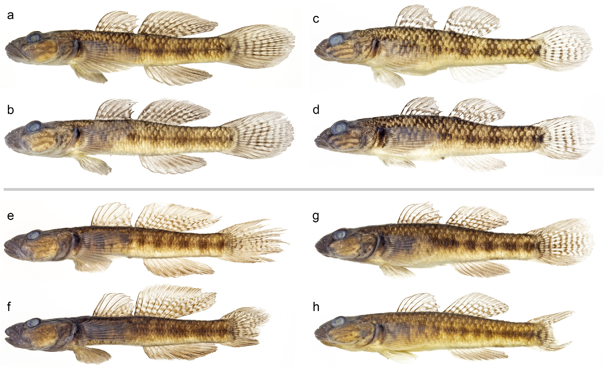
( Figs. 1 View FIGURE 1 , 3 View FIGURE 3 , 6–8 View FIGURE 6 View FIGURE 7 View FIGURE 8 ; Tables 2–6 View TABLE 2 )
Material examined. Thirteen males and 16 females from Cayulo River , Bahile, Puerto Princesa City, Palawan Island in the Philippines .
Holotype. NSMT-P 140093 , male (43.6 mm SL), 1 June 2018, coll. K. Maeda, H. Kobayashi, and H. P. Palla.
Paratypes. NSMT-P 140094 , female (45.7 mm SL), 31 May 2018 coll. K. Maeda, H. Kobayashi, and H. P. Palla; URM-P 49307–49315, 7 males (36.1–46.9 mm SL) and 2 females (37.3–38.9 mm SL), 31 May 2018 coll. K. Maeda, H. Kobayashi, and H. P. Palla; URM-P 49316–49323, 2 males (40.8–44.8 mm SL) and 6 females (35.6–45.2 mm SL), same data as holotype ; WPU-PPC-P 60–64, 5 females (30.8–36.5 mm SL), 31 May 2018 coll. K. Maeda, H. Kobayashi, and H. P. Palla; WPU-PPC-P 65–69, 3 males (43.6–46.1 mm SL) and 2 females (39.6–41.3 mm SL), same data as holotype .
Diagnosis. Pectoral fin with 16 or 17 rays. Nape and posterior part of occipital region covered by cycloid scales; scaled area extending anteriorly to around a vertical line through posterior margin of preopercle or a little posterior to this line. Longitudinal scales 29–34, predorsal scales 5–13. Number of vertebrae 26. Cephalic sensory pore system usually with B´, C, D(S), E, F, H´, K´, L´, M´, N, and O´, but often lacking one or both side(s) of pore C. Transverse rows of sensory papillae on cheek. In preservative, lateral and dorsal sides of body without clear meshlike markings, obscure markings on cheek, and upper half of pectoral fin with dark brown band vertically across proximal part. In life, pectoral-fin base with a bright white vertical band reaching near bottom of fin.
Description. Body nearly cylindrical anteriorly and somewhat compressed posteriorly. Abdomen of male often thin. Head depressed and larger in male than female (head length 32.1–37.4 vs 30.8–33.4% of SL; Fig. 3 View FIGURE 3 ). Eyes located dorsolaterally. Mouth terminal and oblique with thick upper and lower lips. Anterior tips of upper and lower jaws almost even or upper jaw slightly protruding beyond lower jaw. Posterior end of upper jaw always exceeding a vertical line through anterior margin of eye. Mouth larger in male than female (upper-jaw length 13.3–20.9 vs 10.1–12.7% of SL; Fig. 3 View FIGURE 3 ). Anterior nostril short tubular, posterior nostril a pore. Cephalic sensory pore system usually with B´, C, D(S), E, F, H´, K´, and L´ in oculoscapular canal and M´, N, and O´ in preopercular canal, but 9/ 29 specimens lacking one pore C, 7/ 29 specimens lacking both pores C, 4/ 29 specimens lacking one pore E, 1/ 29 specimen having two pores E on one side, 4/ 29 specimens having pore G on one side, 1/ 29 specimen having pores G on both sides, and 5/ 29 specimens lacking one pore N. One specimen having combined pores K´ and L´ on one side (to be a groove without roof). Arrangement of cutaneous sensory papillae of head shown in Fig. 7 View FIGURE 7 . Cheek having two longitudinal rows of papillae and 3–6 transverse rows both between eye and upper longitudinal row and between upper and lower longitudinal rows. Vertebrae 10+16=26 (n=10), P-V 3/II II I I 0/9 (n=10).
All males had damage to the caudal fin, except holotype; 10/ 16 female specimens with damaged caudal fin ( Figs. 1 View FIGURE 1 , 3 View FIGURE 3 ). Other fins of male and female sometimes damaged also. First dorsal fin with six spines supported by six pterygiophores. Second dorsal fin usually with one spine and eight soft rays. First and second dorsal-fin bases separated each other by a small interval (0.2–4.4% of SL). First dorsal fin rounded, usually almost semi-circular; posterior tips of fin (tips of fourth to sixth spines) not reaching second dorsal fin origin (N=9), just touching base of second dorsal-fin spine (n=12), or exceeding base of second dorsal-fin spine but not reaching base of first soft ray of second dorsal fin (n=8). Anal fin usually with one spine and eight soft rays, but 4/ 29 specimens with one spine and seven soft rays. Caudal fin usually with 17 segmented rays but 3/ 29 specimens with 16 segmented rays, including 11 (n=1), 12 (n=6), 13 (n=15), or 14 (n=22) branched rays (unknown in four specimens due to fin damage); posterior margin rounded in non-damaged individuals. Male having larger second dorsal and anal fins than female (second dorsal-fin length 33.8–43.6 vs 29.5–35.3% of SL and anal-fin length 30.2–38.3 vs 26.7–32.7% of SL), and nondamaged male with larger caudal fin than female ( Fig. 3 View FIGURE 3 ). Pectoral fin with 16 or 17 rays ( Table 2 View TABLE 2 ). Pelvic fin with one spine and five soft rays; pelvic fins joined together to form a cuplike disk with fleshy bilobed frenum. Posterior tip of pelvic fin located below middle of first-dorsal-fin base.
Ctenoid scales covering lateral side of body and dorsal and ventral sides of caudal peduncle. Nape and posterior part of occipital region covered by cycloid scales; scaled area extending anteriorly around vertical line through posterior margin of preopercle (above pore H´) or to a little posterior to this line (above area between pores H´ and K´). Other regions in head naked. Belly and first dorsal-fin base covered with cycloid scales. A few cycloid scales also present on trunk behind pectoral fin, along bases of second dorsal, anal, and caudal fins, and on proximal part of caudal fin. Pectoral-fin base and breast (prepelvic area) probably naked (at least invisible without staining by arizarin red; see Suzuki et al. 2016). Longitudinal scales 29–34 (usually 30 or 31), transverse scales 8–10 (usually 9 or 10), transverse scales in caudal peduncle always 7, and predorsal scales 5–13 ( Tables 3–5).
Colour in preservative ( Fig. 1 View FIGURE 1 ): Background of head and body brown dorsally and laterally and cream ventrally. Snout with obscure dark brown stripe connecting upper lip and eye. Cheek often with two obscure, dark brown intermittent lines. Posterior margin of opercle fringed by dark brown vertical band. Pectoral-fin base blackish except cream or pale brown bottom. Four to eight dark brown transverse bars or round blotches aligned along lateral midline of body. First and second dorsal fins pale grey with 3–7 brown horizontal stripes on proximal and middle parts, but anterior half of first dorsal fin lacking this marking. Anal fin greyish brown but light brown proximally. Caudal fin pale grey or greyish brown with 6–11 dark brown vertical bands, but dorsal and ventral parts lacking these bands. Pectoral fin pale greyish brown with a dark brown vertical band proximally on upper half. Pelvic fin greyish brown.
Colour in life ( Fig. 8 View FIGURE 8 ): Background of head and body olive green or yellowish grey. Snout with reddish brown stripe connecting upper lip and eye. Cheek with two reddish brown longitudinal intermittent lines and several reddish brown spots. Posterior margin of opercle fringed by reddish-brown vertical band. Pectoral-fin base black or dusky with a large greyish blue blotch anteriorly, and with a bright white vertical band posteriorly. Four to eight, dark brown transverse bars or round blotches aligned along lateral midline of body. Irregularly arranged dark brown markings dorsally on body. Centre of lateral scales tinged with sky blue. First and second dorsal fins light yellowish brown with 3–7 dark brown horizontal stripes on proximal and middle parts and reddish brown on distal part, but anterior half of first dorsal fin reddish brown without clear marking. Anal fin reddish brown or orange but yellowish proximally. Caudal fin reddish brown dorsally and ventrally and whitish with 6–11 dark reddish-brown transverse bands on middle part. Pectoral fin translucent but proximal parts of some ventral rays white. Pelvic fin reddish brown proximally and light grey distally.
Distribution. Rhinogobius tandikan is endemic to Palawan Island. It was found only in the Cayulo River, Bahile, northern part of Puerto Princesa City. The Cayulo River is a small stream (length approximately 4 km including 1 km of an estuary) flowing into Ulugan Bay on South China Sea-side of the island. The stream system is surrounded by mountains of about 200–400 m above sea level. Cayulo Falls, composed of a series of small waterfalls, is located at the middle eaches of this stream (10°02'12"N 118°45'03"E). Rhinogobius tandikan was found at both the lower and upper reaches of this waterfall, as well as pools in the middle of the falls. The new species was abundant in this site with many other goby species, cyprinids, and a jungle perch species.
Etymology. Tandikan is a local name of Palawan peacock-pheasant, Polyplectron napoleonis Lesson , known as a symbol of Puerto Princesa City. The new goby species is named Rhinogobius tandikan , as it has blue markings on the body, suggestive of the plumage of the Palawan peacock-pheasant, and as the type locality is located in the northern part of Puerto Princesa City. The new specific name is a noun in apposition.
Comparison. Rhinogobius similis Gill, 1859 is the only species previously known in the genus having transverse rows of sensory papillae on cheek ( Suzuki et al., 2016), and our two new species share this unique feature with R. similis . However, the two species in Palawan are distinguished from R. similis in having fewer rays in the pectoral fin (14–17 vs 18–19), predorsal squamation composed of cycloid scales (vs ctenoid scales), anterior-most dorsal scale located around a vertical line through posterior margin of the preopercle or more posteriorly (vs dorsal squamation reaching anteriorly near the posterior margin of the eye), and proximal part of the caudal fin covered by cycloid scales (vs by ctenoid scales).
Rhinogobius estrellae shares many characters with R. tandikan , but the former differs from the latter by having fewer pectoral-fin rays (usually 15 vs 16–17; Table 2 View TABLE 2 ), fewer scale counts (longitudinal scales 27–31 vs 29–34, predorsal scales 3–11 vs 5–13; Tables 3, 5), and colourations of the body. In preservative, R. estrellae has dark brown mesh-like markings on the lateral and dorsal sides of the body (vs rather uniformly brown in R. tandikan ), three dark brown stripes on the snout and cheek (vs often obscure), and a dark brown vertical band on the proximal part of the upper and middle pectoral-fin rays except lower 2–6 rays (vs this band restricted to upper half of the fin). In life, R. estrellae has a more reddish body than the yellowish or olive green R. tandikan , and the former has a bright white vertical band restricted to the upper and middle parts of the pectoral-fin base while this band reaches nearly the bottom of the fin in R. tandikan . Arrangements of the cephalic sensory pore system are basically the same between the two species, but R. estrellae more often lacks pore E on one or both sides than in R. tandikan (12/24 vs 5/29) while R. tandikan more often lacks pore C on one or both sides than in R. estrellae (16/29 vs 7/24).
In addition to the transverse rows of sensory papillae on cheek (present vs absent), there are many differences between the two new species from Palawan and three other Philippine species, R. carpenteri Seale, 1910 , R. bucculentus ( Herre, 1927) , and R. philippinus ( Herre, 1927) , such as numbers of the vertebrae (26 vs 27–29), first-dorsalfin pterygiophores (usually 6 vs usually 7), first-dorsal-fin spines (usually 6 vs usually 7 in R. bucculentus and 6 or 7 in R. carpenteri and R. philippinus ), pectoral-fin rays (14–17 vs 17–20), having fewer scales in the longitudinal scales (27–34 vs 35–43), in the transverse scales (8–10 vs 12–18), and in the transverse scales in caudal peduncle (7 vs 9–11), and presence of scales on belly behind the pelvic fins (vs absent in R. carpenteri and R. bucculentus ). The latter three species are distributed in Luzon, northern Philippines (see Discussion).
Molecular phylogenetic analysis. In the ML phylogenetic tree using the aligned 16,409 bp of mitochondrial genomes ( Fig. 9 View FIGURE 9 ), the species in Rhinogobius were divided into two robust clades. One clade included R. similis and the two new species from Palawan, while all other species belonged to another clade. Six specimens of R. estrellae and four specimens of R. tandikan formed each well-supported monophyletic clade and were sister species to each other. Rhinogobius similis was a sister to this pair.


No known copyright restrictions apply. See Agosti, D., Egloff, W., 2009. Taxonomic information exchange and copyright: the Plazi approach. BMC Research Notes 2009, 2:53 for further explanation.