
Anthaxia (Anthaxia) magnanii, Baiocchi, Daniele, 2011
|
publication ID |
https://doi.org/ 10.5281/zenodo.278056 |
|
DOI |
https://doi.org/10.5281/zenodo.3509056 |
|
persistent identifier |
https://treatment.plazi.org/id/637087DE-F839-7147-84AE-9501FC8CC411 |
|
treatment provided by |
Plazi |
|
scientific name |
Anthaxia (Anthaxia) magnanii |
| status |
sp. nov. |
Anthaxia (Anthaxia) magnanii View in CoL n. sp.
(Figs. 1,3,4,13,14,15,31,37,39,45,51,57,60,63,64)
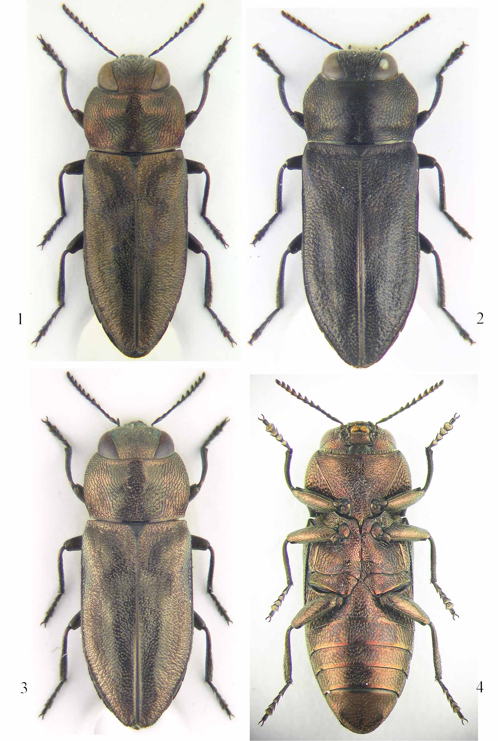
Description of holotype 3 ( Fig. 1 View FIGURES 1 – 4 ). Body suboval, rather convex; length: 6.0 mm, maximum width at humeral height: 2.5 mm, length/width ratio: 2.4 times longer than wide; dorsal colouration brilliant bronze with slight coppery tinge on head, pronotum; discal area of pronotum with two longitudinal, indistinct, blurred maculae, interrupted in frontal 1/2; legs, antennae, ventral surface of same bronze colour as rest of the body; last five antennomeres yellowish at respective base.
Head ( Fig. 13 View FIGURES 13 – 18 ) slightly narrower than anterior pronotal margin; eyes not projecting beyond outline of head; vertex flat, rather narrow (0.4 times as wide as width of head), with an evident dark median line; frons ( Fig. 31 View FIGURES 31 – 38 ) wide, slightly depressed in middle; inner ocular margins S-shaped, moderately converging on vertex, diverging on lower frontal portion; sculpture of vertex very weak, shallow in middle, strongly stretched near the upper ocular margins, consisting of irregular cells, with finely microsculptured brilliant bottom and tiny setigerous punctures; sculpture of frons areolate, consisting of deep irregular cells, rather oblong on mid area, with slightly microsculptured bottom and large, flat, central grain; sculpture along inner ocular margins deep, longitudinally stretched; frons covered with long, yellowish, recumbent pubescence, slightly divergent on upper frontal portion, convergent near lower ocular margin; pubescence of vertex subtle, short, hardly visible; clypeus feebly prominent, flattened frontally, lateral margins subparallel, anterior margin smooth, deeply roundly incised.
Antennae ( Fig. 39 View FIGURES 39 – 50 ) slender, 1.2 times longer than middle pronotal length; antennomeres 6 – 11 yellowish at base; scape finely club-shaped; pedicel oval, 1.4 times longer than wide; antennomere 3 subcilyndrical, 1.1 times longer than pedicel; antennomere 4 subconical; antennomeres 5 – 10 sharply triangular, slightly longer than wide, slender at base, with sinuate inner margin; last antennomere elliptically elongate.
Pronotum ( Fig. 13 View FIGURES 13 – 18 ) transverse, 1.7 times wider than long, more convex on anterior 1/2, with maximum width and shallow trasversal depression at midlength; anterior margin rather deeply emarginate, with feeble central lobe; anterior angles acute; lateral margins regularly arched, distinctly narrowed before the obtuse posterior angles; posterior margin slightly narrower than elytral base, moderately arched backward; lateroposterior depressions wide, shallow; sculpture areolate and regularly polygonal only on anterior angles; lateroposterior corner cells irregular, longitudinally prolonged, separated by dark, thick borders; middle discal area rather glossy, with sculpture consisting of large, contiguous, irregularly lengthened cells, bordered by long furrows, very shallow on mid anterior portion, deeper in the mid posterior 1/2, forming a curvilinear, exarate pattern, hosting multiple, tiny, setigerous punctures; whole pronotal surface very finely microsculptured, sparsely covered with very short, whitish pubescence.
Scutellum ( Fig. 13 View FIGURES 13 – 18 ) dull black, subtrapezoidal, wider than long, more convex on posterior 1/2, with finely microreticulated surface.
Elytra subparallel, 1.9 times longer than wide, basal 2/3 rather convex, apical 1/3 more flat, feebly, arcuately tapering to separately, obtusely, subrounded apex; basal transverse depressions wide, slightly deeper at humeral angles, not reaching the scutellum; humeral swellings normally developed; one rounded, shallow, postscutellar depression at the basal 1/3, and longitudinal depressions on the posterior portion of each elytron; lateral elytral groove complete, shallow, narrow, weakly wider at humeral height and along part of apical 1/3; epipleura broad, not complete, expiring before the apex; surface coarsely sculptured at base and along outer edges, smoother, brighter along the slightly raised elytral suture; pubescence transparent, very short and sparse.
Ventral surface ( Fig. 4 View FIGURES 1 – 4 ). Anterior margin of prosternum straight; prosternal process rather wide, with lateral sides poorly incurved and posterior apex moderately long; posterior end of central metasternal suture shortly, narrowly divergent; protrocantheres unarmed; mesotrochanters with obtuse posterior spine; metatrochanters with acute posterior spine; sculpture of sternum variolate, consisting of cells of varying depth, with tiny central pore; sculpture of ventral abdominal surface extremely shallowly variolate, with very fine basal microreticulation; coxae and ventral face of legs finely microreticulated; ventral pubescence sparse, short and transparent.
Anal ventrite (Fig. 351 wider than long; preapical area flat, depressed, with fairly rounded, smooth, strongly raised apical edge; lateral margins weakly serrate.
Legs. Foretibiae slightly curved at base; foretarsomeres 1 – 4 equal, tarsomere 5 longer; mesotibiae straight, inner margin weakly incised, feebly serrate on distal 1/3; mesotarsomeres 1 – 4 equal, tarsomere 5 longer; metatibiae ( Fig. 45 View FIGURES 39 – 50 ) straight, flattened distally, inner margin slightly incised, serrate on apical 1/4; metarsomeres 2 – 4 equal, shorter than 1, tarsomere 5 as long as 1; tarsal claws thick, distinctly incurved, smoothly enlarged at base, brown with darker tips.
Aedeagus strongly bottle-shaped, 4.9 times longer than wide, widest at midlength; basal 1/2 and apical margins yellowish, rest of anterior 1/2 very dark; phallobasis subparallel, parameres ( Fig. 14 View FIGURES 13 – 18 ) subcylindrical at base, anterior 1/2 strongly narrowed, apical lobes feebly enlarged, sharply pointed, slightly convergent, setigerous portion lengthened, hidden within the apicolateral membranaceous margin; median lobe ( Fig. 15 View FIGURES 13 – 18 ) subparallel, 9 times longer than wide, distinctly convex at base, apical 1/4 mildly tapered, apex slightly narrowed, acute but not sharp, anterior portion of dorsal surface feebly depressed in middle, slightly rugulose, posterior portion smoother, preapical portion of lateral margins very finely serrate, basal apodemes 1/3 of total length.
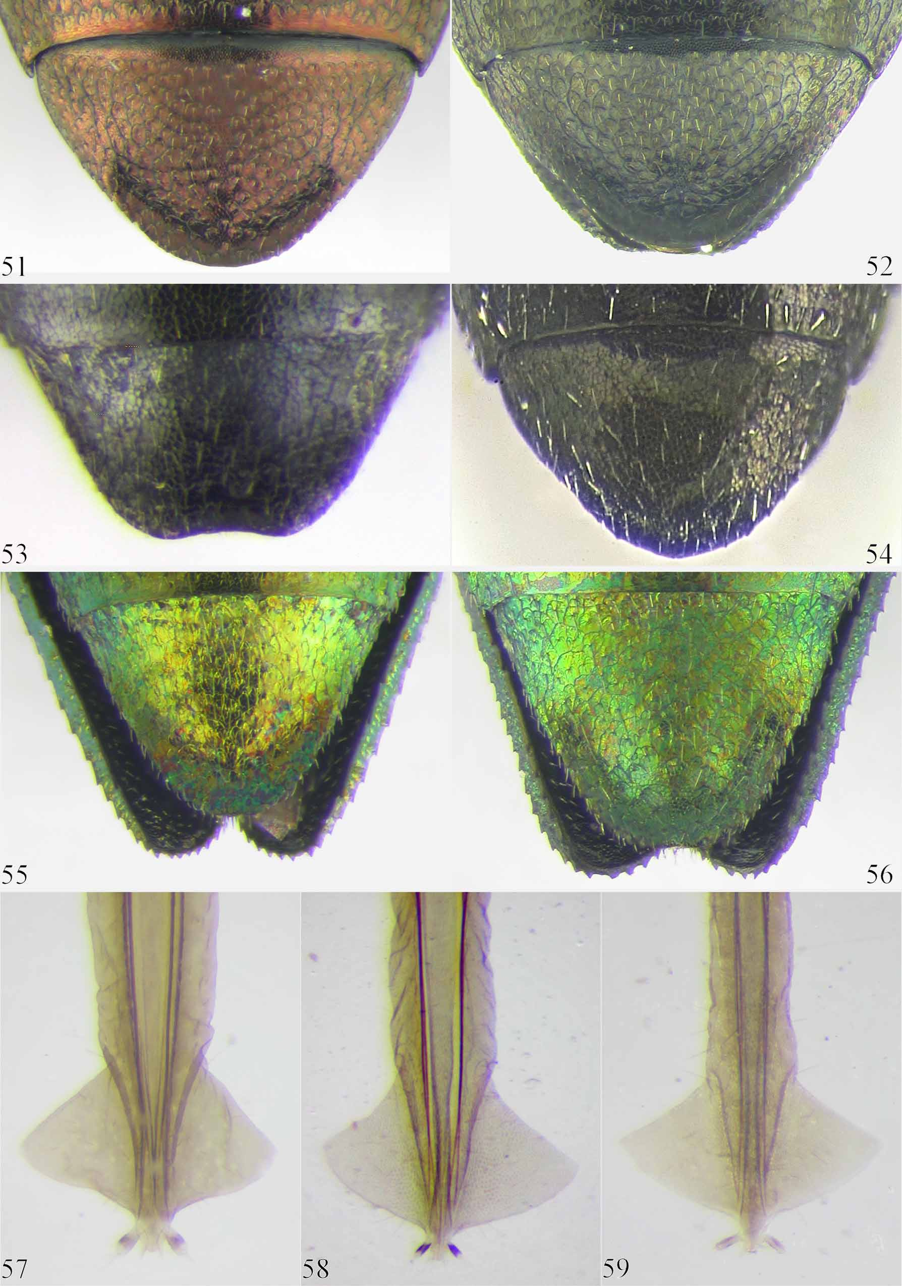
Variation. Medium-sized species (4.1 – 7.2 mm); body suboval, rather convex; the size of males ranges from 4.3 mm x 1.7 mm to 6.9 mm x 2.8 mm (holotype: 6.0 mm x 2.5 mm), while the females vary from 4.1 mm x 1.7 mm to 7.2 mm x 3.0 mm (allotype: 6.0 mm x 2.5 mm). Sexual dichroism absent, except for the colouration of antennae, which in the female are completely dark bronze ( Fig. 3 View FIGURES 1 – 4 ); the female also differs with the following features: lower part of inner ocular margins usually less divergent; meso- and metatibiae with smooth inner margins; posterior margin of anal ventrite slightly angulated at apex. Ovipositor of the allotype illustrated ( Fig. 57 View FIGURES 51 – 59 ). Concerning the morphological characters, A. magnanii has proved to be poorly variable, although convexity and sculpture of pronotun can be more or less pronounced, as well as the depth of the median frontal depression; in several specimens the widest point of the body is located on the posterior 1/2 of the elytra; the shape of male genitalia is rather constant, but in some specimens the subcylindrical basal portion of parameres is slightly longer; concerning the colouration, a weak variability is present in the extension of the basal yellow colouration of male antennomeres; some specimens show a deeper coppery tinge on head and pronotum, while in others it can be slightly more greenish; generally, the colouration of the body may be somewhat lighter or darker, but never so dark as in closely related species.
Specimens examined. Holotype, 3: Iran, ( Fārs), 2050 m., 7 km W Dašt-e Arjan, 29°38’N 51°54’E, 27 – 29.IV.2006, Baiocchi leg. / ex larva Fraxinus sp. 28.III.2007; allotype, Ƥ: Iran, ( Fārs), 2050 m., 7 km W Dašt-e Arjan, 29°38’N 51°54’E, 27 – 29.IV.2006, Baiocchi leg. / ex larva Fraxinus sp. 20.III.2007; paratypes: Iran, ( Fārs), 2050 m., 7 km W Dašt-e Arjan, 29°38’N 51°54’E, 12.V.2005, Baiocchi leg. (1 Ƥ); Iran, ( Fārs), 2050 m., 7 km W Dašt-e Arjan, 29°38’N 51°54’E, 27 – 29.IV.2006, Baiocchi leg. / ex larva Fraxinus sp. 16 – 28.III.2007 (23 3 25 ƤƤ); Iran, ( Fārs), 2050 m., 7 km W Dašt-e Arjan, 29°38’N 51°54’E, 25.IV.2007, Baiocchi leg. / ex larva Fraxinus sp. 4 – 20.III.2008 (5 3 9 ƤƤ); Iran, ( Fārs), 2050 m., 7 km W Dašt-e Arjan, 29°38’N 51°54’E, 25.IV.2007, Baiocchi leg. / ex larva Fraxinus sp. II.2009 (19 3 19 ƤƤ); Iran, ( Fārs), 2050 m., 7 km W Dašt-e Arjan, 29°38’N 51°54’E, 4 – 6.V.2008, Baiocchi leg. / ex larva Fraxinus sp. 5 – 29.III.2009 (26 3 49 ƤƤ); Iran, ( Fārs), 2050 m., 7 km W Dašt-e Arjan, 29°38’N 51°54’E, 4 – 6.V.2008, Baiocchi leg. / ex larva Fraxinus sp. 17 – 29.III.2010 (3 3 5 ƤƤ); Iran, ( Fārs), 2050 m., 7 km W Dašt-e Arjan, 29°38’00’’N 51°54’50.7’’E, 1 – 3.V.2009, Baiocchi leg. (7 3 2 ƤƤ); Iran, ( Fārs), 2050 m., 7 km W Dašt-e Arjan, 29°38’00’’N 51°54’50.7’’E, 1 – 3.V.2009, Baiocchi leg. / ex larva Fraxinus sp. 4 – 17.III.2010 (2 3 3 ƤƤ); Iran, ( Fārs), m. 2050, 7 km. W Dašt-e Arjan, N29°38’00” E51°54’50.7”, 27.IV.2006, D. Gianasso leg. (1 3); Iran, ( Fārs), m. 2050, 7 km. W Dašt-e Arjan, N29°38’00” E51°54’50.7”, 27.IV.2007, D. Gianasso leg. (1 Ƥ); Iran, ( Fārs), m. 2050, 7 km. W Dašt-e Arjan, N29°38’00” E51°54’50.7”, 27.IV.2007, D. Gianasso leg. / ex larva Fraxinus rotundifolia 11.II–16.III.2008 (2 3 3 ƤƤ); Iran, ( Fārs), m. 2050, 7 km. W Dašt-e Arjan, N29°38’00” E51°54’50.7”, 4 – 6.V.2008, D. Gianasso leg. (2 ƤƤ); Iran, ( Fārs), m. 2050, 7 km. W Dašt-e Arjan, N29°38’00” E51°54’50.7”, 4 – 6.V.2008, D. Gianasso leg. / ex larva Fraxinus rotundifolia 26.I – 12.III.2009 (40 3 47 ƤƤ); Iran, ( Fārs), m. 2050, 7 km W Dašt-e Arjan, N29°38’00” E51°54’50.7”, 4 – 6.V.2008, D. Gianasso leg. / ex larva Fraxinus rotundifolia [adult dead in pupal cell], 3.V.2009 (1 ex.); Iran, ( Fārs), m. 2050, 7 km. W Dašt-e Arjan, N29°38’00” E51°54’50.7”, 1 – 3.V.2009, D. Gianasso leg. (3 3 2 ƤƤ); Iran, ( Fārs), m. 2050, 7 km. W Dašt-e Arjan, N29°38’00” E51°54’50.7”, 1 – 3.V.2009, D. Gianasso leg. / ex larva Fraxinus rotundifolia 28. II.2010 (1 3); Iran – Fārs, m. 2.055, 7 km W. Dašt-e Arjan, 25.IV.2007, leg. G. Magnani (2 3 2 ƤƤ); Iran – Fārs, m. 2.050, 7 km. W. Dašt-e Arjan, 25.IV.2007, leg. G. Magnani / ex larva Fraxinus sp. 08.III.2008 (2 3 1 Ƥ); Iran – Fārs, m. 2.050, 7 km. W. Dašt-e Arjan, 25.IV.2007, leg. G. Magnani / ex larva Fraxinus sp. 10.III.2008 (2 3 1 Ƥ); Iran – Fārs, m. 2.050, 7 km. W. Dašt-e Arjan, 25.IV.2007, leg. G. Magnani / ex larva Fraxinus sp. 15.III.2008 (4 3 5 ƤƤ); Iran – Fārs, m. 2.050, 7 km. W. Dašt-e Arjan, 25.IV.2007, leg. G. Magnani / ex larva Fraxinus sp. 18.III.2008 (1 3); Iran – Fārs, m. 2.050, 7 km. W. Dašt-e Arjan, 25.IV.2007, leg. G. Magnani / ex larva Fraxinus sp. 20.III.2009 (1 3 1 Ƥ); Iran – Fārs, m. 2.050, 7 km. W. Dašt-e Arjan, 25.IV.2007, leg. G. Magnani / ex larva Fraxinus sp. [adults dead in pupal cell], IV.2010 (9 3 7 ƤƤ).
The holotype, allotype and some paratypes are deposited in D. Baiocchi collection (Rome, Italy); paratypes in the following collections: Centro Iberoamericano de la Biodiversidad (CIBIO) of the University of Alicante ( Spain); National Musem of Prague (Czech Rep.), Plant Pests and Diseases Research Institute (Tehran, Iran), Zoological Institute - Russian Academy of Sciences (St. Petersburg, Russia), C. L. Bellamy (Sacramento, U.S.A.), S. Bílý (Prague, Czech Rep.), J. De La Rosa (Madrid, Spain), D. Gianasso (Castelnuovo Don Bosco, Italy), M. Gigli (Rome, Italy), F. Izzillo (Naples, Italy), M. Kafka (Neratovice, Czech Rep.), V. Kubáň (Brno, Czech Rep.), A. Liberto (Rome, Italy), G. Magnani (Cesena, Italy), H. Mühle (Munich, Germany), M. Niehuis (Albersweiler, Germany), G. Novak (Wien, Austria), and M. Škorpík (Lukov, Czech Rep.).
Comments. Despite some morphological and bionomical affinities with A. bicolor , we consider A. magnanii to be more closely related to A. (Anthaxia) chaerodrys Szallies, 2001 ( Fig. 2 View FIGURES 1 – 4 ), a relict species with a peculiar distribution, and known so far only from Turkey ( Szallies 2001) and Spain ( Arnáiz Ruiz & Bercedo Páramo 2007 a, b; De La Rosa 2007). Both species show an overall dark colouration resembling that of A. (Anthaxia) vladivostokana Obenberger, 1938 and A. (Anthaxia) majzlani Bílý, 1991 , the two easternmost species of the A.dimidiata-fulgurans species-group. We agree with the opinion of Bílý (1991) according to whom, although showing a rather different pattern of pronotal sculpture, A. majzlani and A. vladivostokana represent a separate complex of dark species within their species-group, in which we consider are to be included also A. chaerodrys and A. magnanii .
At the time Obenberger gave his definition of the A. dimidiata species-group ( Obenberger 1917), all the above mentioned taxa were still unknown and, for the same reason, most of them had neither been included in the A.dimidiata-fulgurans species-group as defined by Bílý (Bílý 1984) and Niehuis (Niehuis 1990); since then, further new species have been described, and the group is in the need of a new, more detailed revision.
Anthaxia magnanii View in CoL n. sp. Anthaxia chaerodrys Szallies, 2001 View in CoL
Body ( Fig. 1 View FIGURES 1 – 4 ) suboval, brilliant light bronze, usually with a Body ( Fig. 2 View FIGURES 1 – 4 ) subparallel, more angularly shaped, uniformely slight more coppery tinge on head and pronotum dark brown, with very faint purple tinge on pronotal lateral margins, elytral base and along a short basal portion of the elytral suture
Head ( Fig. 13 View FIGURES 13 – 18 ) slightly narrower than anterior pronotal mar- Head ( Fig. 16 View FIGURES 13 – 18 ) distinctly narrower than anterior pronotal margin; vertex 0.4 times as wide as width of head; pubescence of gin; vertex narrower, 0.3 times as wide as width of head; frons yellowish; upper lobe of eye acutely rounded; frons pubescence of frons withish; upper lobe of eye more obtusely ( Fig. 31 View FIGURES 31 – 38 ) with bright copper colouration; anterior margin of rounded; frons ( Fig. 32 View FIGURES 31 – 38 ) with strong purple tinge, greenish at clypeus distinctly more deeply emarginate lower ocular angles; anterior margin of clypeus weakly emar-
ginate
Antennae ( Fig. 39 View FIGURES 39 – 50 ) 1.2 times longer than middle pronotal Antennae ( Fig. 40 View FIGURES 39 – 50 ) 1.1 times longer than middle pronotal
length; scape with equal lateral sides; antennomeres 5 – 10 length; scape with inner lateral side strongly gibbous, outer slender, acutely triangular shaped; last antennomere more side slightly bent outward; antennomeres 5 – 10 more compact, elliptically elongate distinctly trapezoidal; last antennomere subrhomboidal
Pronotum ( Fig. 13 View FIGURES 13 – 18 ) less transverse, 1.7 times wider than Pronotum ( Fig. 16 View FIGURES 13 – 18 ) more transverse, 1.8 times wider than long, long, widest at midlength; lateral margins uniformely arched, widest at anterior 1/3; lateral margins distincly angulated at distinctly sinuate before posterior angles; lateroposterior anterior 1/3, more directly tapered posteriorly; lateroposterior depressions smaller, extended for 1/3 of the pronotal length; depressions wider, extended for 1/2 of the pronotal length; disdiscal sculpture deeper, distinctly curvilinear, consisting of cal sculpture shallower, less clearly curvilinear, consisting of larger, contiguous, irregularly lengthened cells, bordered by smaller, irregularly polygonal, areolate cells, bordered by shaldeep furrows low furrows
Elytra tapereded on distal 1/3; inner portion of basal depres- Elytra tapered on distal 1/4; inner portion of basal depression sion shallow; humeral swellings well developed; apex (Fig. deep; humeral swellings poorly developed; apex ( Fig. 38 View FIGURES 31 – 38 ) 37) not incurved downward; apical lateromarginal punctures slightly bent downward, deeply punctured along the apicolatabsent eral margins
Apical portion of foretibiae slightly sinuate externally; inner Apical portion of foretibiae straight externally; inner margin of margin of mesotibiae weakly incised, feebly serrate on distal mesotibiae straight, slightly serrate on distal 1/3; metatibiae 1/3; metatibiae ( Fig. 45 View FIGURES 39 – 50 ) flattened distally, inner margin ( Fig. 46 View FIGURES 39 – 50 ) thicker, inner margin straight, acutely serrate on distal incised, slightly serrate on apical 1/4; mesotarsomere 1 1/3; mesotarsomere 1 much longer than 2; metatarsomere 1 slightly longer than 2; basal metatarsomere 1.5 times longer twice as long as 2
than 2
Aedeagus: narrowed portion of parameres ( Fig. 14 View FIGURES 13 – 18 ) longer Aedeagus: narrowed portion of parameres ( Fig. 17 View FIGURES 13 – 18 ) shorter (1/ (nearly 1/2 of parameral length); dorsal surface of median 3 of parameral length); dorsal surface of median lobe ( Fig. 18 View FIGURES 13 – 18 ) lobe ( Fig. 15 View FIGURES 13 – 18 ) more distinctly rugulose on anterior portion less rugulose on anterior portion
Some of the distinctive morphological features of the A. dimidiata-fulgurans species-group, in this new species seem to be poorly developed or even completely absent and, although characters as the partial yellow colouration of male antennomeres, the typical bottleneck shape of anterior portion of the aedeagus, and the peculiar pronotal sculpture, undoubtly set A. magnanii View in CoL in this group, the lateroposterior depressions of pronotum more shallow than in close species, and the absence of deep punctures along the lateroapical elytral margins ( Fig. 37 View FIGURES 31 – 38 ), denote that it is one of the most derived species within it.
The character states that differentiate A. magnanii View in CoL from A. chaerodrys View in CoL , are listed in table 1.
Bionomy and distribution. At present A. magnanii View in CoL has been collected only in its “locus typicus” ( Fig. 60, 63 View FIGURES 60 – 63 ), but its peculiar timid and hiding behaviour, shared also with its closest species A. (Anthaxia) chaerodrys Szallies, 2001 View in CoL , ( Fig. 2 View FIGURES 1 – 4 ) suggests that it might be more widespread than it actually seems; nearly all known specimens of both species were reared exclusively from branches of Fraxinus View in CoL species, while only few specimens have been caught on yellow flowers of Asteraceae View in CoL , and few others were found walking on fallen dead branches, or hiding under bark ( Szallies 2001; De La Rosa pers. comm.).
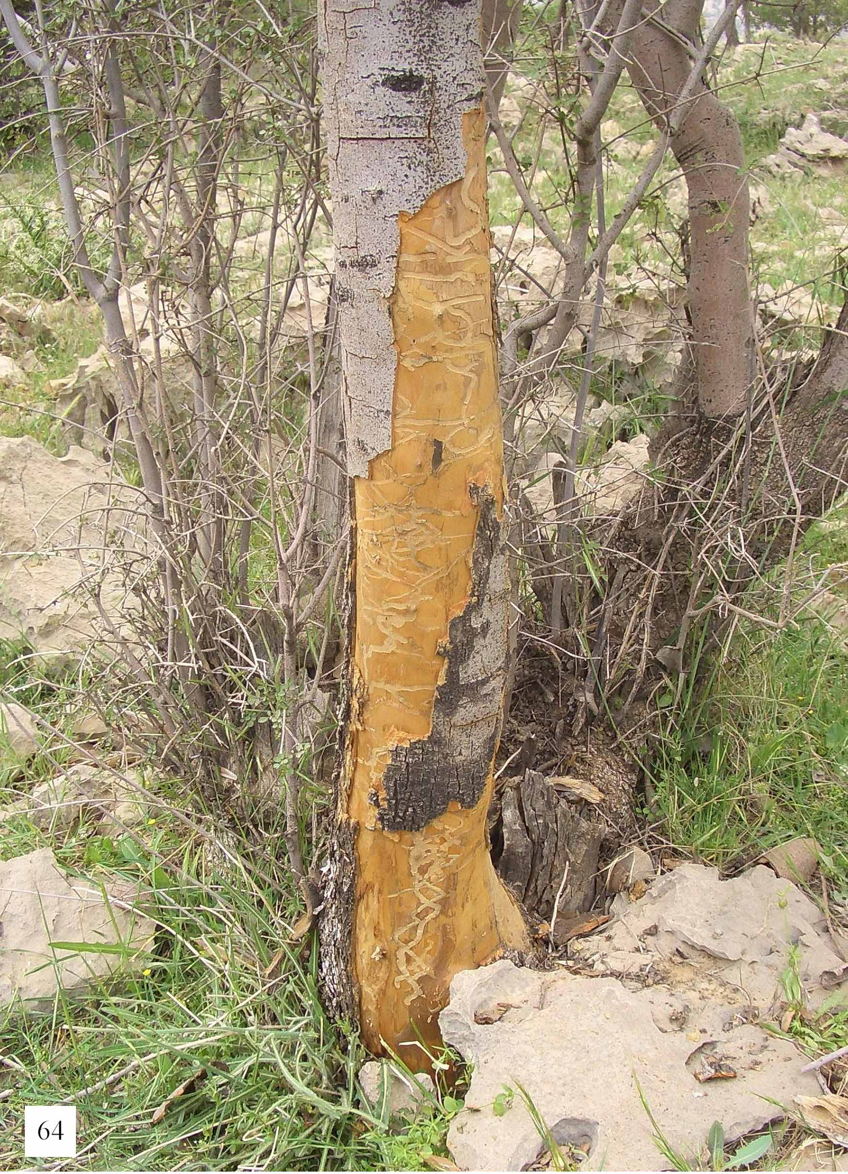
The larva of A. magnanii View in CoL develops preferably in the trunk and in large size branches of its host plant, boring long, tortuous galleries under the bark ( Fig. 64 View FIGURE 64 ); the larval development takes two years, and the imago spends the second winter as adult in its pupal cell, ready to emerge in early spring.
Other species of Anthaxia View in CoL which emerged from samples of Fraxinus View in CoL collected in the same locality are the following: A. (Anthaxia) bicolor Faldermann, 1836 View in CoL , the rare A. (Anthaxia) morgani Théry, 1925 View in CoL (both last two species belonging to the A. dimidiata-fulgurans species-group, like A. magnanii View in CoL ) and few specimens of A. (Haplanthaxia) farah Bílý, 1983 View in CoL , here reported for first time from Fraxinus View in CoL , and probably generated by stray females, since its main host plant ( Pistacia View in CoL sp.) in this locality is found only at a lower altitude.
Etymology. This new species is dedicated to our friend Gianluca Magnani, a keen expert of Buprestidae , with whom we have enjoyed so many entomological adventures.


No known copyright restrictions apply. See Agosti, D., Egloff, W., 2009. Taxonomic information exchange and copyright: the Plazi approach. BMC Research Notes 2009, 2:53 for further explanation.
|
Kingdom |
|
|
Phylum |
|
|
Class |
|
|
Order |
|
|
Family |
|
|
Genus |
Anthaxia (Anthaxia) magnanii
| Baiocchi, Daniele 2011 |
Anthaxia chaerodrys
| Szallies 2001 |
Anthaxia (Anthaxia) chaerodrys
| Szallies 2001 |
A. (Anthaxia) chaerodrys
| Szallies 2001 |
A. (Haplanthaxia) farah Bílý, 1983
| Bily 1983 |
A. (Anthaxia) morgani Théry, 1925
| Thery 1925 |
A. (Anthaxia) bicolor
| Faldermann 1836 |