
Arthrostoma supriatnai, Dewi & Purwaningsih & Hasegawa, 2023
|
publication ID |
https://doi.org/ 10.11646/zootaxa.5353.1.7 |
|
publication LSID |
lsid:zoobank.org:pub:195E5266-9F40-4232-9538-7B8CD08BF58B |
|
DOI |
https://doi.org/10.5281/zenodo.8434991 |
|
persistent identifier |
https://treatment.plazi.org/id/32C47586-E20B-441D-872E-2D0E361922CD |
|
taxon LSID |
lsid:zoobank.org:act:32C47586-E20B-441D-872E-2D0E361922CD |
|
treatment provided by |
Plazi |
|
scientific name |
Arthrostoma supriatnai |
| status |
sp. nov. |
Arthrostoma supriatnai sp. nov.
( Ancylostomatidae : Ancylostomatinae : Ancylostomatini: Arthrocephalinii)
( Figs. 1a–l View FIGURES 1 ; 2a–d View FIGURES 2 )
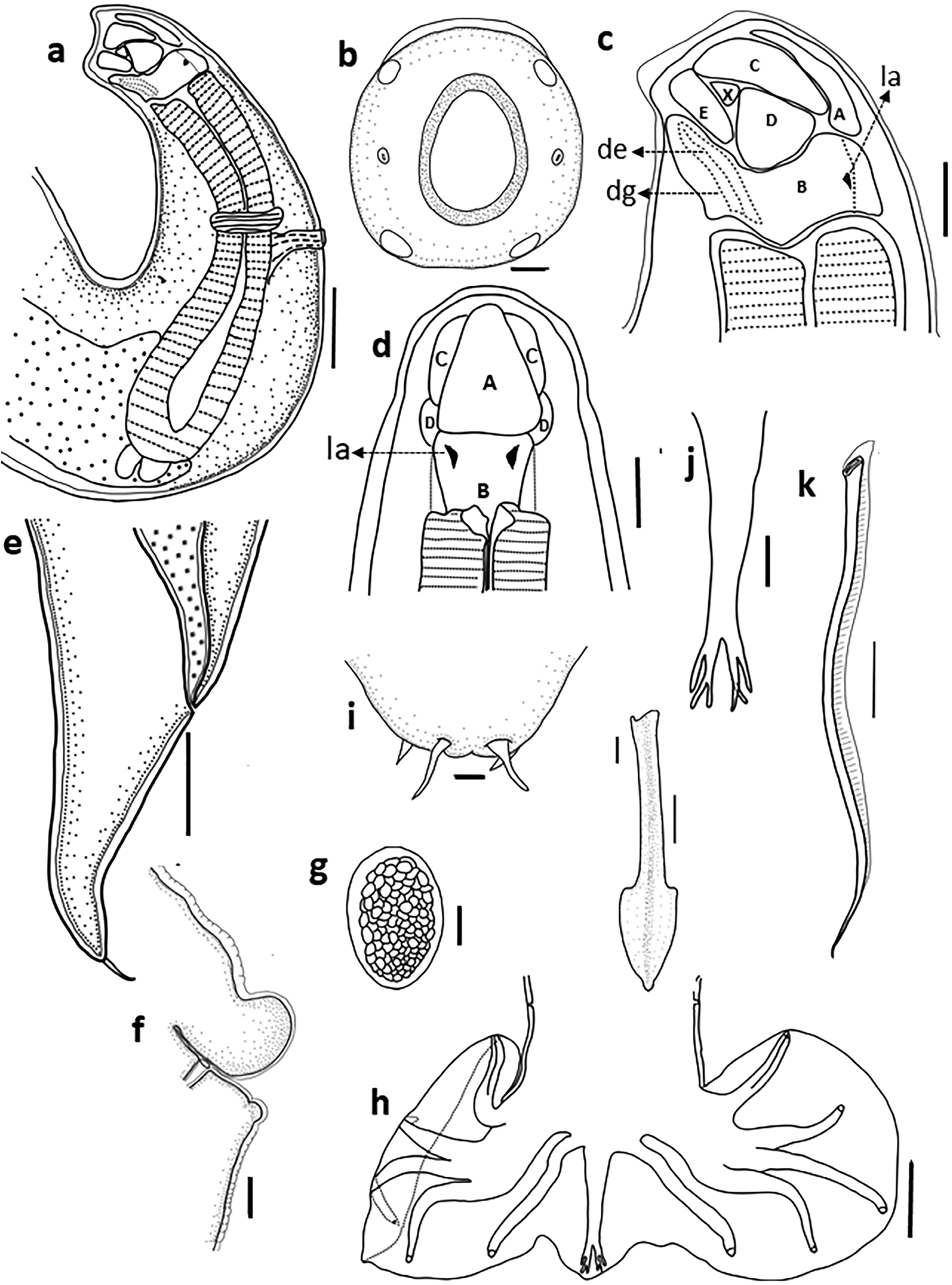
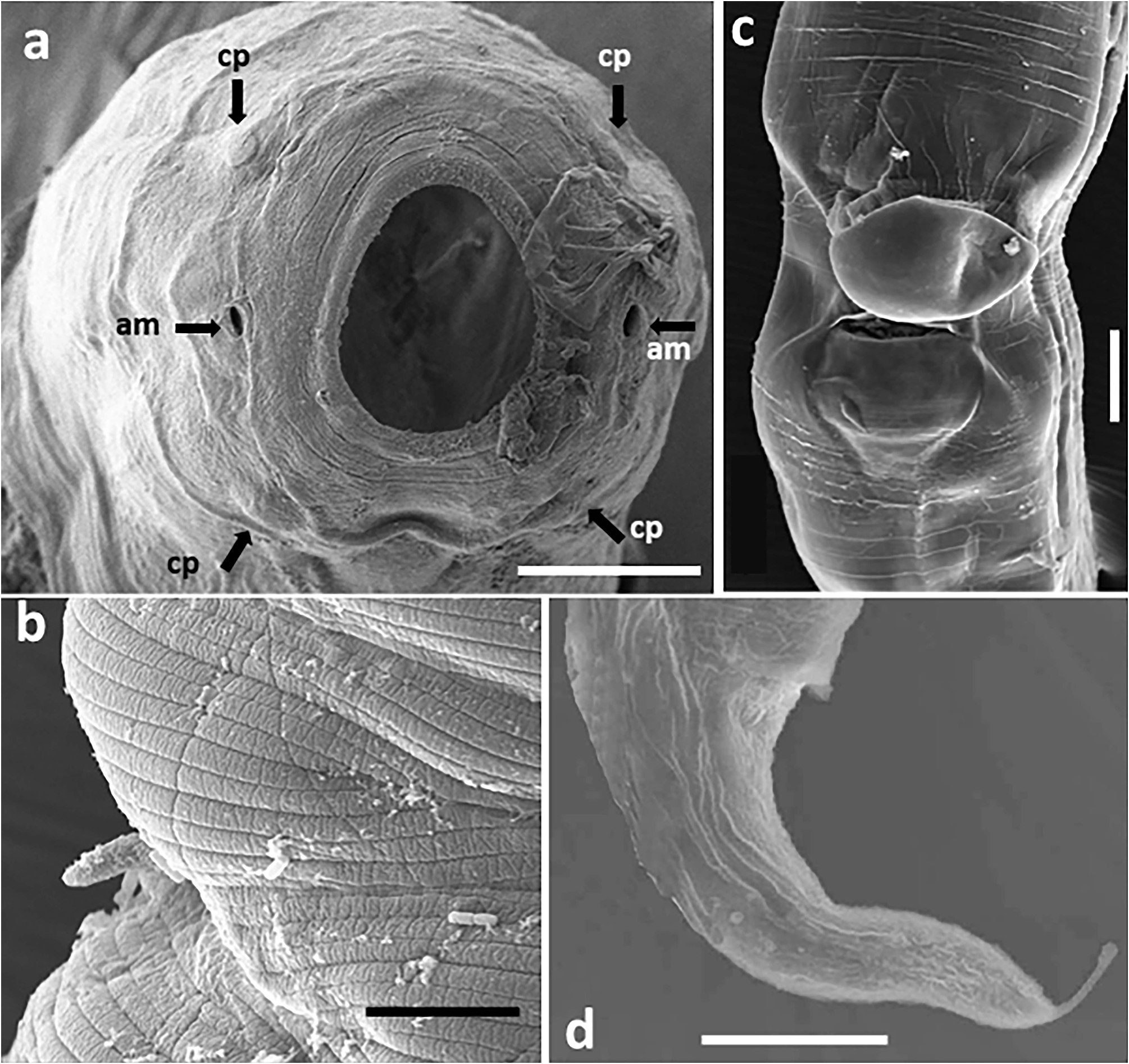
General: Small and stout worm, cuticle transversely striated with striae 2.5–2.9 (2.7) apart. Anterior end curved dorsally. Mouth opening directed antero-dorsally, surrounded by four papillae and two amphids ( Fig. 1b View FIGURES 1 , 2a View FIGURES 2 ). Buccal capsule pyriform in lateral view, composed of ten articulating sclerotized plates ( Figs. 1a, c, d View FIGURES 1 ), consisting of one large basal plate (B), two lateral plates (D), two laterodorsal (E), two lateroventral (C), one large triangular ventral plate (A), and two additional lateral plates (X). Mouth lacking teeth or cutting plates, but with one pair of ventral lancets ( Figs. 1a, c View FIGURES 1 ). Dorsal esophageal gland well developed with duct passing in dorsal gutter ( Fig. 1c View FIGURES 1 ). Esophagus club shaped, slightly swollen posteriorly, terminating in a lobed valve at junction with intestine; intestinal diverticulum present ( Fig 1a View FIGURES 1 ). Cervical papillae finger-shaped ( Fig. 2b View FIGURES 2 ).
Male: Total length 2.92 (2.72–3.11) mm; maximum width at level of posterior end of esophagus 304 (282–320), buccal capsule 101 (99–102) deep, 64 (63–66) wide. Nerve ring, excretory pore, and cervical papillae 311 (306– 318), 318 (305–330), and 418 (396–435) from anterior end, respectively. Esophagus length 515 (480–590), width 118 (111–124). Copulatory bursa symmetrical with large lateral lobes and small dorsal lobe, prebursal papillae small ( Fig. 1h View FIGURES 1 ). Ventral rays stout, parallel, close together, cleft at middle of length. Lateral rays divergent from each other, externolateral shortest, mediolateral almost same length with posterolateral. Externodorsal rays derived from base of dorsal ray. Dorsal ray long, bifurcated near distal end, each branch bifid into outer and inner offshoots, and each inner offshoot divided again apically ( Fig.1j View FIGURES 1 ). Simple genital cone present with one pair of tiny dorsal raylets and one pair of ventral cuticular appendages. Spicules 372 (350–390) long, equal and similar, slender tubular, transversely striated alae, with sharply pointed tip ( Fig. 1k View FIGURES 1 ). Gubernaculum arrow-shaped, 104 (102–105) long ( Fig. 1l View FIGURES 1 ).
Female: Total length 3.51 (3.28–3.77) mm; maximum width 323 (310–336) at level of vulva. Buccal capsule 120 (119–122) deep. Nerve ring, excretory pore, and cervical papillae 349 (315–405), 367 (339–430), and 476 (463–497) from anterior end, respectively. Esophagus length 675 (600–690), width 137 (123–147). Vulva at 1.44 (1.35–1.61) mm from posterior end, with large and protruding prevulval flap and postvulval spherical swelling ( Figs. 1f View FIGURES 1 , 2c View FIGURES 2 ). Vagina short, uteri opposed. Tail 360 (324–390) long, tapering to elongate, sharp terminal spike ( Figs. 1e View FIGURES 1 , 2d View FIGURES 2 ). Eggs not embryonated in uteri, oval, thin–shelled, 62 (60–65) x 39 (30–50) ( Fig. 1g View FIGURES 1 ).
No known copyright restrictions apply. See Agosti, D., Egloff, W., 2009. Taxonomic information exchange and copyright: the Plazi approach. BMC Research Notes 2009, 2:53 for further explanation.