
Nesodiprion tsugae Togashi, 2001
|
publication ID |
https://doi.org/10.11646/zootaxa.4007.4.2 |
|
publication LSID |
lsid:zoobank.org:pub:6EEDD233-746F-4A04-84CD-565540676C60 |
|
DOI |
https://doi.org/10.5281/zenodo.5669552 |
|
persistent identifier |
https://treatment.plazi.org/id/643687C2-D858-FFB2-FF7B-FE1CF2B4F811 |
|
treatment provided by |
Plazi |
|
scientific name |
Nesodiprion tsugae Togashi, 2001 |
| status |
|
Nesodiprion tsugae Togashi, 2001
Figs 1A–D View FIGURES 1 A – K , 5A–C View FIGURES 5 A – I , 7A View FIGURES 7 A – J , 8A View FIGURES 8 A – J , 9A View FIGURES 9 A – L , 11A–B View FIGURES 11 A – L , 12A–B View FIGURES 12 A – M , 13A–B View FIGURES 13 A – K , 14D–E View FIGURES 14 A – Y , 15A–B, 16A, 17A–B, 18A
Nesodiprion tsugae Togashi, 2001: 915 ; Taeger et al. 2010: 210; Hara & Smith 2012: 22.
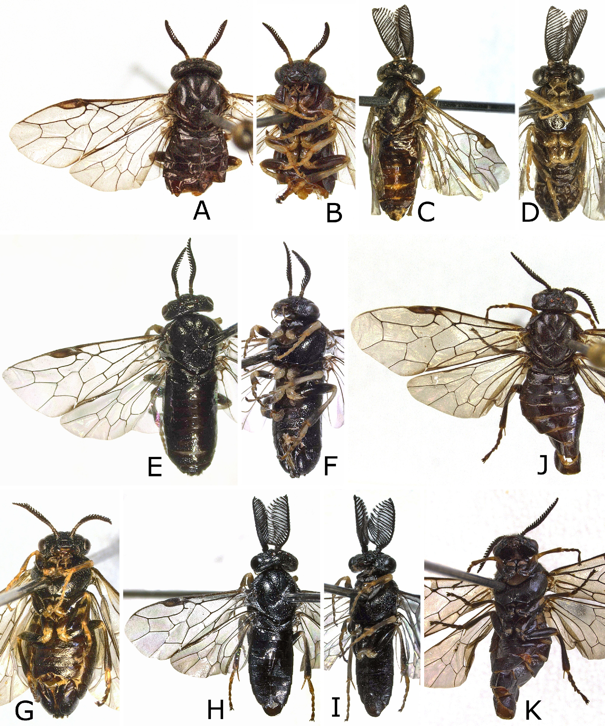
Additional description of female ( holotype). Shiny, without colored metallic reflections ( Figs 1A–B View FIGURES 1 A – K ). Palpi yellow, basally darkened. Tibial spurs brown.
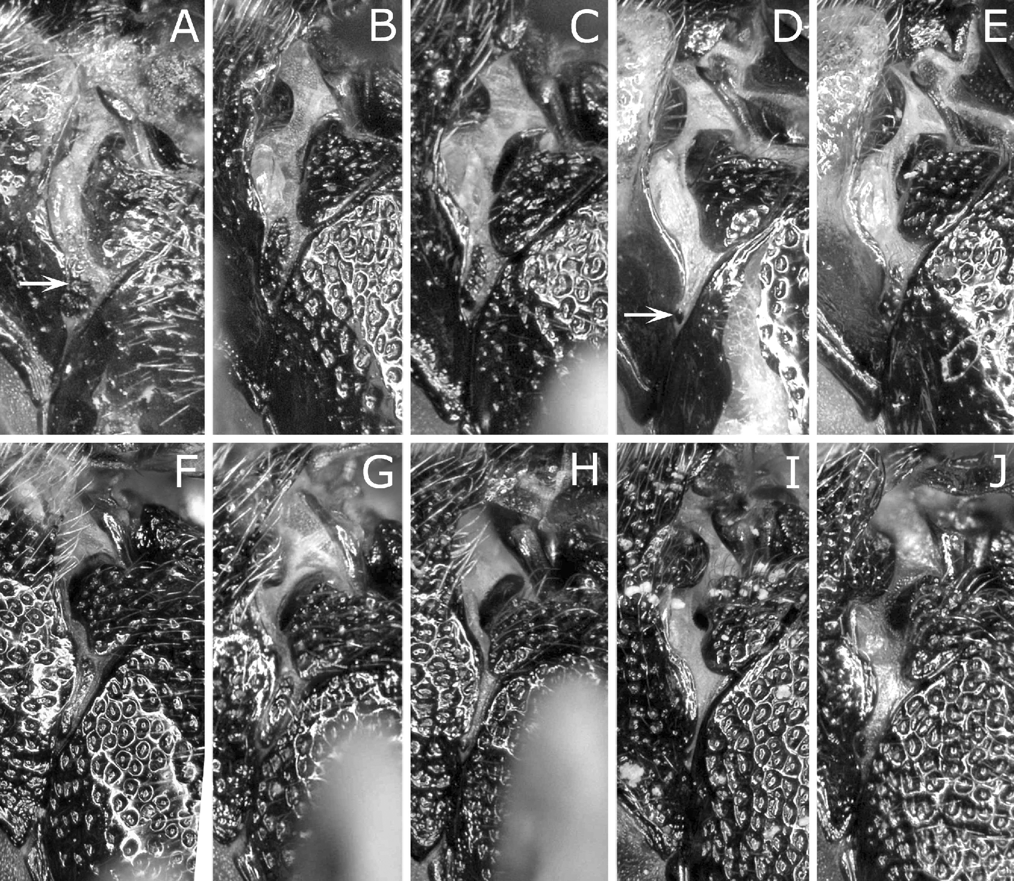
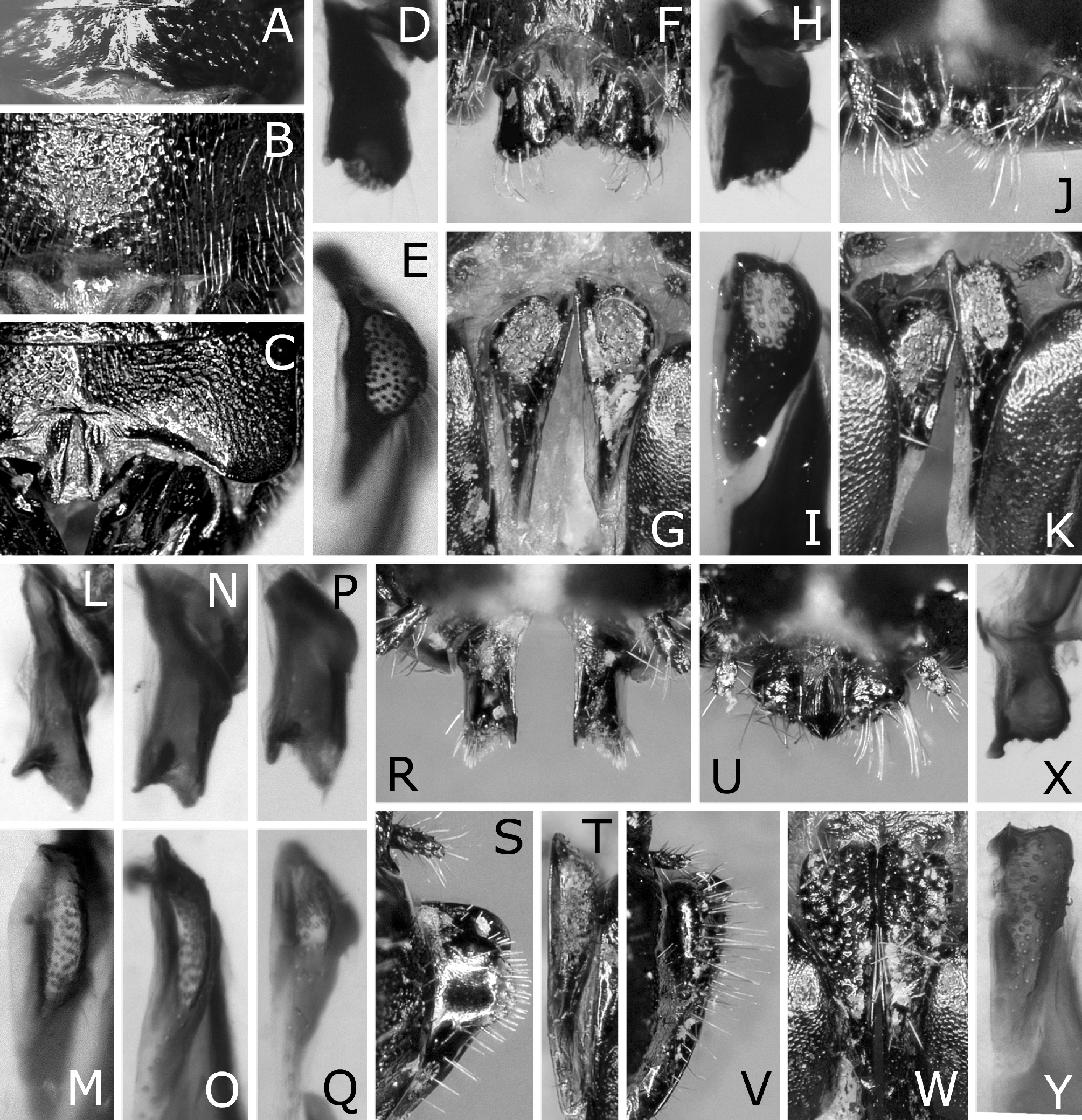
Head and thorax with punctures shallow and ill-defined or fine, partly large and well defined ( Figs 5A–B View FIGURES 5 A – I , 9A View FIGURES 9 A – L ); punctures on dorsal part of mesepisternum large, predominantly contiguous or fused ( Fig. 11A View FIGURES 11 A – L ). First tergum inconspicuously punctured on narrow medial area ( Fig. 13A View FIGURES 13 A – K ).
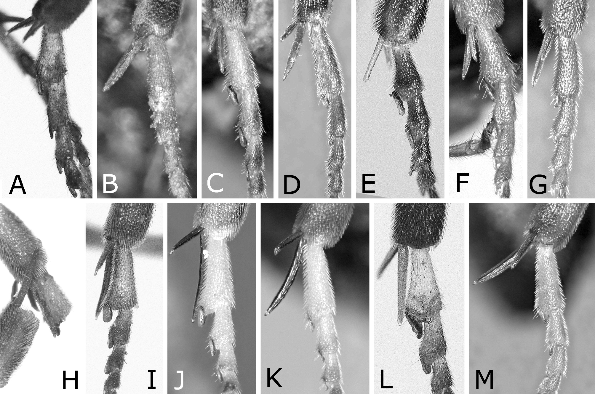
Distance between eyes 1.7 × eye height. Distance between eye and lateral ocellus 1.2 × distance between lateral ocellus and posterior margin of head. First flagellomere in lateral view with length along dorsal margin 1.1 × apical breadth except for ramus and ramus length 1.1 × its flagellomere length ( Fig. 7A View FIGURES 7 A – J ). Anterior edge of mesoscutellum angled about 100° ( Fig. 9A View FIGURES 9 A – L ). In hind leg in lateral view, first tarsomere length 1.1 × tibia breadth ( Fig. 12A View FIGURES 12 A – M ). In hind wing, section of vein 1A between crossveins a and cu-a 0.9 × as long as width of cell 1A.
Lance as in Figs 15A–B. Lancet (Fig. 16A) slightly concave on ventral margin in outline, with length from apex to ventral end of first row of spines 3.7 × maximum width and apex narrowly rounded; first (most basal) row of spines (ctenidium) curved, and its ventral end widely separated from ventral margin of lancet; first annulus without serrula; second row of spines sinuated; all serrulae posteriorly angularly convex; distance between serrulae of second and third annuli (d in Fig. 16A) about 2 × length of serrula of second annulus (s in Fig. 16A).
Description of male (hitherto undescribed). Length 5.0 mm ( Figs 1C–D View FIGURES 1 A – K ). As in female, but differing as follows except for usual sexual differences.
Punctures on head relatively distinct ( Fig. 5C View FIGURES 5 A – I ); those on dorsal part of mesepisternum predominantly shallow and ill defined ( Fig. 11B View FIGURES 11 A – L ); dorsum of abdomen apically shallowly punctured (in holotype, female, apex of abdomen missing).
Distance between eyes 1.5 × eye height. Postocellar area with width 1.9 × length. Ratio of distances between eye and lateral ocellus, between lateral ocelli and between lateral ocellus and posterior margin of head 1.0:1.0:0.7; ratio of distances between eye and lateral ocellus and between lateral ocellus and posterior margin of head 1.4:1.0 ( Fig. 5C View FIGURES 5 A – I ). Distance between torulus and eye 1.1 × distance between toruli. Malar space 1.0 × width of median ocellus. Antenna with 24 antennomeres. Anterior edge of mesoscutellum angled about 110°. In hind leg in lateral view, first tarsomere length 1.3 × tibia breadth ( Fig. 12B View FIGURES 12 A – M ). In hind wing, section of vein 1A between crossveins a and cu-a 1.2 × as long as width of cell 1A. Subgenital plate in ventral view with apical margin gently rounded ( Fig. 1D View FIGURES 1 A – K ).
Genital capsule ( Figs 14A–B View FIGURES 14 A – Y ) wide in dorsal or ventral view. Parapenis in ventral view wide, apically rounded. Harpe in ventral view with medial margin nearly straight, basally gently rounded, and apex narrowly rounded. Valviceps in dorsal view narrow and straight, in lateral view ( Fig. 18A View FIGURES 18 A – F ) wide, with dorsal margin nearly straight and distinctly convex near apex.
Material examined. Holotype ( Figs 1A–B View FIGURES 1 A – K ): ♀, “Togakushi, 6-V '85 Em. [emerged], Aut. [autumn] ' 84 L. [larva], H. Kojima leg. [Kometsuga (in Japanese, = Tsuga diversifolia )]” ( NSMT). Other material examined: JAPAN, HONSHU— 1♂, “[Togakushi], 6?V?1985 Em., Aut. ' 84 L., [Kometsuga] H. Kojima leg.” ( NSMT).
The data of the male is identical with that of the holotype. The collector, H. Kojima, sent two females and two males reared from a group of gregarious larvae on Tsuga diversifolia collected at Togakushi to the late Prof. Okutani at Kobe University on June 1985 (Kojima, personal communication, 2014). The holotype and the male are unquestionably part of those four specimens and siblings.
Distribution. Japan: Honshu.
Host plant. Pinaceae : Tsuga diversifolia (Maxim.) Mast.
Life history. A larval group was collected in autumn in a mountainous region in Honshu. Larvae are yellow orange and gregariously feed on needles (Kojima, personal communication, 2014, 2015).
Remarks. This species is very similar to N. kojimai sp. nov. below. For their differences, see under the remarks of the latter species and the key below.


| NSMT |
National Science Museum (Natural History) |
No known copyright restrictions apply. See Agosti, D., Egloff, W., 2009. Taxonomic information exchange and copyright: the Plazi approach. BMC Research Notes 2009, 2:53 for further explanation.
|
Kingdom |
|
|
Phylum |
|
|
Class |
|
|
Order |
|
|
Family |
|
|
Genus |
Nesodiprion tsugae Togashi, 2001
| Hara, Hideho & Smith, David R. 2015 |
Nesodiprion tsugae
| Hara 2012: 22 |
| Taeger 2010: 210 |
| Togashi 2001: 915 |