
Fagineura togashii Hara, 2022
|
publication ID |
https://doi.org/ 10.11646/zootaxa.5116.2.3 |
|
publication LSID |
lsid:zoobank.org:pub:EBF12EE4-4675-45AC-80C1-CFA61FD0C297 |
|
DOI |
https://doi.org/10.5281/zenodo.6369217 |
|
persistent identifier |
https://treatment.plazi.org/id/65401F59-C00C-FF89-FF6A-2F9EFBAAF9EC |
|
treatment provided by |
Plazi |
|
scientific name |
Fagineura togashii Hara |
| status |
nom. nov. |
Fagineura togashii Hara , nom. nov.
( Figs 2G–N View FIGURE 2 , 3A–K View FIGURE 3 , 6F, G, N, R View FIGURE 6 , 7C, F, L, R, S View FIGURE 7 , 8F, I, Q View FIGURE 8 , 9C, K View FIGURE 9 , 10D, E View FIGURE 10 , 12A, B View FIGURE 12 , 14C, I, J, O, P View FIGURE 14 )
Fagineura quercivora Togashi, 2006: 169 ; Taeger et al. 2010: 405; Tazunoki et al. 2018: 142; Hara 2019: 77; Hara 2020: 82, 339; Liu et al. 2019: 33; Liu et al. 2021: 130. Junior secondary homonym of Fagineura quercivora ( Togashi, 1997) [= Dineura quercivora Togashi, 1997 ].
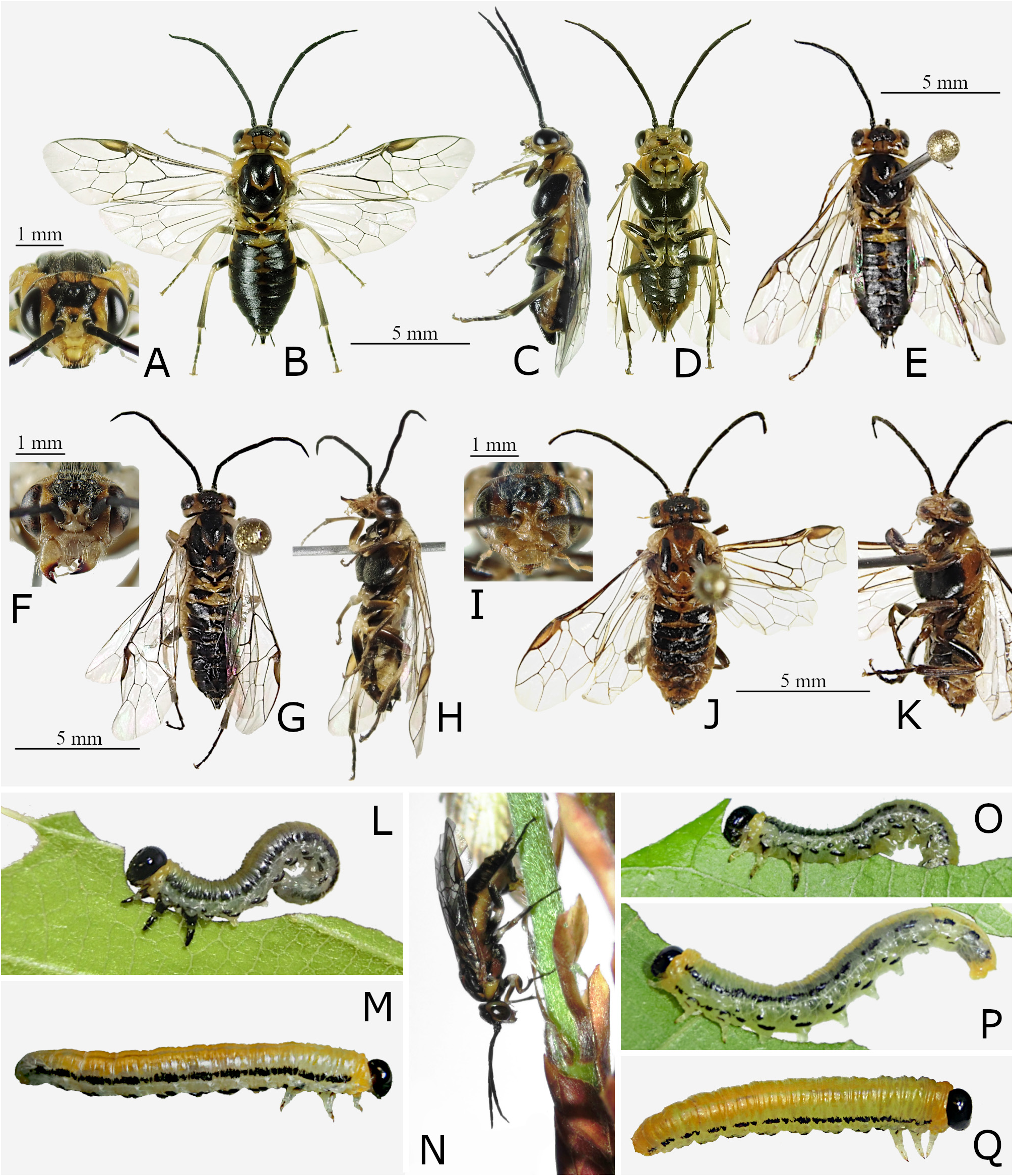
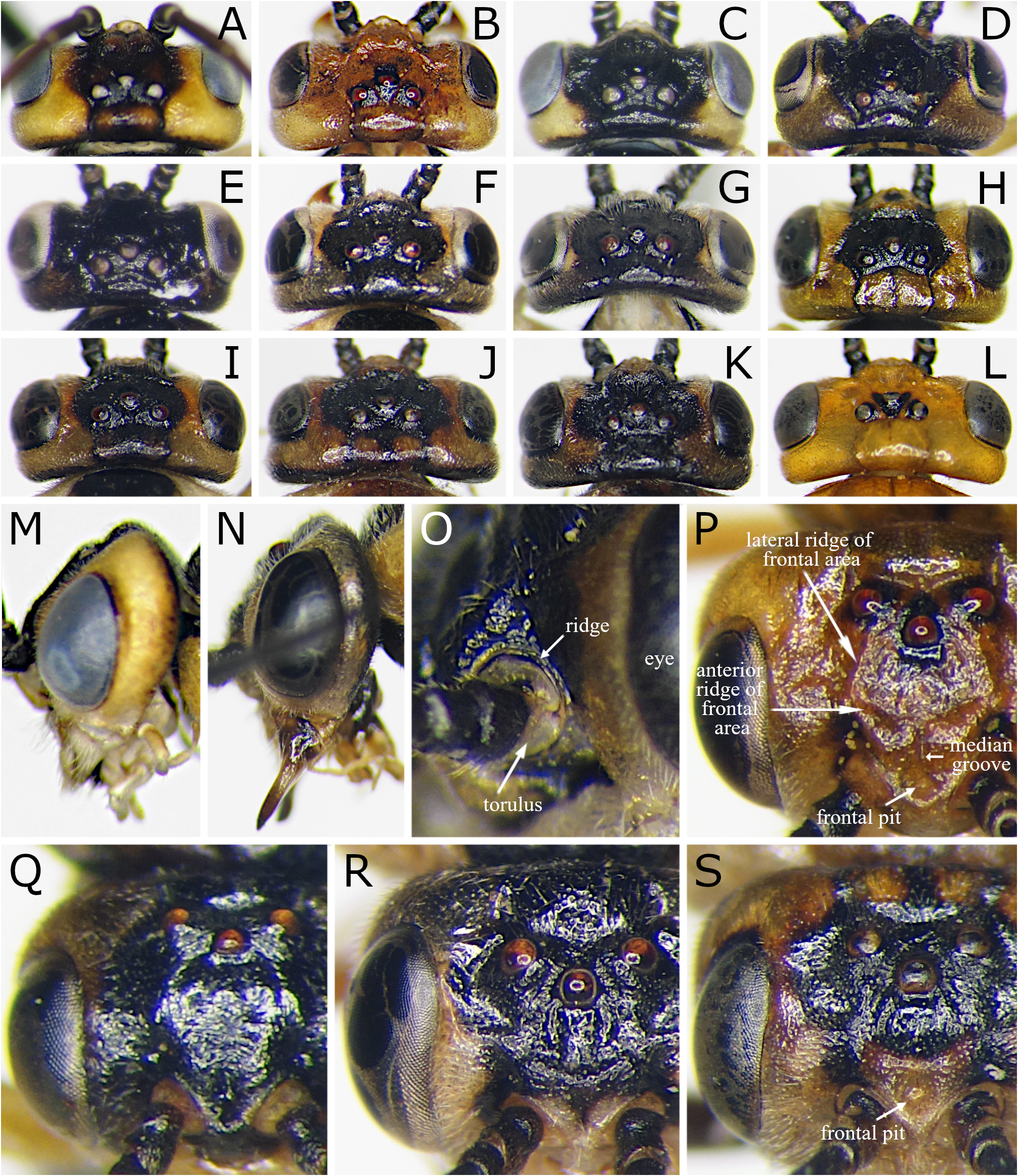
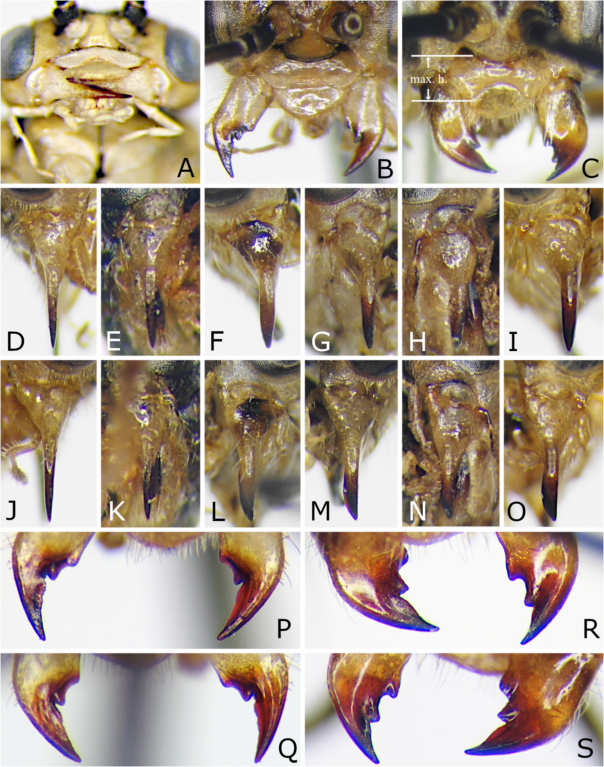
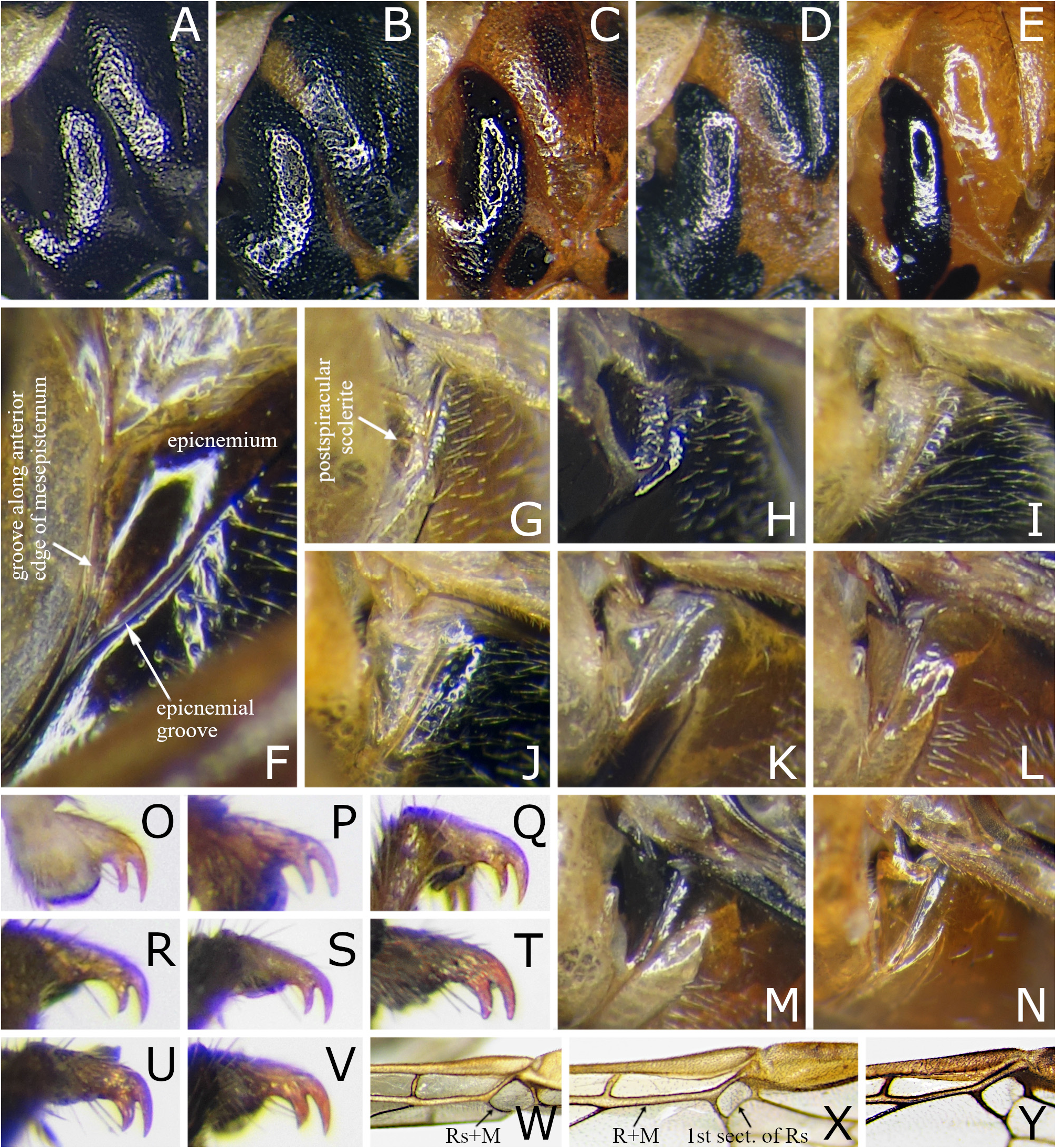
Additional description: female and male. Female Figs 2G–J View FIGURE 2 . Male Figs 2K–N View FIGURE 2 . Length 7.0– 10.5 mm in female, 6.0–7.0 mm in male. Head in dorsal view with length behind eye 0.3–0.5 × eye length in female, 0.3–0.4 × in male ( Figs 6F, G View FIGURE 6 ); length behind lateral ocellus 2.6–3.5 × length of lateral ocellus in female, 1.9–2.2 × in male. OOL: POL:OOCL 0.8–1.0:1.0: 1.1–1.2 in female, 0.8–0.9:1.0: 0.9–1.2 in male. Frontal area with lateral ridge low and anterior ridge well developed ( Fig. 6R View FIGURE 6 ); frontal field with long lateral convexity. Distance between eyes at anterior tentorial pit 1.1–1.2 × major axis of eye in female, 1.0–1.1 × in male ( Figs 2G, K View FIGURE 2 ). Inner edges of eyes nearly parallel. Malar space 0.2–0.3 × as long as median ocellus width. Antenna 2.3–2.4 × as long as head width in female, 2.7–2.8 × in male ( Figs 2H, L View FIGURE 2 ); flagellum tapered; flagellomere 1 0.6–0.7 × as long as major axis of eye in female, 0.7–0.8 × in male; flagellomere 2 1.1–1.2 × as long as flagellomere 1 in both sexes. Mesepisternum barely expanded beside postspiracular sclerite ( Fig. 8I View FIGURE 8 ). Hind tibia with posterior spur 1.0–1.2 × as long as apical breadth of tibia in lateral view. Fore wing without crossvein 2r-rs.
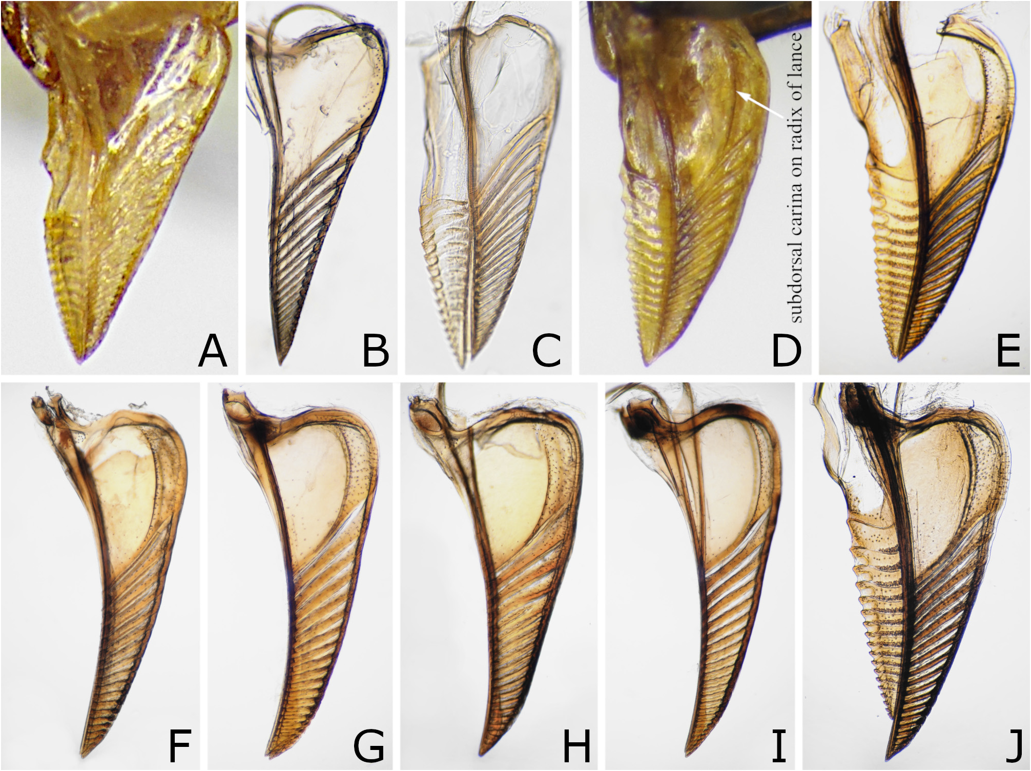
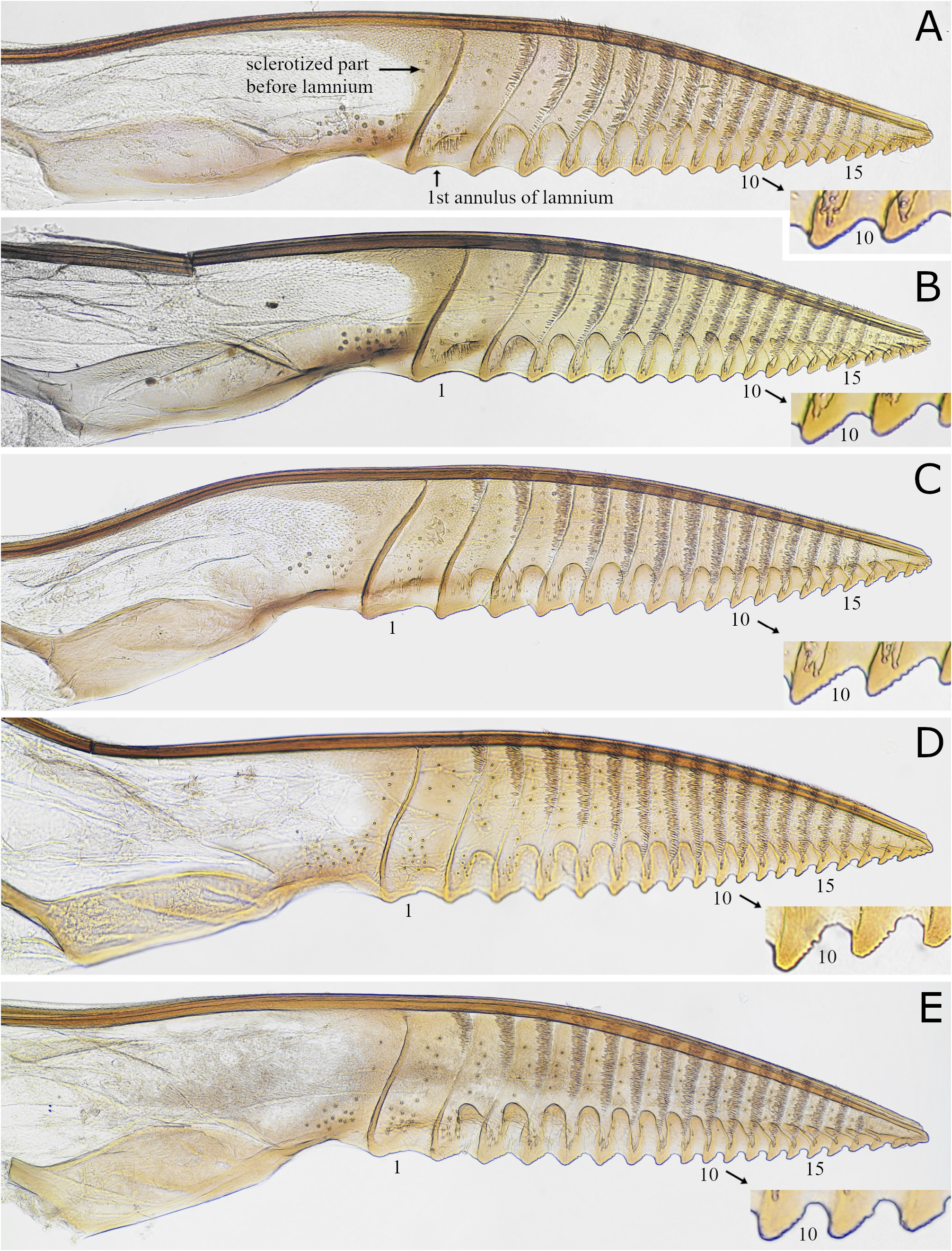
Female abdomen: valvula 3 in dorsal view about twice as wide as cercus, not or very slightly concave near pointed apex ( Fig. 9C View FIGURE 9 ), in lateral view rounded apically, straight or very slightly rounded on dorsal edge and slightly rounded on ventral edge ( Fig. 9K View FIGURE 9 ). Lance with dorsal edge gently rounded ( Figs 10D, E View FIGURE 10 ). Lancet with radix about 0.7 × as long as lamnium ( Figs 12A, B View FIGURE 12 ); lamnium with 19–20 annuli; basal annuli slightly sinuous; middle and apical annuli straight and oblique; annulus 1 and some apical annuli without ctenidium; ctenidia widely separated from each other, each consisting of some irregular transverse rows of setae; basal and middle ctenidia slightly expanded dorsally; basal serrulae with anterior slope as long as posterior slope.
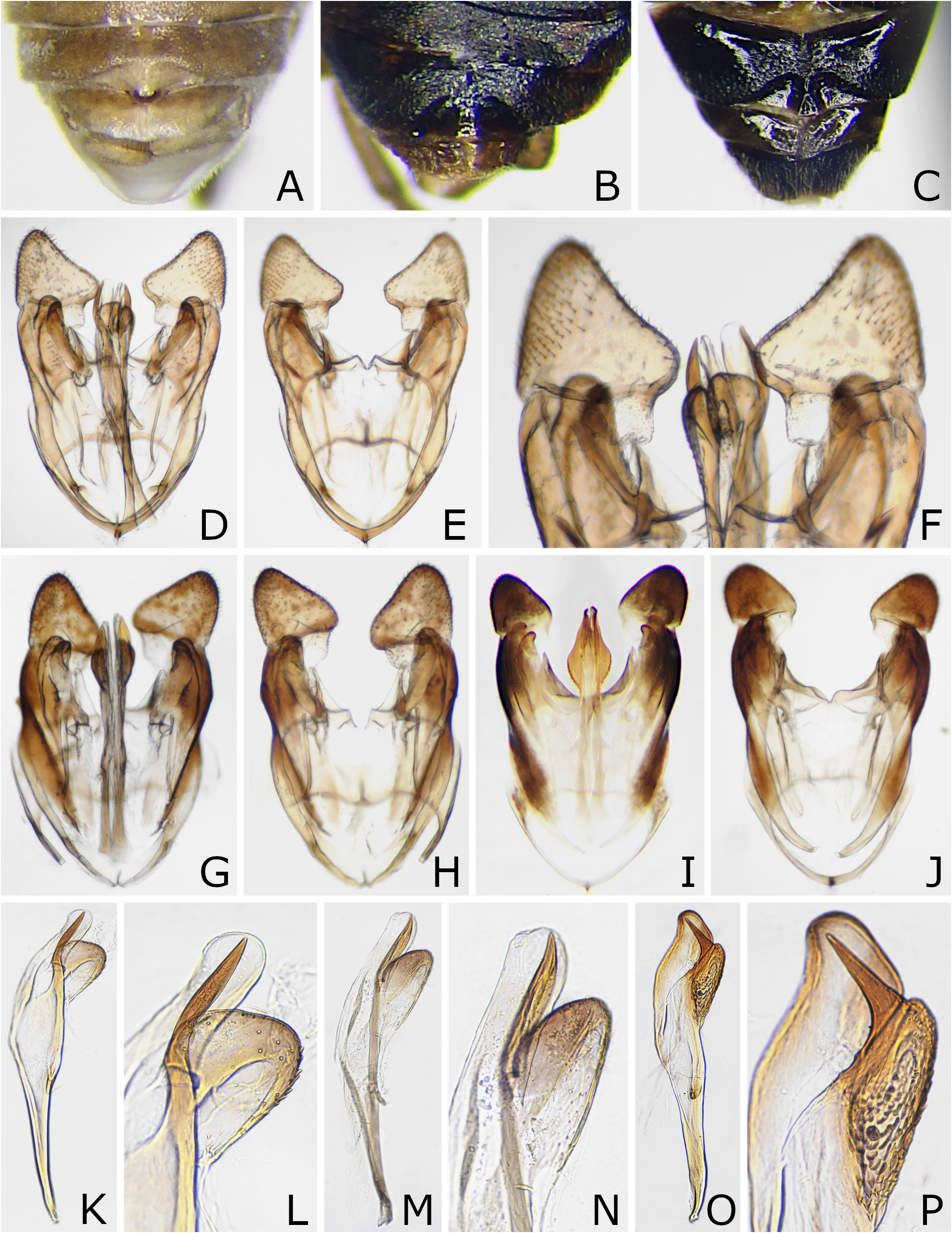
Male abdomen: subgenital plate in ventral view apically rounded or angulated. Genitalia in dorsal and ventral views Figs 14I, J View FIGURE 14 ; parapennis pointed apically, roundly concave on medial edge; harpe about as long as wide, with medial edge distinctly convex at basal third, lateral edge gently rounded, and apex rounded. Penis valve with paravalva swollen with many distinct spinules ( Figs 14O, P View FIGURE 14 ); valvispina located near apex of paravalva, directed posterodorsally.
Head and thorax with punctures minute; interspaces between punctures generally smooth. Mesoscutum mostly covered with setiferous punctures; posterolateral hollow with rugose and granular microsculpture. Mesopostnotum with rugose and granular microsculpture, medially smooth. Metapostnotum mostly smooth. Postspiracular sclerite with many setae. Mesepisternum not glabrous beside postspiracular sclerite ( Fig. 8I View FIGURE 8 ). Katepimeron widely covered with setae. Abdomen microsculptured imbricately, with punctures inconspicuous.
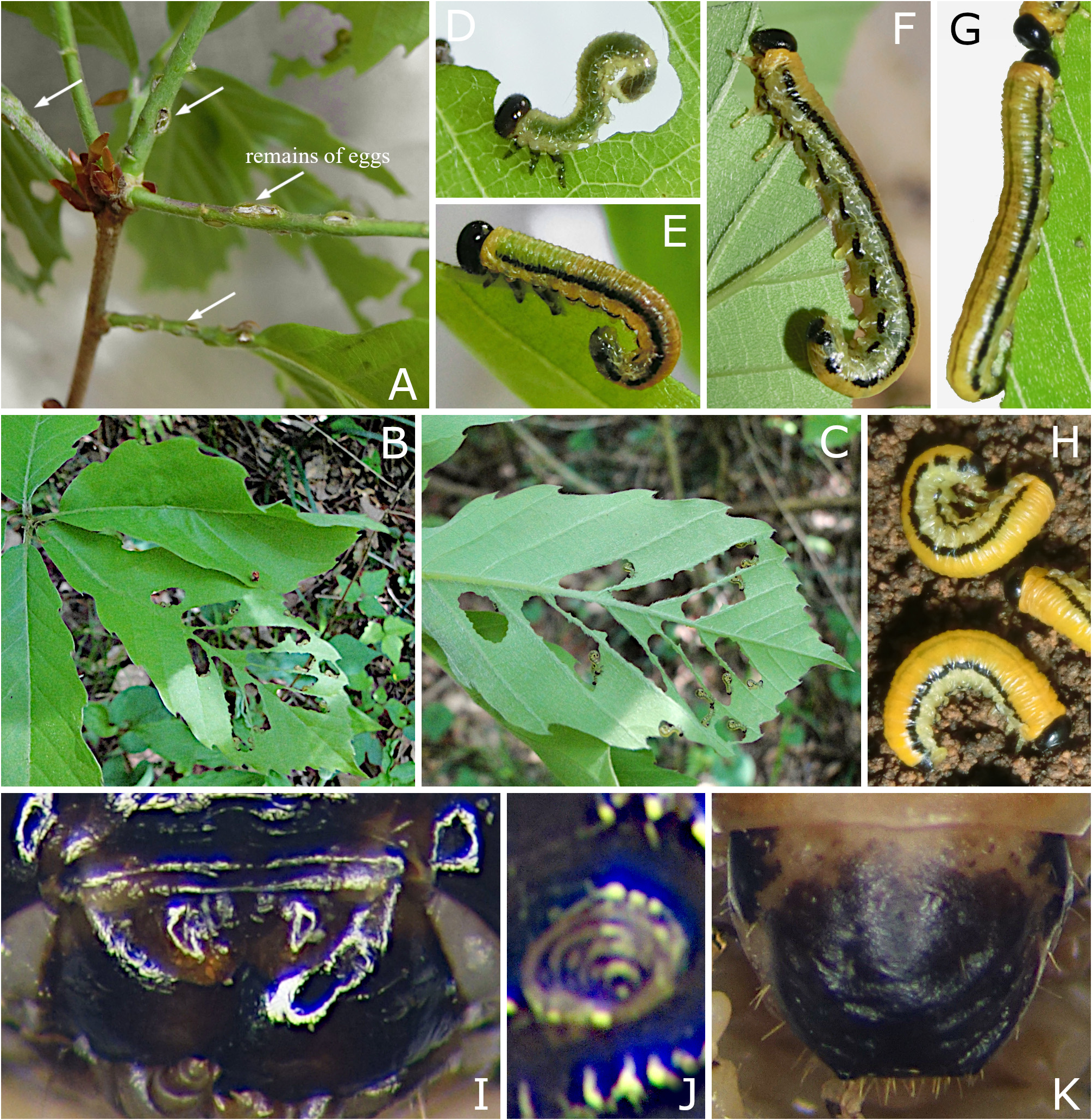
Immature stages. Early instar larva: head and thoracic legs black ( Fig. 3D View FIGURE 3 ); trunk pale greenish yellow, sometimes with inconspicuous dark broken supraspiracular and pleural stripes; setae short. Middle and late instar larvae: head and thoracic legs black ( Fig. 3E View FIGURE 3 ); trunk yellow, with black supraspiracular stripe and black broken pleural stripe; setae short. Final instar larvae: 13–18 mm long; color as in middle instar, but thoracic legs pale yellow, trunk pale grey below supraspiracular stripe and abdominal tergum 10 mostly black ( Figs 3F, G View FIGURE 3 ); supraspiracular stripe often broken on abdominal segment 9; setae inconspicuous; when mature, trunk uniformly bright yellow above supraspiracular stripe and on thoracic segment 1, pale yellow ventrally ( Fig. 3H View FIGURE 3 ); clypeus laterally with one or two setae on each side; mandible with one or two setae; labrum medially notched on ventral margin, with median and submedian grooves ( Fig. 3I View FIGURE 3 ) and one or two setae on both sides; antenna conical with four antennomeres ( Fig. 3J View FIGURE 3 ); palpifer with two setae; stipes with two or three setae; abdominal segments 2–7 and 10 each with proleg; abdominal segments 3–8 each dorsally with six annulets; in abdominal segment 3, annulets 1, 2 and 4 each sometimes with one seta, annulet 4 always with one wart; abdominal tergum 10 without caudal protuberance, apically truncate in dorsal view ( Fig. 3K View FIGURE 3 ). Cocoon: 7–10 mm long, blackish brown, single walled.
Material examined. Paratypes: 2♀ 2♂, “Mt. Funaoka, Tsurugi-machi, Ishikawa Pref., 16. IV. 2004, I. Togashi”, “ Paratype Fagineura quercivora n. sp. ” ( Figs 6F, G, N, R View FIGURE 6 , 7C, F, L, R, S View FIGURE 7 , 8F, I, Q, Y View FIGURE 8 , 9C, K View FIGURE 9 , 10E View FIGURE 10 , 12A View FIGURE 12 , 14I, J, O, P View FIGURE 14 ), kept in the National Museum of Natural History, Smithsonian Institution, Washington, D.C. We have not examined the holotype of Fagineura quercivora Togashi, 2006 . Togashi (2006) wrote “ Holotype and 10 paratypes deposited in the collection of the National Science Museum (Nat. Hist.), Tokyo ”, but there are no type specimens of the species in the museum.
Other material examined: JAPAN: HONSHU: 1♀, Tochigi Pref., Nakagawa, Koisago, 36°47’N 140°11’E, coll. gregarious larvae on Quercus acutissima 6. V. 2016, mat. 16. V., em. 13. IV. 2017, S. Ibuki; 2♀, Tochigi Pref., Nakagawa, Wami, 36°46’N 140°10’E, coll. gregarious larvae on Quercus acutissima , mat. 20. V. 2010, em. 16–19. IV. 2011, S. Ibuki; 3♀, same data but coll. gregarious larvae on Quercus serrata 10. V. 2014, mat. 19. V., em. 21–27. III. 2015; 4♀ 1♂, same data but coll. larvae 3. V. 2019, mat. 11–12. V., em. 22–24. III. 2020 ( Figs 2G–N View FIGURE 2 , 3A, E–H View FIGURE 3 ); 2♀ 4♂, same data but coll. larvae 5. V. 2019, mat. 13–16. V., em. 22–26. III. 2020 ( Figs 3B–D View FIGURE 3 ); 10♀ 1♂, Tochigi Pref., Nakagawa, Wami, coll. larvae on Quercus serrata 8. V. 2020, mat. 16. V., em. 30. III. 2021, S. Ibuki; 3 final instar larvae, same data but coll. larvae 7. V. 2021, fix. 8. V. 2021, S. Ibuki ( Figs 3H–J View FIGURE 3 ); 4♀, Tochigi Pref., Sakura, coll. larvae on Quercus serrata 18. V. 2017, mat. 20–21. V., em. 1. IV. 2018, S. Ibuki ( Fig. 10D View FIGURE 10 ); 6♀, Tokyo Met., “Sirokane Estate, Meguro”, 11. IV. 1947, T. Okutani ― KYUSHU: 3 final instar larvae, Saga Pref., Saga, Dondonnomori, 33°15’N 130°17’E, on Quercus dentata , 30. IV. 2016, M. Tokuda (cited by Tazunoki et al. 2018); 2♀ 2♂, same locality, 20. IV. 2017, S. Fujita & Y. Tazunoki ( Fig. 12B View FIGURE 12 ) (cited by Tazunoki et al. 2018, Hara 2020); 3♀ 2♂, same locality, 1. IV. 2018 ― LOCALITY UNKNOWN: 1♀, “4.24 [haka (in Japanese)]” ― KOREA: 1♀, Gangwon-do, Mt. Samagsan, 9. V. 1990, A. Shinohara; 1♀, Gyeonggi-do, Suwon, V. 1925, Y. Hasegawa; 1♀, same locality, on Quercus , 13. V. 1926, K, Sato; 1♀, same locality and collector, coll. larva on Quercus serrata , em. 20. IV. 1927; 1♀, same locality and collector, 27. IV. 1927; 1♀, same locality and collector, 4. V. 1927; 2♀, same locality and collector, 3. V. 1931; 9♀, same locality and collector, coll. larvae on Quercus acutissima , V. 1932; 1♀, Mt. Chongmasan, 9. V. 1980, A. & N. Shinohara & S. J. Yoon.
Distribution. Japan: Honshu ( Togashi 2006), Kyushu ( Tazunoki et al. 2018). Korea (new record).
Life history. Host plants: Fagaceae : Quercus acutissima Carruth. ( Tazunoki et al. 2018) , Q. crispula Blume var. crispula ( Togashi 2006, under rearing condition), Q. dentata Thunb. ( Tazunoki et al. 2018) , Q. serrata Murray ( Togashi 2006, under rearing condition; present study).
This species has univoltine life cycle and adults occur in early spring ( Togashi 2006, Tazunoki et al. 2018). Eggs are deposited in young shoots ( Fig. 3A View FIGURE 3 ). Larvae are gregarious but they are not in contact with each other ( Fig. 3C View FIGURE 3 ). Young larvae eat the insides of leaves. Old larvae eat leaves from the edges. When larvae reach maturity, they enter into the soil without an extra molt and become cocoons in the soil. Tazunoki et al. (2018) reported severe defoliation by the larvae.
Remarks. Fagineura togashii is very similar to F. fulvistriata . Their differences we noticed are only the condition of the mesepisternum and the coloration as stated in the key, malar space length, the shape of the basal serrulae of a lancet and the larval color. A malar space is 0.2–0.3 × as long as a median ocellus width in the former but 0.5–0.6 × in the latter. The basal serrulae of a lancet have the posterior slope about as long as the anterior slope in the former ( Figs 12A, B View FIGURE 12 ) but the posterior slope longer than the anterior slope in the latter ( Fig. 12C View FIGURE 12 ). In the late instars except for the mature stage, the former larva has the dorsally yellow trunk ( Figs 3E–G View FIGURE 3 ), while the latter larva has the mostly pale grey trunk ( Fig. 4L View FIGURE 4 ).
This species is also similar to F. flavomaculata in color, but it is distinguished from the latter by the mesepisternum dorsally not glabrous and barely expanded ( Fig. 8I View FIGURE 8 ), the lancet annuli markedly oblique ( Figs 12A, B View FIGURE 12 ), the female mesoscutum and mesepisternum entirely black ( Figs 2H–J View FIGURE 2 ), the female abdominal terga 1, 2 yellow ( Fig. 2H View FIGURE 2 ), the female abdominal laterotergites 7, 8 mostly black ( Fig. 2I View FIGURE 2 ) and the trunk of the late instar larva dorsally yellow ( Figs 3E–G View FIGURE 3 ). The latter species has the mesepisternum dorsally glabrous and distinctly expanded ( Fig. 8K View FIGURE 8 ), the lancet with the middle annuli nearly erect ( Figs 12D, E View FIGURE 12 ), the female mesoscutum and mesepisternum partly yellow to brown ( Figs 4G, H View FIGURE 4 ), the female abdominal terga 1, 2 mostly black ( Fig. 4G View FIGURE 4 ), the female abdominal laterotergites 7, 8 yellow ( Fig. 4H View FIGURE 4 ) and the trunk of the late instar larva mostly pale grey ( Figs 4O, P View FIGURE 4 ).


| V |
Royal British Columbia Museum - Herbarium |
| T |
Tavera, Department of Geology and Geophysics |
No known copyright restrictions apply. See Agosti, D., Egloff, W., 2009. Taxonomic information exchange and copyright: the Plazi approach. BMC Research Notes 2009, 2:53 for further explanation.
|
Kingdom |
|
|
Phylum |
|
|
Class |
|
|
Order |
|
|
Family |
|
|
Genus |
Fagineura togashii Hara
| Hara, Hideho & Ibuki, Shinichi 2022 |
Fagineura quercivora
| Liu, M. & Li, Z. & Wei, M. 2021: 130 |
| Hara, H. 2020: 82 |
| Hara, H. 2019: 77 |
| Liu, M. & Li, Z. & Wei, M. 2019: 33 |
| Tazunoki, Y. & Fujita, S. & Hara, H. & Adachi, S. & Kuchiki, F. & Tokuda, M. 2018: 142 |
| Taeger, A. & Blank, S. M. & Liston, A. D. 2010: 405 |
| Togashi, I. 2006: 169 |