
Echiniscus tristis Gąsiorek & Kristensen, 2018
|
publication ID |
https://doi.org/10.11646/zootaxa.4701.1.1 |
|
publication LSID |
lsid:zoobank.org:pub:B1C6B96D-E9BA-4897-A9FD-EE6064BB3179 |
|
DOI |
https://doi.org/10.5281/zenodo.5584476 |
|
persistent identifier |
https://treatment.plazi.org/id/654D87EB-FFE4-494B-37FF-BD60FC6EED76 |
|
treatment provided by |
Plazi |
|
scientific name |
Echiniscus tristis Gąsiorek & Kristensen, 2018 |
| status |
|
Echiniscus tristis Gąsiorek & Kristensen, 2018 View in CoL
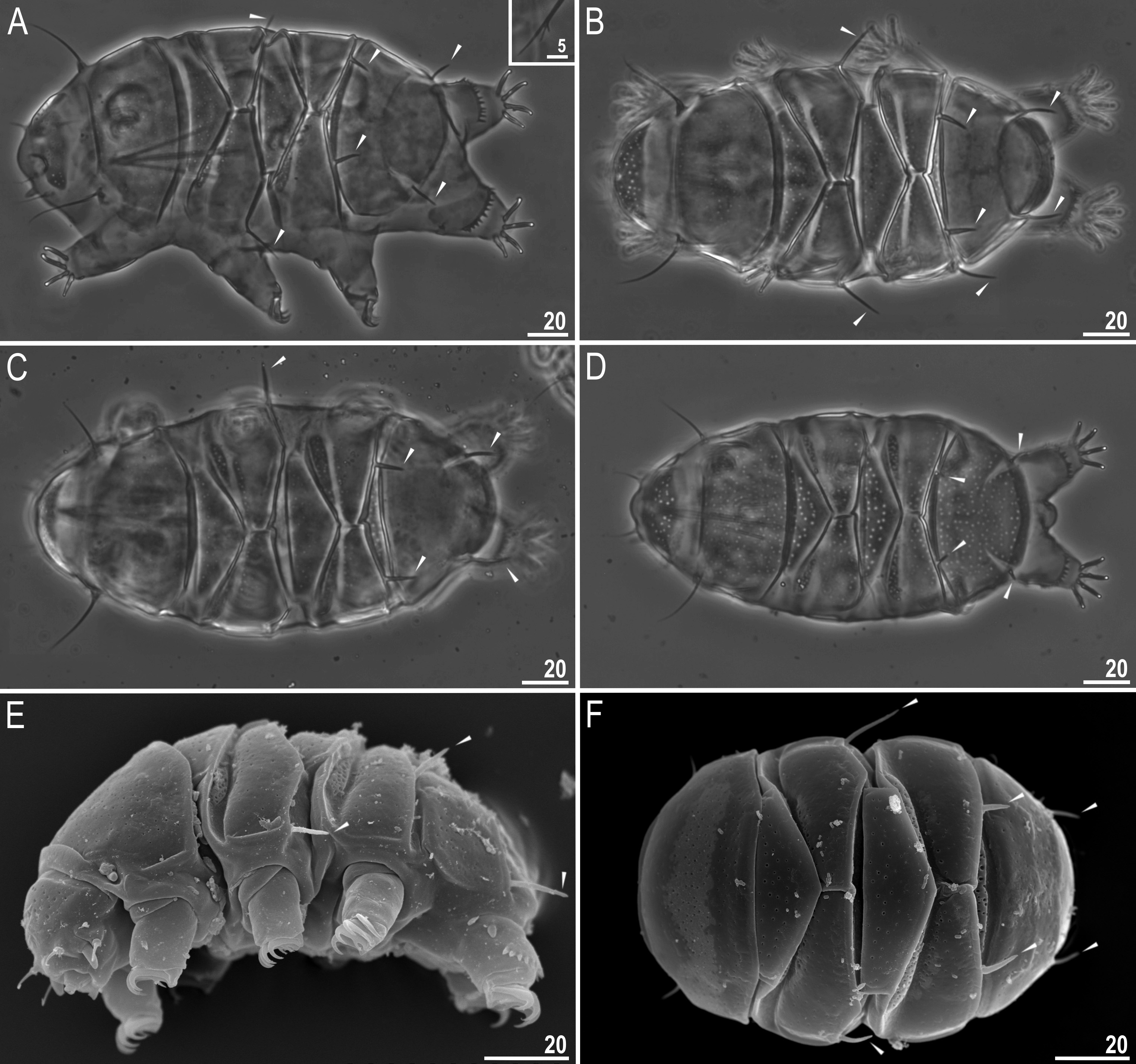
( Tables 4–10 View TABLE 4 View TABLE 5 View TABLE 6 View TABLE 7 View TABLE 8 View TABLE 9 View TABLE 10 , Figs 2–9 View FIGURE 2 View FIGURE 3 View FIGURE 4 View FIGURE 5 View FIGURE 6 View FIGURE 7 View FIGURE 8 View FIGURE 9 )
Material examined: 320 animals mounted on microscope slides in Hoyer’s medium, 20 animals prepared for SEM and 6 prepared for molecular analysis from Madagascar, 22°37’07.7”S, 46°43’14.5”E, ca. 1,187 m asl, Fianarantsoa Province, Ivohibory forest, lichen on quartz rock, coll. Marta Kepel and Andrzej Kepel.
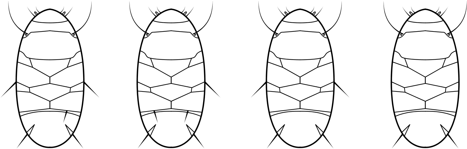
Description of the Madagascan population. Animals (measurements and statistics in Tables 4-8 View TABLE 4 View TABLE 5 View TABLE 6 View TABLE 7 View TABLE 8 ) Females. Body light orange in live specimens and transparent/white after preparation ( Fig. 2 View FIGURE 2 ). Eyes absent or not visible after the preparation. Apart from the head appendages (internal and external cirri and cylindrical cephalic papillae [secondary clavae], the appendage A with clava [primary clava] near its base) present ( Fig. 2 View FIGURE 2 ). Dorsal and lateral appendages in the shape of long and short spines present in positions A-C-Dd- E (mainly in adult females; morphotype 1) or A-Dd- E (mainly in juveniles and larvae, morphotype 2). This means that the general chaetotaxy of adult females of this species is A-(C)- Dd- E. However, single females in the studied population possessed also different chaetotaxy configurations, i.e. A-E, A-C-E, A-C-Cd- Dd- E, A-C-D-Dd- E, but with some spines being present only on one side of the body ( Fig. 2–3 View FIGURE 2 View FIGURE 3 ). See also Morphological variability below.
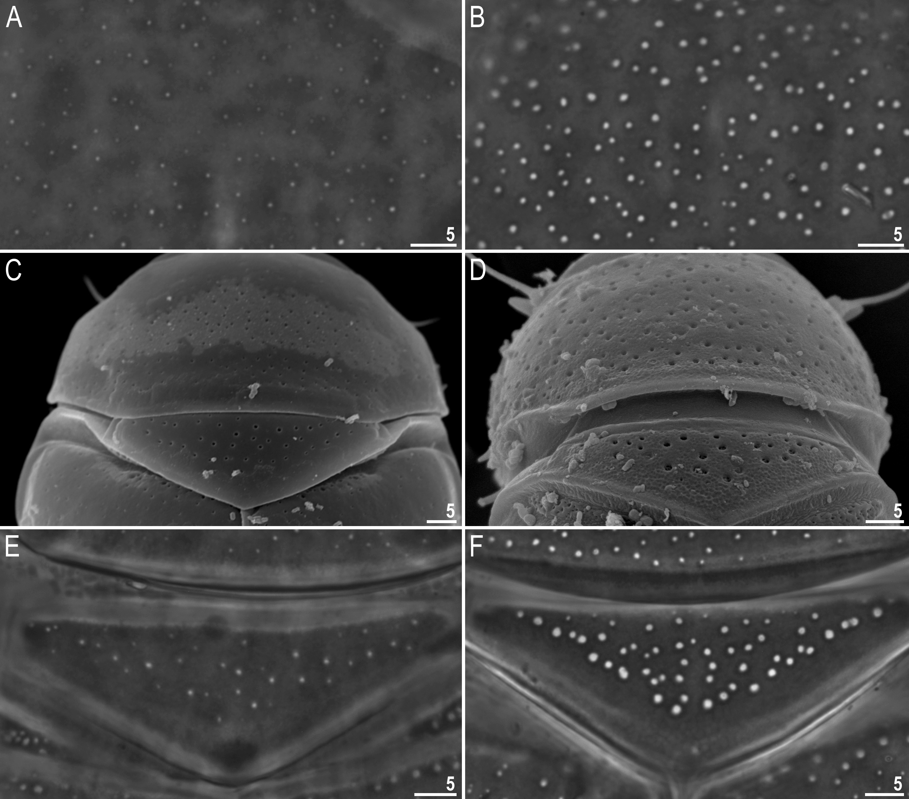
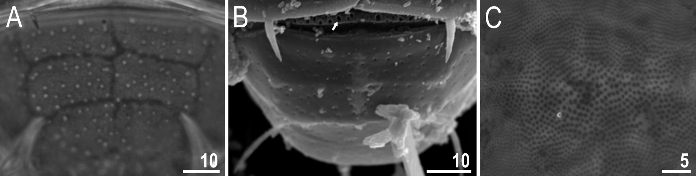
Dorsal plates covered by smaller and larger pores ( Figs 4–6 View FIGURE 4 View FIGURE 5 View FIGURE 6 ). Pores 0.6–2.2 µm in diameter visible as bright/ white dots when focusing down through cuticle ( Figs 4 View FIGURE 4 A–B, E–F; 5A, C–F; 6A). Although pores may differ significantly in the same individual and between specimens, in general they are largest on the head plate, narrow anterior stripe of paired plates and in the place where median plate 3 should be localised ( Figs 5A View FIGURE 5 , 6B View FIGURE 6 arrow). Dorsal plates well developed. Head and scapular plates not faceted. In PCM, lateral portions of the scapular plate appear to be detached from the dorsal plate, forming small additional plates (one on each side of the body) divided from the scapular plate by a thin bright stripe. This false division is caused by a bend of the plate where the cuticle is thinner; in SEM, this pseudo-division is not visible. Paired plates I and II are divided into two parts—narrow anterior stripe and a wider posterior part—by smooth stripe without pores ( Fig. 5 View FIGURE 5 B–D). Median plates 1 and 2 undivided, median plate 3 absent (but cuticle in this place sculptured with pores) ( Fig. 6B View FIGURE 6 ). The terminal plate possesses two notches and in some specimens a characteristic pattern ( Figs 6 View FIGURE 6 A–B). Narrow stripes form four rectangular areas in central part of terminal plate ( Figs 6 View FIGURE 6 A–B). Ventral plates absent, but cuticle possesses tiny and regular granulation (caused by dense top endings of cuticular pillars). Granulation is little larger between the legs (0.9–1.0 µm diameter) than in other parts of the ventral side, 0.4–0.7 µm diameter) ( Fig. 6C View FIGURE 6 ).
A-C-Dd- E A-Dd- E A-C-Cd- D-Dd- E A-C-D-Dd- E A-C-Cd- Dd- E
A-C-Dd- E A-C-Dd- E A-C-E A-E
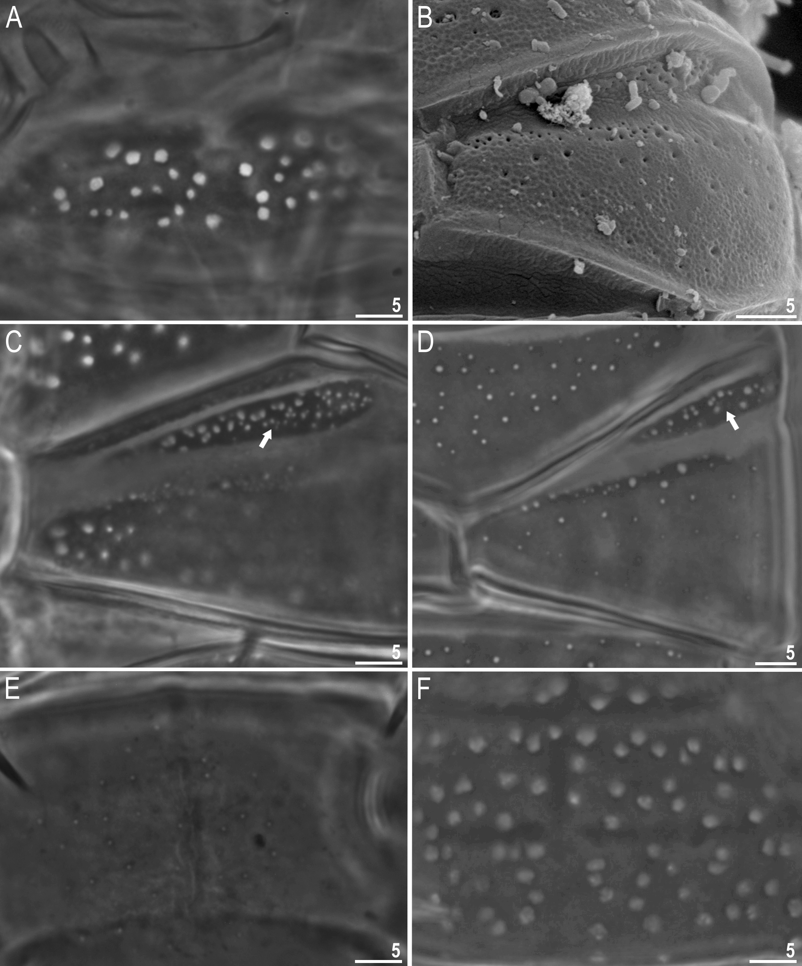
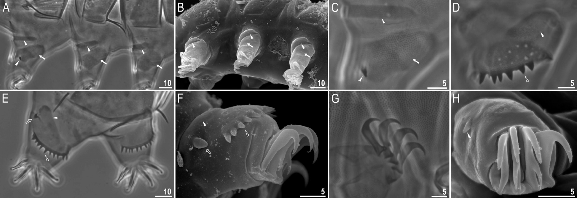
On outer cuticle of legs I–III two clearly visible patches, composed of tiny and regular granulation similar to this on ventral side, are present: a narrow stripe (cuticular fold) in upper part ( Fig. 7 View FIGURE 7 A–C, filled arrowhead) and a large wide stripe below (plate) ( Fig. 7 View FIGURE 7 A–C, filled arrow). Triangular spine on leg I and finger-like papilla on leg IV present ( Fig. 7C View FIGURE 7 , E–F, H, indented filled arrowhead and empty arrow). Dentate collar on legs IV with sculpture similar to those on dorsal plates but with smaller pores, and with eight to twelve sharp, triangular teeth ( Fig. 7 View FIGURE 7 D–F, empty arrowhead). Above the dentate collar a cuticular fold also present ( Fig. 7 View FIGURE 7 D–F, filled arrowhead). External claws of all legs smooth, internal with spurs directed downwards ( Fig. 7 View FIGURE 7 F–H).
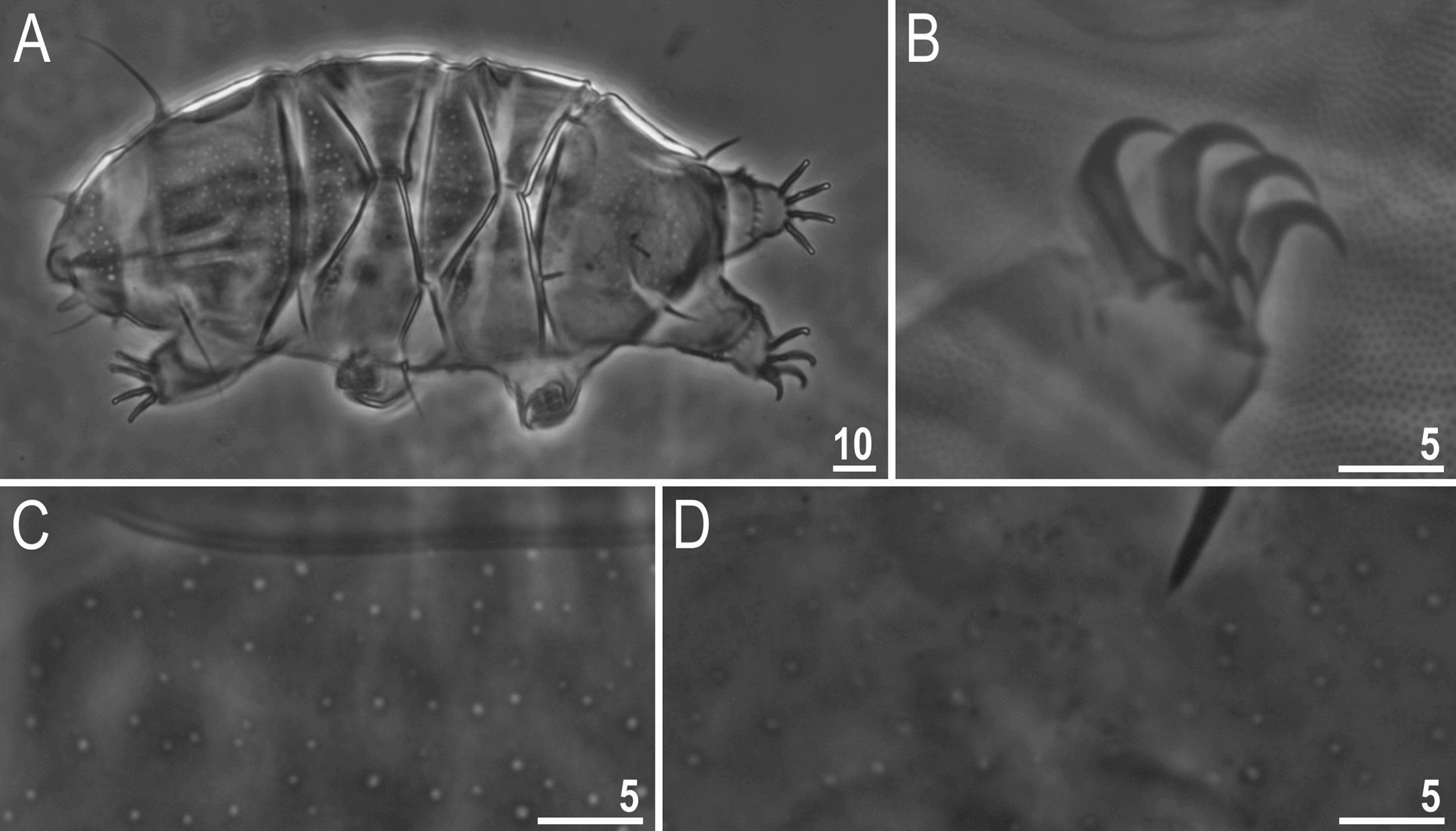
Juveniles ( Fig. 8 View FIGURE 8 ). In general they are similar to adult females. The chaetotaxy types in juveniles are A-C-Dd- E, A-Dd- E, A-C-D-Dd- E, A-C-E, with the most frequent A-Dd- E. For more details see Table 9 View TABLE 9 and Morphological variability below. Terminal plate without characteristic pattern or with pattern only poorly visible. Gonopore absent.
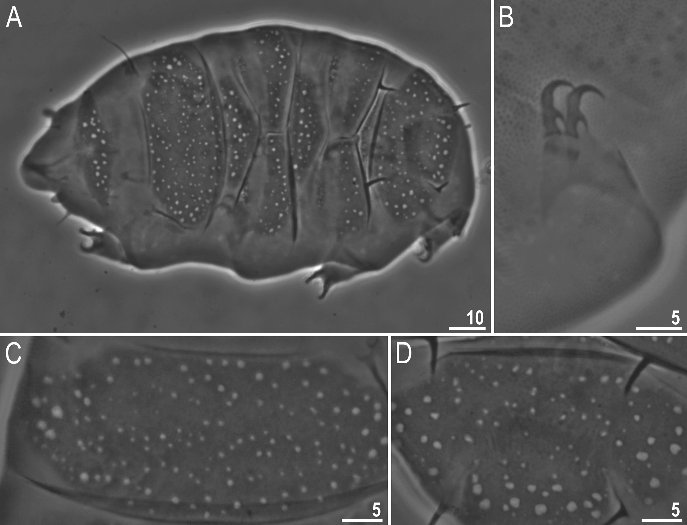
Larvae ( Fig. 9 View FIGURE 9 ). In general they are similar to adult females, but dorsal cuticular pores seem to be larger in comparison to the body size. The chaetotaxy in larvae is always A-Dd- E. For more details see Table 9 View TABLE 9 and Morphological variability below. Terminal plate without the characteristic pattern. All legs with two internal claws. Gonopore and anus absent.
Males. Not found, indicating the species is most likely parthenogenetic.
Eggs. Smooth, light orange and deposited in the exuviae up to 5 in one exuvium.
DNA sequences. We obtained good quality sequences for the applied molecular markers (voucher numbers marked in bold indicate specimens which were analysed using all three markers):
No known copyright restrictions apply. See Agosti, D., Egloff, W., 2009. Taxonomic information exchange and copyright: the Plazi approach. BMC Research Notes 2009, 2:53 for further explanation.
|
Kingdom |
|
|
Phylum |
|
|
Class |
|
|
Order |
|
|
Family |
|
|
Genus |