
Goodeniaphila, Tatarnic, Nikolai J., 2009
|
publication ID |
https://doi.org/10.5281/zenodo.187769 |
|
DOI |
https://doi.org/10.5281/zenodo.6212500 |
|
persistent identifier |
https://treatment.plazi.org/id/655E6C37-BA63-7619-1795-92F4FEBCFD8E |
|
treatment provided by |
Plazi |
|
scientific name |
Goodeniaphila |
| status |
gen. nov. |
Goodeniaphila View in CoL gen. n.
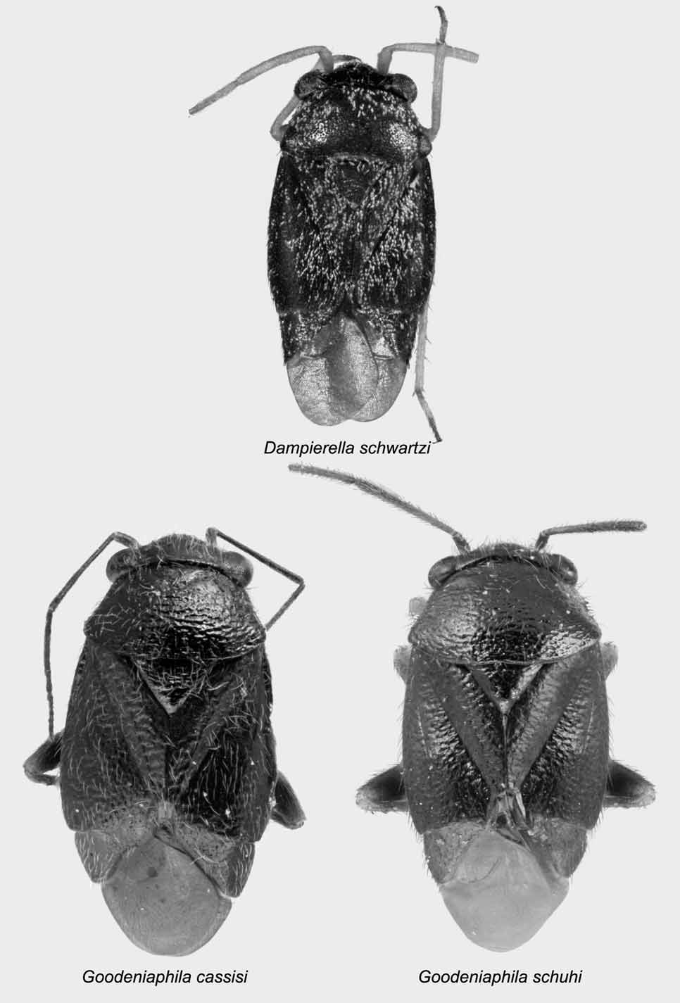
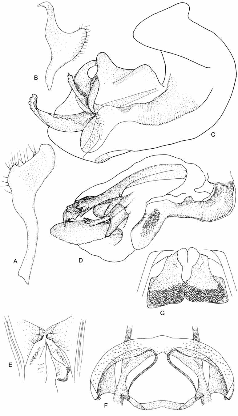
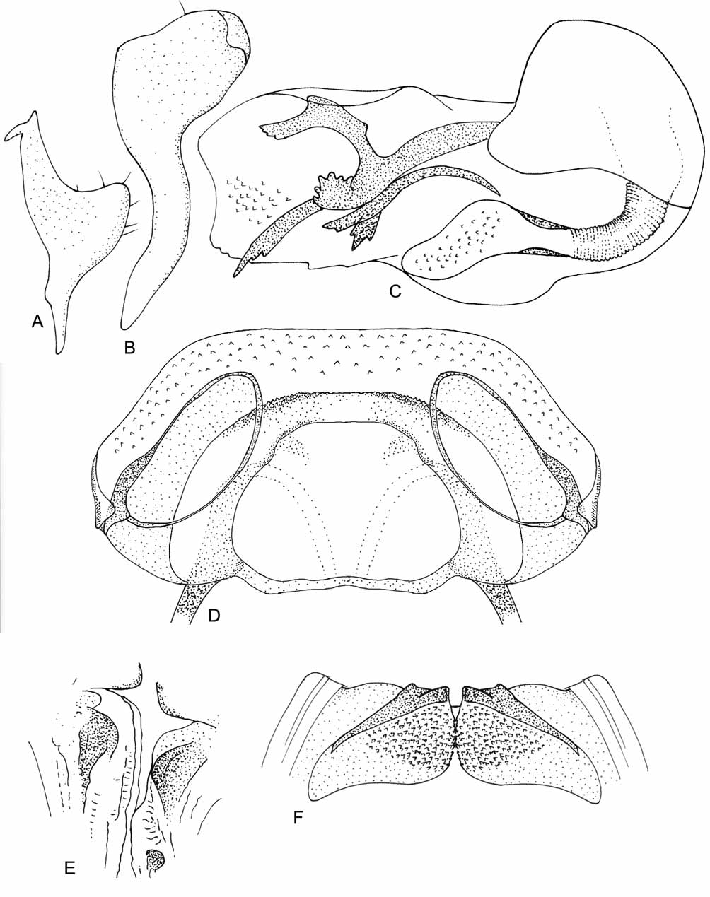
Figures 1 View FIGURE 1 , 4–8 View FIGURE 4 View FIGURE 5 View FIGURE 6 View FIGURE 7 View FIGURE 8
Type species: Goodeniaphila cassisi sp. n., by original designation
Diagnosis: Goodeniaphila is most similar in appearance to Strongylocoris , but is distinguished by the presence of a well-developed metathoracic scent gland external efferent system, slightly olive coloration of the hemelytra, and the male and female genitalia.
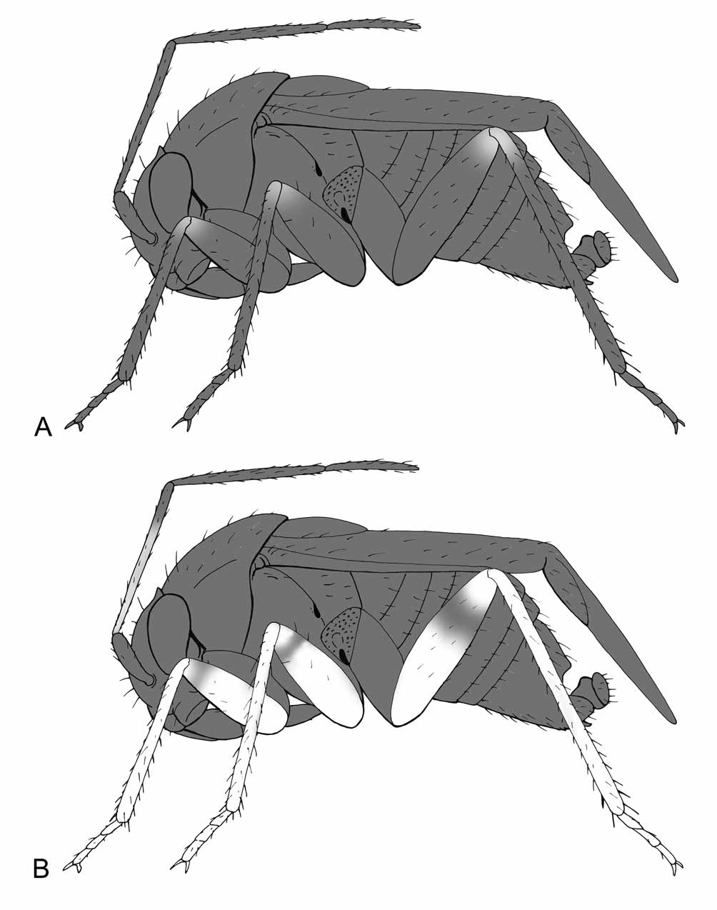
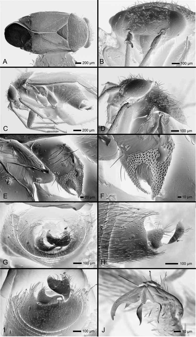
Description: Both sexes macropterous, elongate-oval, body length 2.44–3.27 (Table 1). COLORATION ( Figs. 1 View FIGURE 1 , 5 View FIGURE 5 ): Head, pronotum, and scutellum glossy black, hemelytra faintly dark olive; antennae yellowbrown to black; legs yellow to black. SURFACE AND VESTITURE ( Figs. 1 View FIGURE 1 ; 6): Surface mostly glossy and shining except for dull hemelytra; head smooth, pronotum, scutellum and hemelytra rugulopunctate; body clothed in golden, hairlike setae; hemelytra densely covered with minute golden setae; antennae without spines, tibiae spinose. STRUCTURE: Body elongate-oval. Head ( Figs. 1 View FIGURE 1 , 5 View FIGURE 5 , 6 View FIGURE 6 A–D): Transverse, short, broader than tall; eyes sessile, height of gena slightly less than eye height; vertex convex, with transverse sulcus along posterior margin, posterior margin upturned and carinate, wrapping around pronotum; clypeus angled slightly posteriorly; bucculae small, narrow. Antennae ( Figs. 1 View FIGURE 1 , 5 View FIGURE 5 , 6 View FIGURE 6 B–D): Insertion in line with lower margin of eye; shorter than body length, segment I shorter than eye height, only slightly thicker than segment II; segment IV shorter than III. Labium: Extending to metacoxae, segment I somewhat swollen, subequal in length to gena height; Thorax ( Figs. 1 View FIGURE 1 , 5 View FIGURE 5 , 6 View FIGURE 6 C–F): Pronotum trapezoidal, broad, collar absent, callosite region weakly defined, posterior margin straight, thin, and carinate, extending over mesoscutum; metathoracic spiracle a narrow slit surrounded by evaporative bodies; metathoracic scent gland external efferent system prominent and swollen, peritreme oval, oriented vertically above ostiole, surrounded by evaporative bodies. Hemelytra ( Figs. 1 View FIGURE 1 , 5 View FIGURE 5 , 6 View FIGURE 6 A, C): Lateral margins weakly convex; clavus laterally declivent; medial fracture obsolete; cuneus broad; membrane and cuneus declivent at cuneal fracture; membrane with single enclosed cell, extending beyond abdomen. Legs ( Figs. 1 View FIGURE 1 ; 5, 6C, J): Metafemora moderately incrassate; pretarsi with lamellate, apically convergent parempodia and fleshy pulvilli. Abdomen ( Figs. 5 View FIGURE 5 , 6 View FIGURE 6 C): Elongate-ovoid. Male genitalia ( Figs. 6 View FIGURE 6 G–I, 7A–D, 8A–C): Pygophore opening broad, posterior margin with shallow concavity below left paramere; left paramere broad with short base, sensory lobe prominent, apophysis thin and apically hooked; right paramere longer than left, with sensory lobe, hockey-stick shaped, projects beyond genital opening of pygophore; phallotheca short, heavily contoured and sclerotized, ductus seminis wide, with sclerotized ribbing; secondary gonopore scoop-shaped with faint scalelike texturing; endosoma with assortment of elongate, serrate and platelike spicules, endosoma apically affixed to phallotheca. Female genitalia ( Figs. 7 View FIGURE 7 E–G, 8D–F): Sclerotized rings thin, weakly sclerotized, widely separated or subcontiguous, elongate-elliptical, with lateral margins and adjacent portion of dorsal labiate plate strongly upturned; margin of ventral labiate plate adjoining rami forming a sclerotized rim, lateralmost region of ventral labiate plate joined with rami and sclerotized rings to form paired, medially projecting, sclerotized processes which continue medially to form a sclerotized interramal band; posterior wall bilaterally divided, partly sclerotized, posteriorly with fields of spines; margins of vesibulum mostly bilaterally symmetrical, left side with sclerotized process.
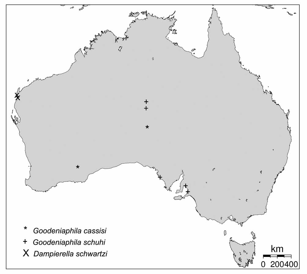
Distribution: This genus is known from two species collected in Western Australia, South Australia and the Northern Territory ( Fig. 4 View FIGURE 4 ).
Included Species:
Host Plant Associations: Both species have been collected exclusively on species belonging to the plant family Goodeniaceae .
Etymology: The name Goodeniaphila reflects the affinity of this genus to plants of the family Goodeniaceae .
No known copyright restrictions apply. See Agosti, D., Egloff, W., 2009. Taxonomic information exchange and copyright: the Plazi approach. BMC Research Notes 2009, 2:53 for further explanation.