
Erythrogonia sexguttata (Fabricius, 1803)
|
publication ID |
https://doi.org/ 10.11646/zootaxa.3872.3.4 |
|
publication LSID |
lsid:zoobank.org:pub:9CCEAC1C-F58A-40E6-8349-D081976E12A5 |
|
DOI |
https://doi.org/10.5281/zenodo.6139016 |
|
persistent identifier |
https://treatment.plazi.org/id/660DF03E-FFB1-FF88-FF65-871E4500FCD8 |
|
treatment provided by |
Plazi |
|
scientific name |
Erythrogonia sexguttata (Fabricius, 1803) |
| status |
|
Erythrogonia sexguttata (Fabricius, 1803) View in CoL
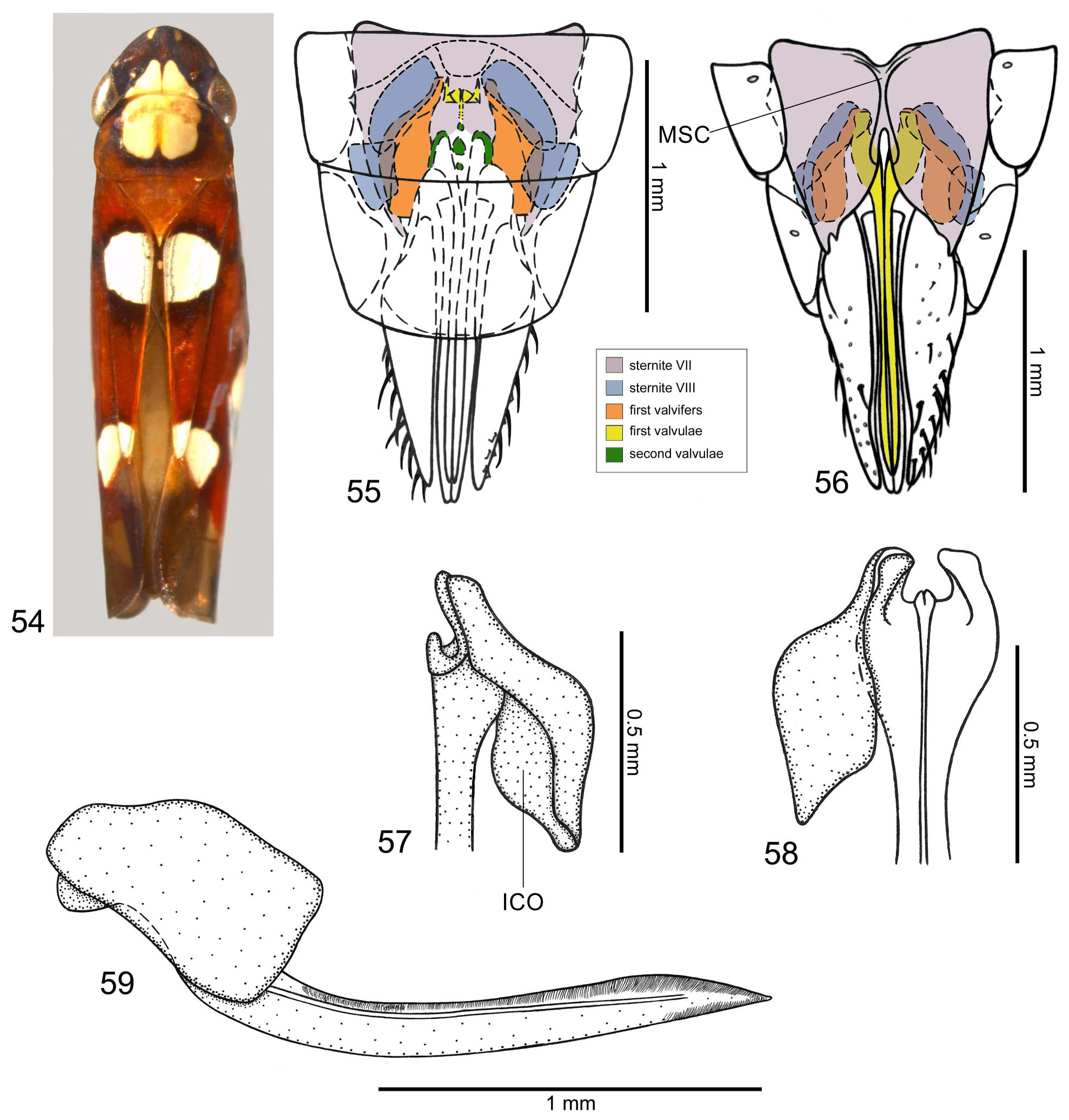
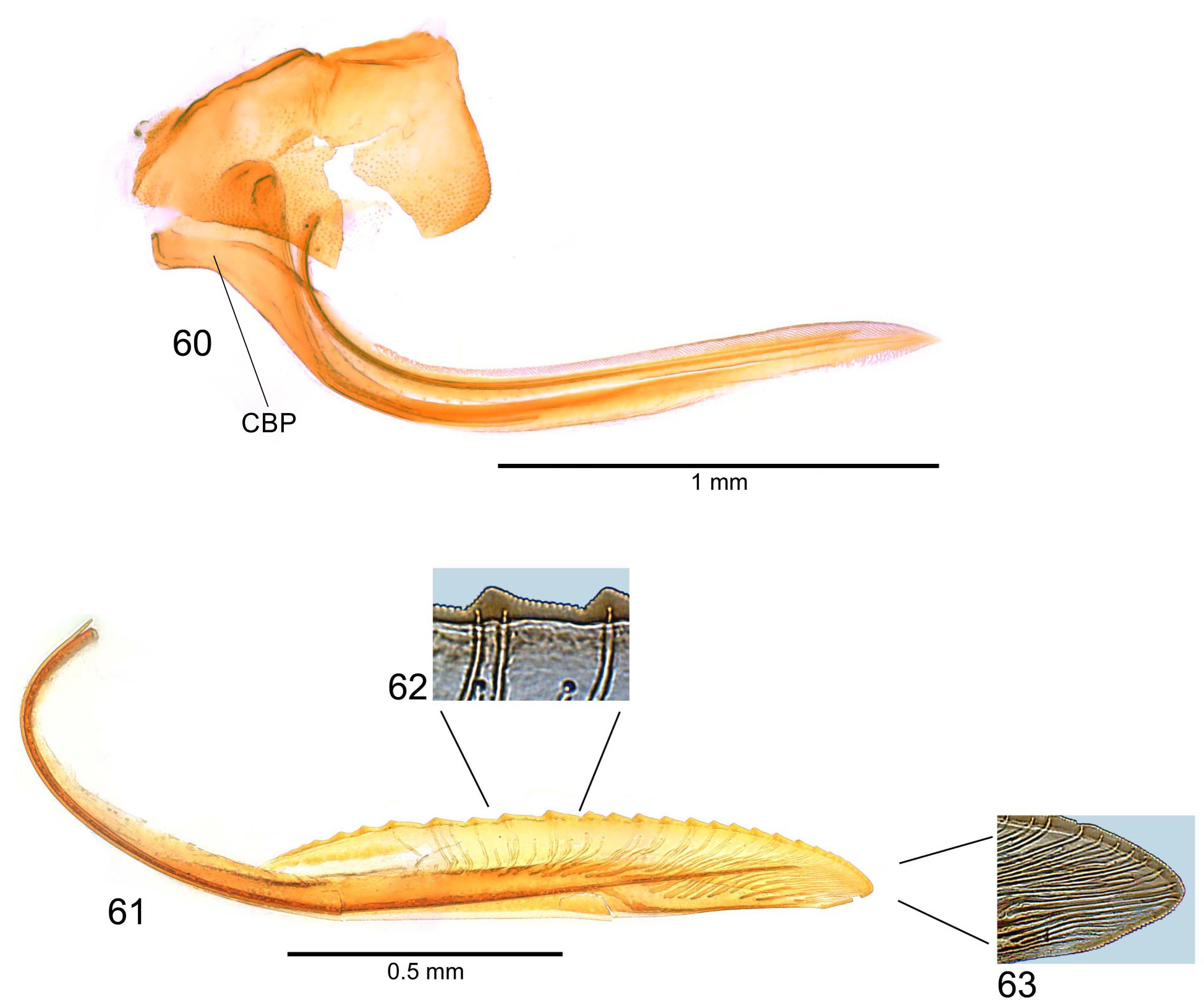
( Figs 54–63 View FIGURES 54 – 59 View FIGURES 60 – 63 )
Length: female 7.3–7.4 mm (n = 2), male 6.4 mm (n = 1).
Diagnosis. Anterior dorsum ( Fig. 54 View FIGURES 54 – 59 ) brown to reddish-brown with triangular, deeply incised yellow to pale yellow macula on crown contiguous to large, shallowly incised macula on pronotum. Forewings ( Fig. 54 View FIGURES 54 – 59 ) reddishbrown with three large yellow to whitish-yellow spots bordered with brown, one on basal half of clavus adjacent to apex of mesonotum, one on median corium third adjacent to costal margin, and one on apical portion of clavus and adjacent area of corium; costal margin bordered with dark brown to black; membrane brown except for unpigmented anterior triangular area. Males with basal half of subgenital plate just a little broader than style; style almost as long as subgenital plate; aedeagus with shaft narrow, curved ventrally and then directed dorsally, apex acute; paraphyses basal portion T-shaped, rami with posterior ends strongly curved ventrally. Female sternite VII ( Fig. 56 View FIGURES 54 – 59 ) with ventral surface longitudinally sulcate and posterior margin deeply incised medially; sternite VIII ( Figs 55 and 56 View FIGURES 54 – 59 ) with two pairs of lateral sclerotized areas; first valvifers ( Figs 57–59 View FIGURES 54 – 59 ) without processes or additional sclerites; second valvulae with three small, longitudinally-aligned sclerites between bases of rami ( Fig. 55 View FIGURES 54 – 59 ).
Female genitalia. Sternite VII ( Fig. 56 View FIGURES 54 – 59 ) with deep median incision on posterior margin; lateroposterior areas with very small projection; lateral margins convergent posteriorly; ventral surface with few irregular striae, without setae; median portion with pair of longitudinal elevations delimiting a deep sulcus ( Fig. 56 View FIGURES 54 – 59 , MSC); internally with median area connected dorsally to sternite VIII. Sternite VIII ( Figs 55 and 56 View FIGURES 54 – 59 ), in dorsal view, with two pairs of lateral sclerotized areas located externally to first valvifers. Pygofer moderately produced posteriorly; posterior margin narrowly rounded; macrosetae on posterior two-thirds. First valvifers ( Figs 57–59 View FIGURES 54 – 59 ), in lateral view, large, subrectangular, without processes or additional sclerites; in dorsal view, convergent anteriorly and deeply concave internally ( Fig. 57 View FIGURES 54 – 59 , ICO). First pair of valvulae ( Figs 57–60 View FIGURES 54 – 59 View FIGURES 60 – 63 ), in anterior view, with outer margin of basal portion enlarged, semicircular, bearing small lobiform projection; in lateral view, distinctly curved basally ( Fig. 60 View FIGURES 60 – 63 , CBP), basal portion angulate in relation to remaining of blade; sculptured areas as in E. phoenicea and E. calva ; apex acute; ventral interlocking device as in E. phoenicea and E. calva . Second pair of valvulae ( Fig. 61 View FIGURES 60 – 63 ) broadened beyond basal curvature, narrowing slightly towards narrowly rounded apex; ventral margin approximately rectilinear; preapical prominence inconspicuous ( Fig. 61 View FIGURES 60 – 63 ); with approximately 25 teeth; teeth ( Figs 61 and 62 View FIGURES 60 – 63 ), denticles ( Figs 62 and 63 View FIGURES 60 – 63 ), and ducts ( Figs 62 and 63 View FIGURES 60 – 63 ) as in E. phoenicea and E. calva ; dorsal and ventral dentate apical portions with about same size ( Fig. 63 View FIGURES 60 – 63 ); valvulae with three small, longitudinally-aligned sclerites between bases of rami ( Fig. 55 View FIGURES 54 – 59 ). Gonoplacs as in E. phoenicea and E. calva .
Distribution. Venezuela, Guiana, French Guiana, Brazil (states of Pará, Mato Grosso, Bahia, Goiás, Mato Grosso do Sul, and Rio de Janeiro), Peru, Bolivia, Argentina.
Material examined. Argentina: province of Salta: two males and one female ( DZRJ). Brazil: state of Bahia: two females ( MNRJ); state of Goiás: one female ( MNRJ).
| MNRJ |
Museu Nacional/Universidade Federal de Rio de Janeiro |
No known copyright restrictions apply. See Agosti, D., Egloff, W., 2009. Taxonomic information exchange and copyright: the Plazi approach. BMC Research Notes 2009, 2:53 for further explanation.
|
Kingdom |
|
|
Phylum |
|
|
Class |
|
|
Order |
|
|
InfraOrder |
Cicadomorpha |
|
Family |
|
|
Genus |