
Erythrogonia phoenicea (Signoret, 1853)
|
publication ID |
https://doi.org/ 10.11646/zootaxa.3872.3.4 |
|
publication LSID |
lsid:zoobank.org:pub:9CCEAC1C-F58A-40E6-8349-D081976E12A5 |
|
DOI |
https://doi.org/10.5281/zenodo.6139008 |
|
persistent identifier |
https://treatment.plazi.org/id/660DF03E-FFB8-FF80-FF65-82AF479DFC04 |
|
treatment provided by |
Plazi |
|
scientific name |
Erythrogonia phoenicea (Signoret, 1853) |
| status |
|
Erythrogonia phoenicea (Signoret, 1853) View in CoL
( Figs 1–16 View FIGURES 1 – 9 View FIGURES 10 – 16 )
Length: female 8.6–8.7 mm (n = 2), male 8.0– 8.4 mm (n = 2).
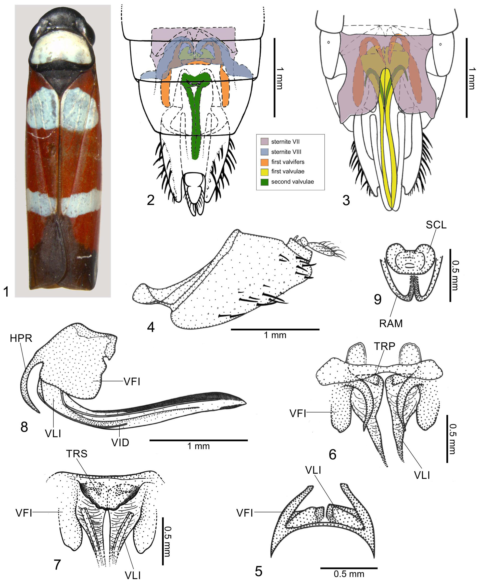
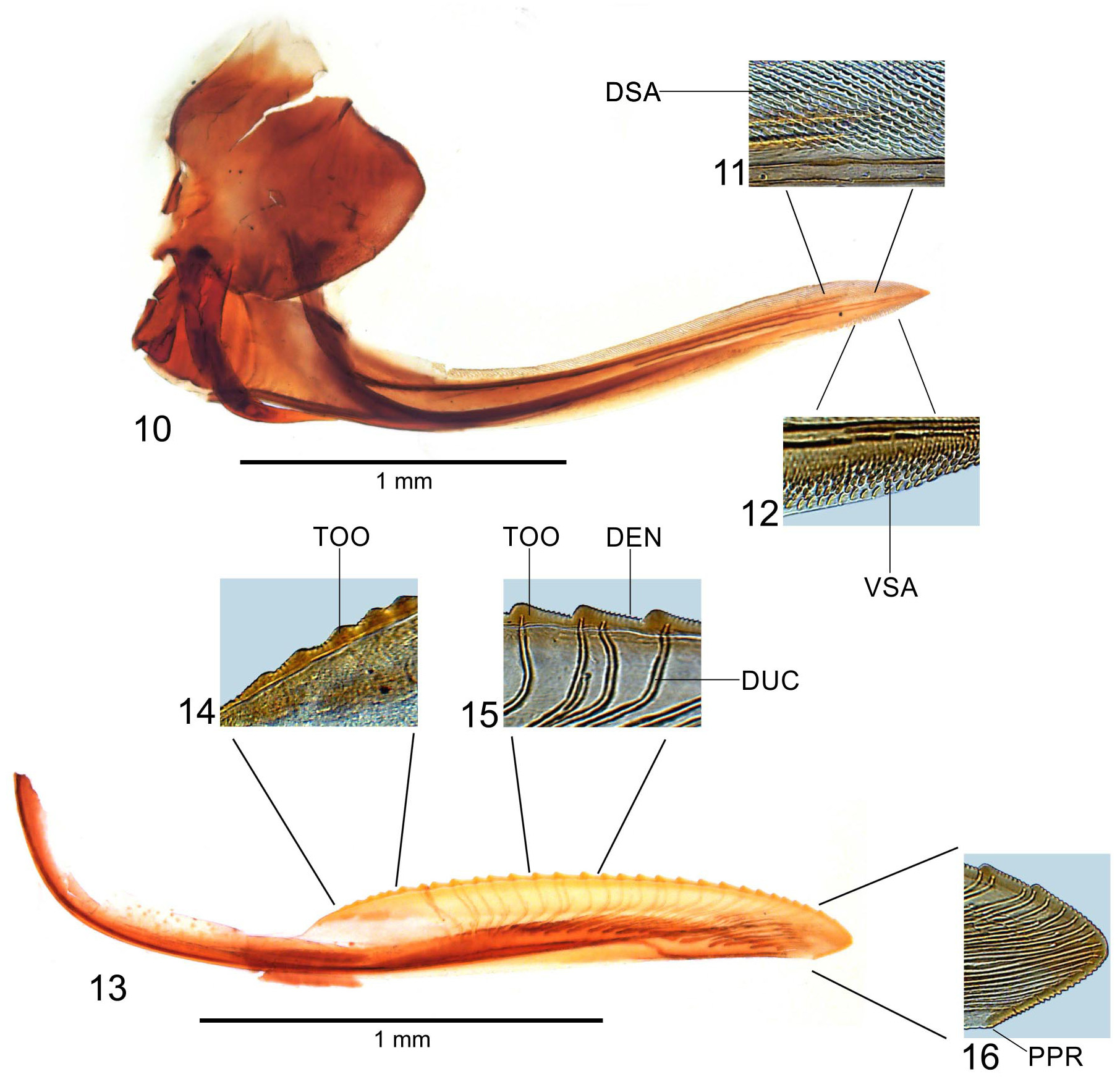
Diagnosis. Anterior dorsum ( Fig. 1 View FIGURES 1 – 9 ) dark brown to black with large white to ivory macula covering most of pronotum. Forewings ( Fig. 1 View FIGURES 1 – 9 ) mostly red with large macula basally on clavus and corium and transverse band extending from claval apex to costal margin, white to bluish-white. Male genitalia with styles long, apical portion turned outwards; aedeagus directed posterodorsally and with pair of apicodorsal spiniform processes directed anteriorly; paraphyses with T-shaped base, rami paired, elongate, with acute apices, each one, in ventral view, with foot-shaped process basally. Female with deep triangular emargination medially on posterior margin of sternite VII ( Fig. 3 View FIGURES 1 – 9 ); sternite VIII ( Fig. 2 View FIGURES 1 – 9 ) with pair of distinct sclerotized areas; first valvifer ( Figs 5–8 View FIGURES 1 – 9 , 10 View FIGURES 10 – 16 ) large and with hooklike process; area between first valvifers ( Fig. 7 View FIGURES 1 – 9 ) with broad triangular sclerite; first pair of valvulae ( Figs 8 View FIGURES 1 – 9 , 10–12 View FIGURES 10 – 16 ) with basiventral portion bearing small triangular projection; bases of second valvulae ( Fig. 9 View FIGURES 1 – 9 ) with laterally expanded sclerite between rami.
Female genitalia. Sternite VII ( Fig. 3 View FIGURES 1 – 9 ) with posterior margin convex or truncate on each side of median, deep triangular emargination, lateral portion slightly emarginated, forming small projection; lateral margins distinctly emarginate medially and serrated posteriorly; ventral surface irregularly striate, without setae, internally with narrow median area connected dorsally to sternite VIII. Sternite VIII ( Fig. 2 View FIGURES 1 – 9 ), in dorsal view, with pair of large sclerotized areas above first valvifers and bases of first valvulae; with dorsal region (including the sclerotized areas and membranous parts linking these areas) well developed and connected lateroposteriorly with tergite VIII. Pygofer ( Fig. 4 View FIGURES 1 – 9 ) moderately produced posteriorly; posterior margin narrowly rounded, macrosetae on posterior portion and extending anteriorly along ventral margin. First valvifers ( Figs 5–8 View FIGURES 1 – 9 , 10 View FIGURES 10 – 16 ), in lateral view, large, subquadrangular, anterior margin with hooklike process directed ventrally ( Fig. 8 View FIGURES 1 – 9 , HPR); in caudal view, connected medially to each other by broad triangular sclerite ( Fig. 7 View FIGURES 1 – 9 , TRS) with ventral margin crenulate. Basal portion of first pair of valvulae ( Figs 5–7 View FIGURES 1 – 9 ), in lateral view, enlarged and subrectangular, forming angle with remainder of blade; basiventral portion bearing small triangular projection ( Fig. 6 View FIGURES 1 – 9 , TRP); sculptured areas ( Figs 11 and 12 View FIGURES 10 – 16 ) mostly scalelike and distributed along almost all dorsal margin and on apical portion of ventral margin (with more linear processes on basal portion of dorsal sculptured area and with more separated scales on ventral sculptured area); ventral margin convex except for small preapical concavity; apex acute; ventral interlocking device ( Fig. 8 View FIGURES 1 – 9 , VID) located on basal half of blade, extending along ventral margin for most of its length, distal portion directed dorsally. Second pair of valvulae ( Fig. 13 View FIGURES 10 – 16 ) broadened beyond basal curvature, narrowing gradually towards narrowly rounded apex; ventral margin approximately rectilinear; preapical prominence ( Fig. 16 View FIGURES 10 – 16 , PPR) inconspicuous; with approximately 30 mostly triangular continuous teeth (basal teeth more rounded; Fig. 14 View FIGURES 10 – 16 ), extending from expanded basal portion to apical portion of blade; most teeth ( Fig. 15 View FIGURES 10 – 16 ) with steep, smaller ascending portion and gradually declivous, larger descending portion; denticles distributed on teeth and on apical portion ( Figs 14–16 View FIGURES 10 – 16 ; except on apex) of blade (dorsal dentate apical portion smaller than ventral one); blade with ducts attaining teeth or terminating below them ( Fig. 15 View FIGURES 10 – 16 ) and extending to apex ( Fig. 16 View FIGURES 10 – 16 ); with ovoid and medially concave sclerite between bases of rami ( Fig. 9 View FIGURES 1 – 9 , SCL). Gonoplacs with basal half distinctly narrow in comparison with apical half, abruptly expanded on median portion; apical half with few setae; apex rounded.
Distribution. Brazil (states of Minas Gerais, Rio de Janeiro, and São Paulo).
Material examined. Brazil: state of Minas Gerais: one female ( MNRJ); state of Rio de Janeiro: three males and four females ( MNRJ).
| MNRJ |
Museu Nacional/Universidade Federal de Rio de Janeiro |
No known copyright restrictions apply. See Agosti, D., Egloff, W., 2009. Taxonomic information exchange and copyright: the Plazi approach. BMC Research Notes 2009, 2:53 for further explanation.
|
Kingdom |
|
|
Phylum |
|
|
Class |
|
|
Order |
|
|
InfraOrder |
Cicadomorpha |
|
Family |
|
|
Genus |