
Erythrogonia calva (Taschenberg, 1884)
|
publication ID |
https://doi.org/ 10.11646/zootaxa.3872.3.4 |
|
publication LSID |
lsid:zoobank.org:pub:9CCEAC1C-F58A-40E6-8349-D081976E12A5 |
|
DOI |
https://doi.org/10.5281/zenodo.6139010 |
|
persistent identifier |
https://treatment.plazi.org/id/660DF03E-FFBE-FF82-FF65-866B459DF868 |
|
treatment provided by |
Plazi |
|
scientific name |
Erythrogonia calva (Taschenberg, 1884) |
| status |
|
Erythrogonia calva (Taschenberg, 1884) View in CoL
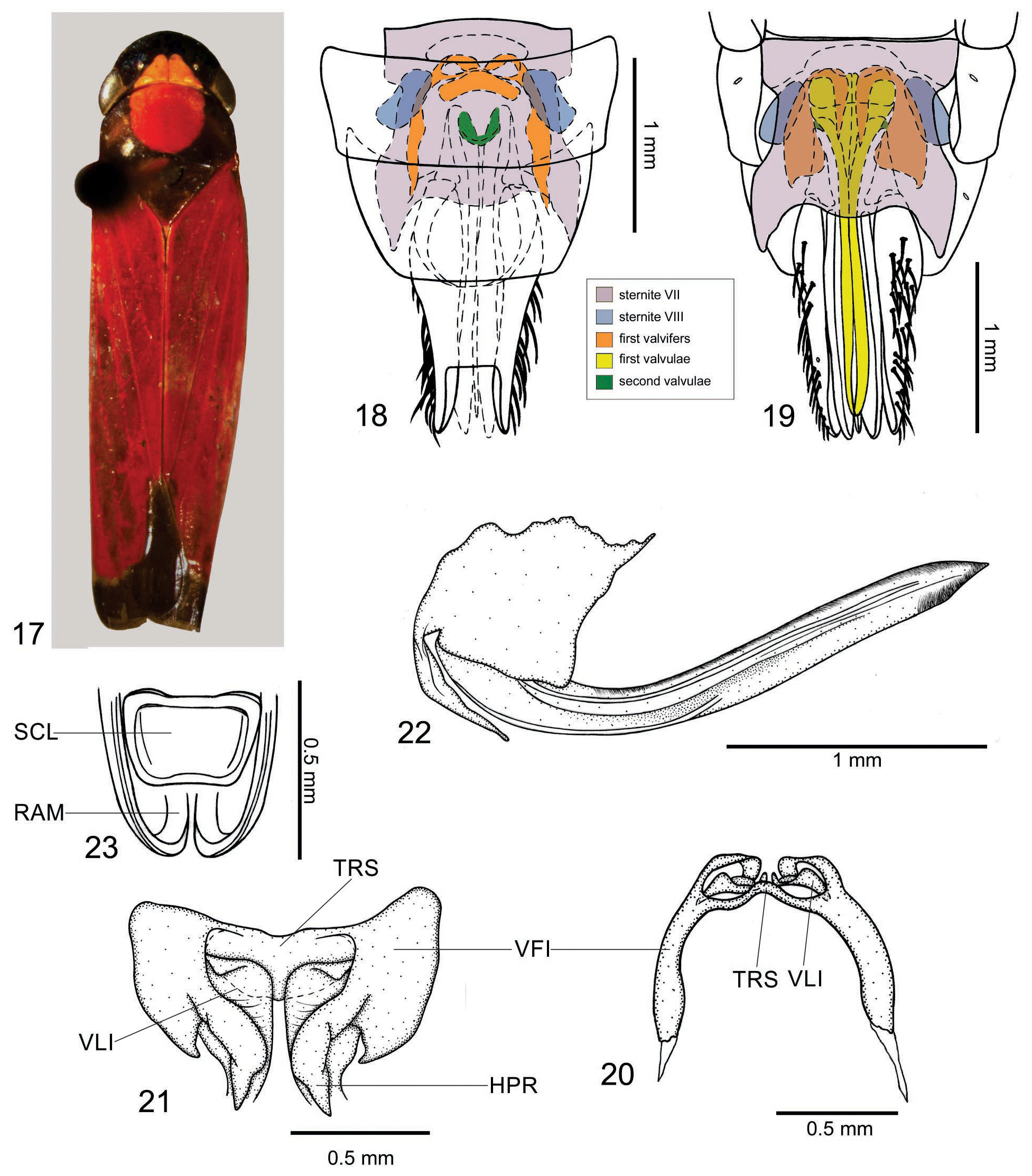
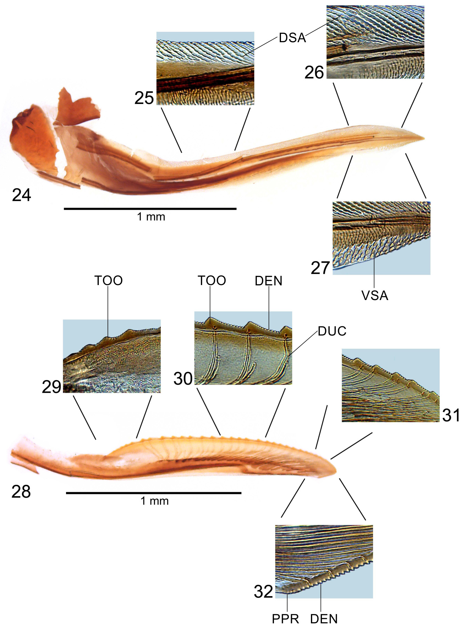
( Figs 17–32 View FIGURES 17 – 23 View FIGURES 24 – 32 )
Length: female 7.8–8.2 mm (n = 2), male 8.0– 8.2 mm (n = 2).
Diagnosis. Anterior dorsum ( Fig. 17 View FIGURES 17 – 23 ) brown to dark brown with yellow or yellowish-red triangular macula on crown contiguous to oval median macula on pronotum. Forewings ( Fig. 17 View FIGURES 17 – 23 ) red with costal margin brown to dark brown, without maculae; membrane dark brown. Male pygofer with pair of small spiniform processes dorsoapically; subgenital plates with distal half strongly narrowed; aedeagal shaft slightly curved dorsally and then ventrally on apical portion, apex with pair of spiniform processes directed anterodorsally; paraphyses with rami long, sinuous, slightly exceeding pygofer apex. Female sternite VII ( Fig. 19 View FIGURES 17 – 23 ) with posterior margin broadly emarginate and with slight median lobe, posterolateral portions forming triangular projections, basal portion with large inner lobe; sternite VIII ( Figs 18 and 19 View FIGURES 17 – 23 ) formed by pair of lateral sclerites; first valvifers ( Figs 20–22 View FIGURES 17 – 23 ) large, with hooklike process, connected to each other by broad triangular sclerite ( Fig. 21 View FIGURES 17 – 23 ; a dashed line depicts the ventral margin of the sclerite); first valvulae ( Figs 22 View FIGURES 17 – 23 and 24 View FIGURES 24 – 32 ) with basiventral margin bearing small triangular projection; bases of second valvulae ( Fig. 23 View FIGURES 17 – 23 ) with distinct, medially concave sclerite between rami.
Female genitalia. Sternite VII ( Fig. 19 View FIGURES 17 – 23 ) with posterior margin broadly emarginate and with slight median lobe, posterolateral portions forming triangular projections; lateral margins expanded on apical half; lateral portions of posterior margin and almost all lateral margin serrated; ventral surface with median longitudinal carina, weakly striate, without setae; basal portion forming large inner lobe. Sternite VIII ( Figs 18 and 19 View FIGURES 17 – 23 ), in dorsal view, with pair of sclerites positioned laterally and externally above first valvifers. Pygofer well produced posteriorly; posterior margin narrowly rounded; macrosetae on posterior portion and extending anteriorly along ventral margin. First valvifers ( Figs 20–22 View FIGURES 17 – 23 ), in lateral view, large and quadrangular, anterior margin with hooklike process directed ventrally ( Fig. 21 View FIGURES 17 – 23 , HPR); posteroventral corner with blunt projection; valvifers connected internally to each other by broad triangular sclerite on dorsal portion ( Figs 20 and 21 View FIGURES 17 – 23 , TRS; a dashed line depicts the ventral margin of the sclerite in Fig. 21 View FIGURES 17 – 23 ). Basal portion of first pair of valvulae ( Fig. 21 View FIGURES 17 – 23 ), in ventral view, broadened, with distinct outer projection; basiventral margin bearing small triangular projection; sculptured areas ( Figs 25–27 View FIGURES 24 – 32 ) mostly scalelike and distributed along almost all dorsal margin and on apical portion of ventral margin (with more linear tegumentary processes on basal portion of dorsal sculptured area and with more separated scales on ventral sculptured area); ventral margin convex except for small preapical concavity; apex acute; ventral interlocking device ( Fig. 22 View FIGURES 17 – 23 ) located on basal half of blade, extending along ventral margin for most of its length, distal portion directed dorsally. Second pair of valvulae ( Fig. 28 View FIGURES 24 – 32 ) broadened beyond basal curvature, narrowing slightly towards narrowly rounded apex; ventral margin approximately rectilinear; preapical prominence ( Fig. 32 View FIGURES 24 – 32 ) inconspicuous; with approximately 27 mostly triangular continuous teeth (basal teeth more rounded; Fig. 29 View FIGURES 24 – 32 ), extending from expanded basal portion to apical portion of blade; most teeth ( Fig. 27 View FIGURES 24 – 32 ) with steep, smaller ascending portion and gradually declivous, larger descending portion; denticles ( Figs 30–32 View FIGURES 24 – 32 ) distributed on teeth and on apical portion (except on apex) of blade (dorsal and ventral dentate apical portions with about same size); blade with ducts attaining teeth or terminating below them and extending to apex ( Figs 30–32 View FIGURES 24 – 32 ); with distinct, medially concave sclerite between bases of rami ( Fig. 23 View FIGURES 17 – 23 , SCL). Gonoplacs with basal half distinctly narrow in comparison with apical half, abruptly expanded on median portion; apical half with few setae; apex rounded.
Distribution. Venezuela, Brazil (states of Paraná and Santa Catarina), Argentina.
Material examined. Brazil: state of Paraná: one male and three females ( DZUP); state of Santa Catarina: one male ( MNRJ). One male and one female without locality label ( MNRJ).
No known copyright restrictions apply. See Agosti, D., Egloff, W., 2009. Taxonomic information exchange and copyright: the Plazi approach. BMC Research Notes 2009, 2:53 for further explanation.
|
Kingdom |
|
|
Phylum |
|
|
Class |
|
|
Order |
|
|
InfraOrder |
Cicadomorpha |
|
Family |
|
|
Genus |