
Costanana cifi, Domahovski & Cavichioli, 2023
|
publication ID |
https://doi.org/ 10.5852/ejt.2023.889.2245 |
|
publication LSID |
lsid:zoobank.org:pub:52E11625-6E26-4E4F-8B15-05195087BAB4 |
|
DOI |
https://doi.org/10.5281/zenodo.8269463 |
|
persistent identifier |
https://treatment.plazi.org/id/8714AD76-D0F8-4A68-89CD-902504BBB98B |
|
taxon LSID |
lsid:zoobank.org:act:8714AD76-D0F8-4A68-89CD-902504BBB98B |
|
treatment provided by |
Felipe |
|
scientific name |
Costanana cifi |
| status |
sp. nov. |
Costanana cifi View in CoL sp. nov.
urn:lsid:zoobank.org:act:8714AD76-D0F8-4A68-89CD-902504BBB98B
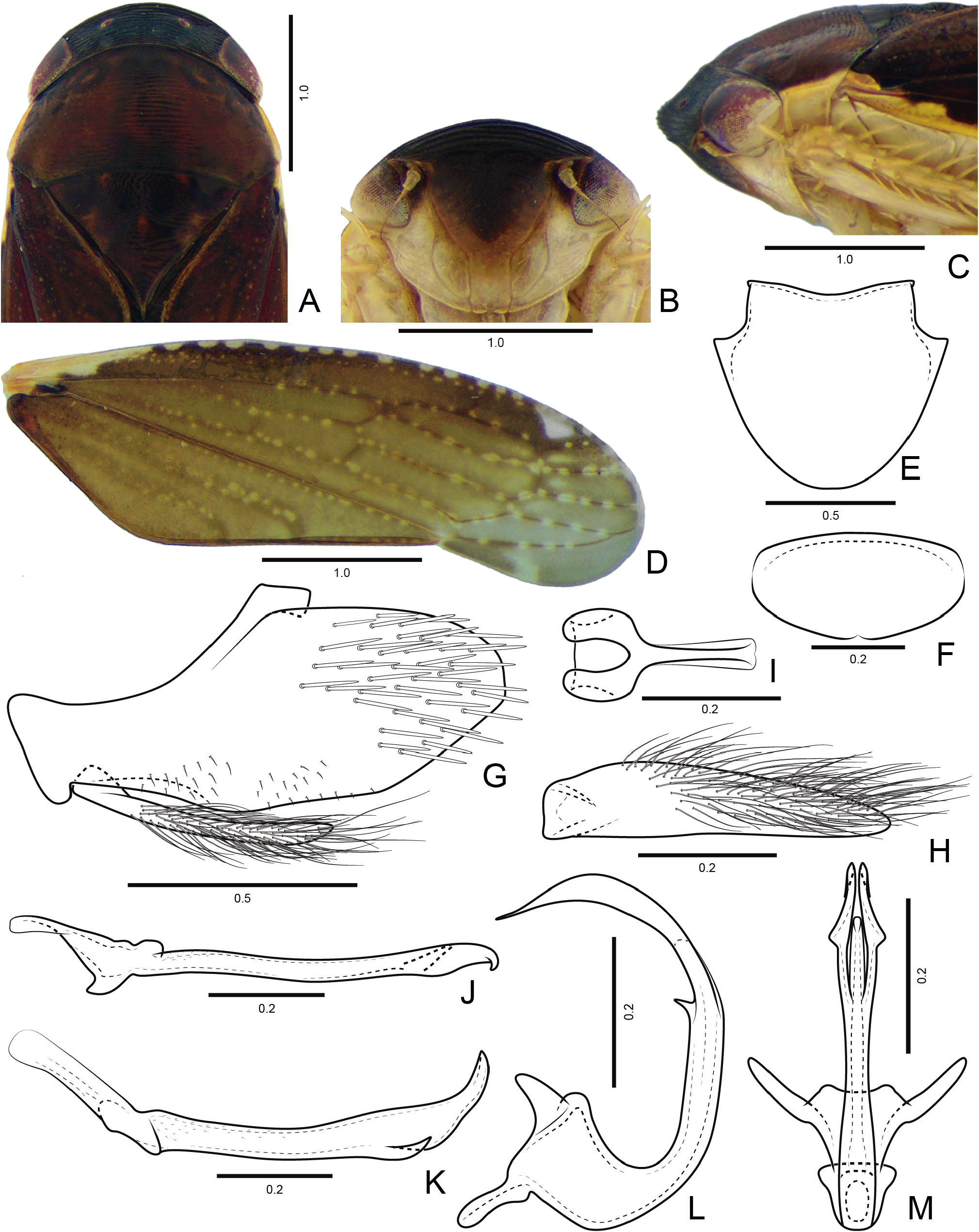
Figs 4– 5 View Fig View Fig , 11E–F View Fig
Diagnosis
Coloration ( Fig. 11E View Fig ) of crown black, pronotum brown with inconspicuous black maculae, forewing costal margin with sequence of small yellow spots and a small depigmented (white) macula on apical third. Style ( Fig. 4K View Fig ) without dentiform ventral process near base of blade; apical third with short slender process overlapping dorsal apex. Aedeagus ( Fig. 4L–M View Fig ) without processes near base; shaft with short subapical spiniform process on dorsal margin and pair of long subapical processes almost as long as shaft length, directed dorsally and curved anterad.
Etymology
The new species name is an anagram of the initials C.I.I.F. and refers to the Centro de Identificação de Insetos Fitófagos, Departamento de Zoologia of Universidade Federal do Paraná, that sampled many insects in Paraná State in the year 1985.
Material examined
Holotype BRAZIL • ♂; “S. [São] José dos Pinhais - PR [Paraná] \ Brasil (BR 277 Km 54) \ 23.III.1985 \ C.I.I.F. (Luminosa)”; DZUP 215484 View Materials .
Paratypes BRAZIL – Paraná • 1 ♀; “ Morretes - PR [Paraná] Brasil \ (IAPAR) [ Instituto Agronômico do Paraná ] \ 31.III.1985 \ C.I.I.F. [Centro de Identificação de Insetos Fitófagos] (Luminosa)”; DZUP 215487 View Materials • 1 ♀; same collection data as for preceding, except “ 06.IV.1985 ”; DZUP 215488 View Materials .
Description
MEASUREMENTS. Holotype male: total length 5.2 mm. Paratype female (n = 2) 5.6–5.8 mm.
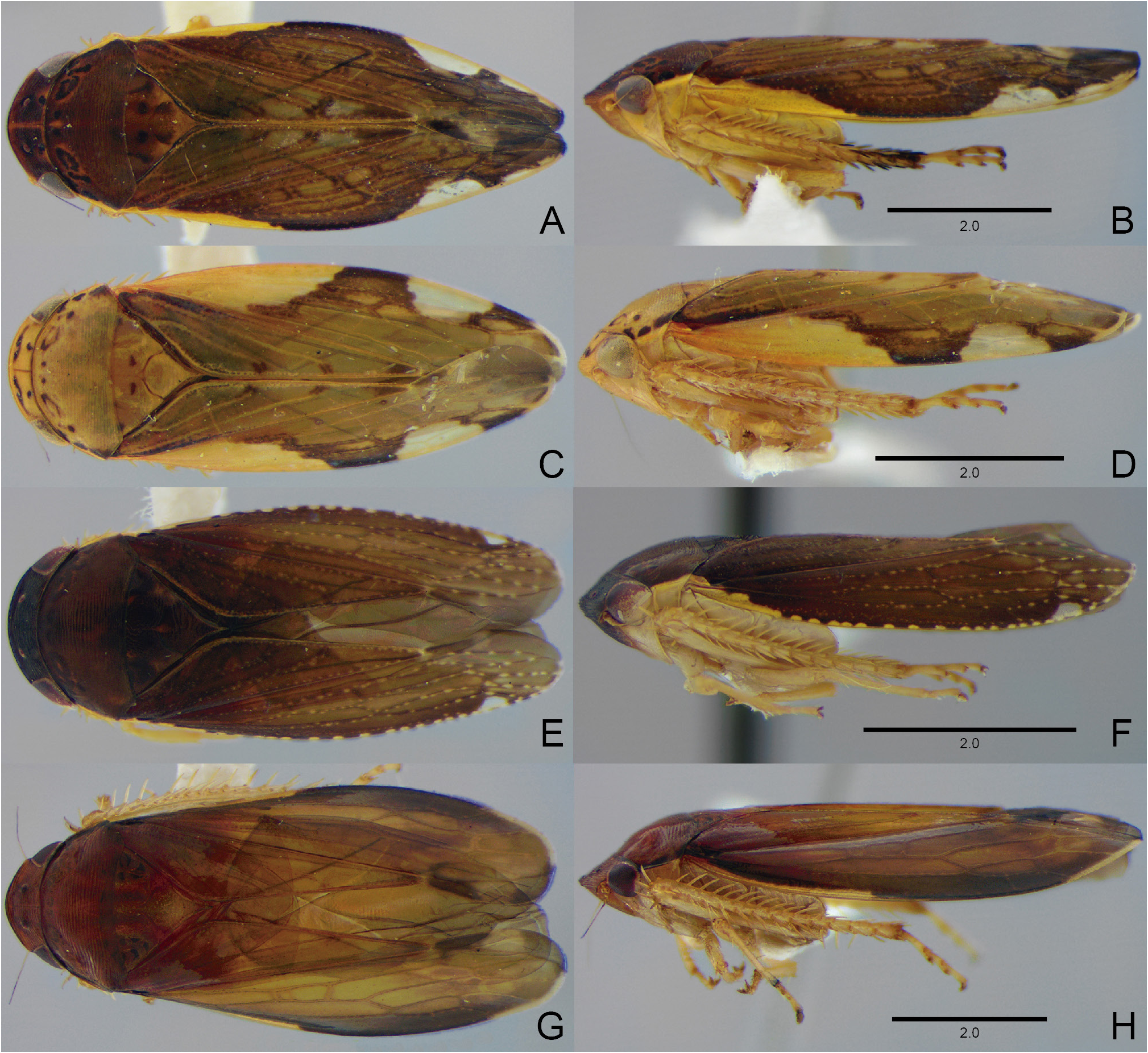
COLORATION. Dorsal portion of head and thorax reddish-brown ( Figs 4A, C View Fig , 11E View Fig ); ventral portion yellow ( Figs 4B–C View Fig , 11F View Fig ). Crown ( Fig. 4A View Fig ) black; eyes and ocelli red. Pronotum ( Fig. 4A View Fig ) with inconspicuous irregular black maculae near anterior margin; lateral margin yellow. Mesonotum with small yellowish spots near anterior margin and scutoscutellar suture. Face ( Fig. 4B View Fig ) with frons almost completely black, except with lateral and ventral margins brownish; gena black between eye and frons. Forewing ( Fig. 4D View Fig ) costal margin with small slender yellow macula on anterior fifth, sequence of 13–14 yellow spots and a small depigmented (white) macula at apex of outer apical cell; veins brown with several yellowish spots. Legs ( Figs 4C View Fig , 11F View Fig ) yellow, without black markings.
STRUCTURE. Head in dorsal view ( Fig. 4A View Fig ): median length slightly shorter than interocular width; transocular width of head 8.5 tenths humeral width of pronotum; in lateral view ( Fig. 4C View Fig ), with anterior margin thick, with 7–8 well developed carinae. Forewing ( Fig. 4D View Fig ) with inner discal cell not distinctly shorter than outer discal cell; apex rounded. Profemur AV row with 3–4 setae restricted to basal half and PV row with 1 apical seta. Protibia PD row with 3 long setae and apical PD 1 seta developed; PV row developed, with 6–7 setae increasing in thickness and length towards apex. Metatibia rows PD, AD, and AV with 18–19, 12, and 13–14 macrosetae, respectively. Metatarsomere I apex with 3–4 platellae. Metatarsomere II apex with 2 apical platellae. Other characteristics as in the generic description.
MALE TERMINALIA. Sternite VIII ( Fig. 4E View Fig ) 1.2× as wide as long; lateral margins excavated at base, forming a triangular projection; apex broadly rounded. Valve ( Fig. 4F View Fig ) 2× as wide as long; posterior margin rounded, slightly emarginated medially. Pygofer ( Fig. 4G View Fig ), in lateral view, 2.2× as long as maximum height; ventral margin slightly rounded; posterodorsal margin rounded; posteroventral margin straight; external surface near ventral margin with short filiform setae; macrosetae distributed on apical half; apex subtruncated. Subgenital plate ( Fig. 4G View Fig ), in lateral view, short, slightly surpassing half length of pygofer; in ventral view ( Fig. 4H View Fig ), 4.5× as long as wide; inner margin straight; external margin broadly rounded; ventral surface and external margin with long filiform setae; apex tapered and weakly sclerotized. Connective ( Fig. 4I View Fig ) Y-shaped; arms weakly produced laterally; stalk narrow, about as long as maximum width of arms; median keel weakly developed. Style ( Fig. 4J View Fig ), in dorsal view, with outer lobe reduced; in lateral view ( Fig. 4K View Fig ), blade strongly elongated, almost straight; ventral margin not serrated, with short slender process at apical third, directed posterad, weakly curved dorsally and overlapping dorsal apex; apex tapered and curved dorsally. Aedeagus ( Fig. 4L–M View Fig ) preatrium developed; dorsal apodeme strongly produced, with divergent arms; shaft elongated curved dorsally; dorsal margin with subapical spiniform process; pair of long subapical processes almost as long as shaft length, directed dorsally and curved anterad, with apex tapered and sharply acute.
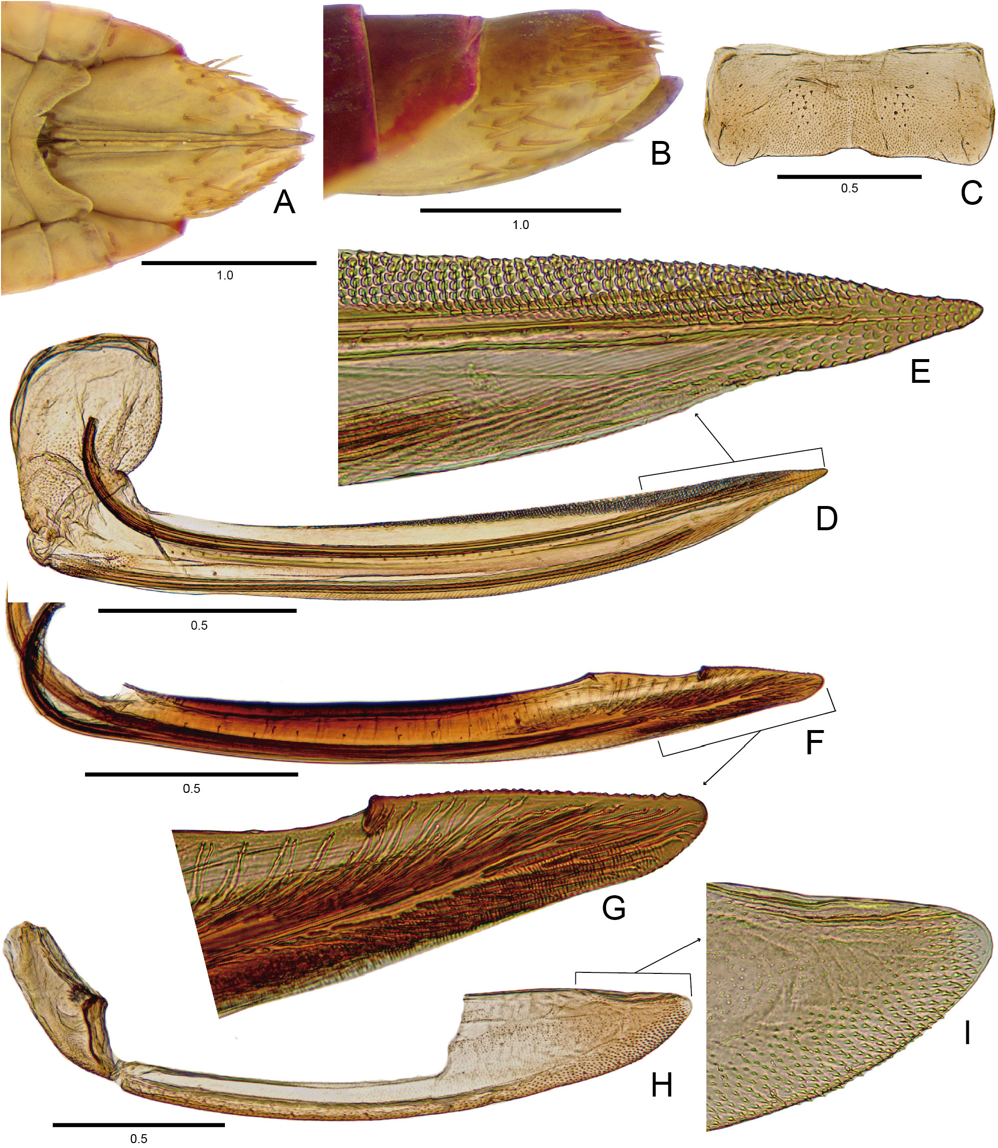
FEMALE TERMINALIA. Sternite VII ( Fig. 5A, C View Fig ), in ventral view, 2.4× as wide as long; posterolateral angles rounded and weakly produced posterad; posterior margin almost straight and slightly excavated medially. Internal sternite VIII membranous. Pygofer ( Fig. 5A–B View Fig ), in lateral view, approximately 2× as long as high; macrosetae distributed on posteroventral quadrant; apex rounded. First valvifer ( Fig. 5D View Fig ) subelliptic, slightly higher than wide. First valvula ( Fig. 5D View Fig ) 10× as long as high; ventral interlocking device long, reaching apical fifth; dorsal sculptured area areolate; apical portion ( Fig. 5E View Fig ) gradually tapering to apex; apex acute and areolate. Second valvula ( Fig. 5F View Fig ) 10.5× as long as high; dorsal protuberance reduced and acute, localized before midlength of blade; apical portion ( Fig. 5G View Fig ) of dorsal margin with a larger tooth followed by small denticles, approximately regular in shape and size; ventroapical margin with minute rounded denticles; apex rounded. Second valvifer ( Fig. 5H View Fig ), in lateral view, approximately 2.5× as high as wide. Gonoplac ( Fig. 5H View Fig ) 5.4× as long as high; dorsoapical margin straight, longer than one-third of gonoplac length; ventral margin evenly rounded, with a few short setae; outer surface ( Fig. 5I View Fig ) with many integumentary denticles; rounded apex.
Remarks
Costanana cifi sp. nov. is similar in coloration to C. nupera comb. nov. in having the costal margin of the forewing with a small and slender yellow macula on the anterior fifth followed by a sequence of small yellowish spots and a larger white macula at the apex of the outer apical cell ( Figs 4D View Fig , 20A View Fig ). Costanana cifi can be separated from the other species of Costanana by the unusual shape of the aedeagus, with a short subapical spiniform process on the dorsal margin and a pair of long subapical processes directed dorsally and curved anterad ( Fig. 4L View Fig ).
No known copyright restrictions apply. See Agosti, D., Egloff, W., 2009. Taxonomic information exchange and copyright: the Plazi approach. BMC Research Notes 2009, 2:53 for further explanation.
|
Kingdom |
|
|
Phylum |
|
|
Class |
|
|
Order |
|
|
Family |
|
|
Genus |