
Nygolaimoides zarrinensis, Asl, Ebrahim Zahedi, Niknam, Gholamreza, Jabbari, Habibeh & Pena-Santiago, Reyes, 2016
|
publication ID |
https://doi.org/ 10.11646/zootaxa.4150.1.3 |
|
publication LSID |
lsid:zoobank.org:pub:4E232C98-222C-4C4A-B4AD-7B17F500EBB4 |
|
DOI |
https://doi.org/10.5281/zenodo.5669907 |
|
persistent identifier |
https://treatment.plazi.org/id/674687A9-F56D-0359-30EE-F976DC33F8D2 |
|
treatment provided by |
Plazi |
|
scientific name |
Nygolaimoides zarrinensis |
| status |
sp. nov. |
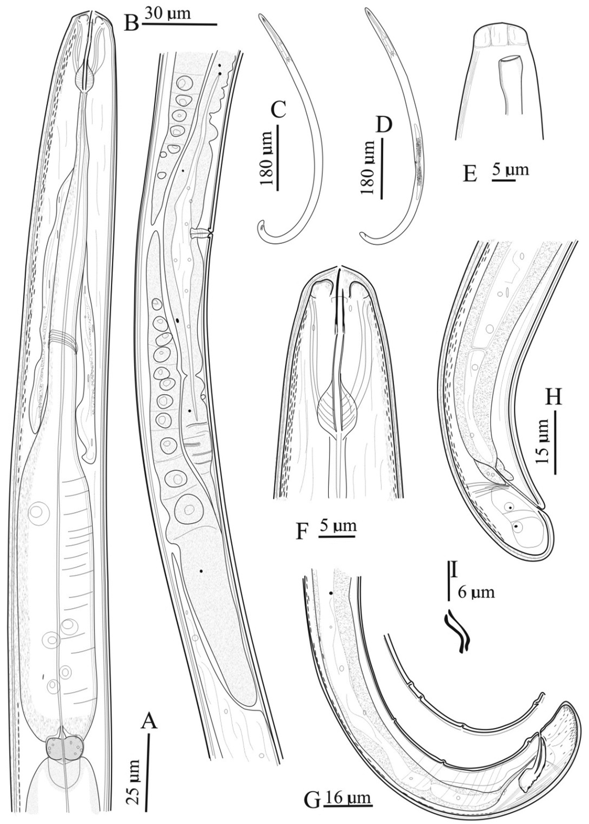
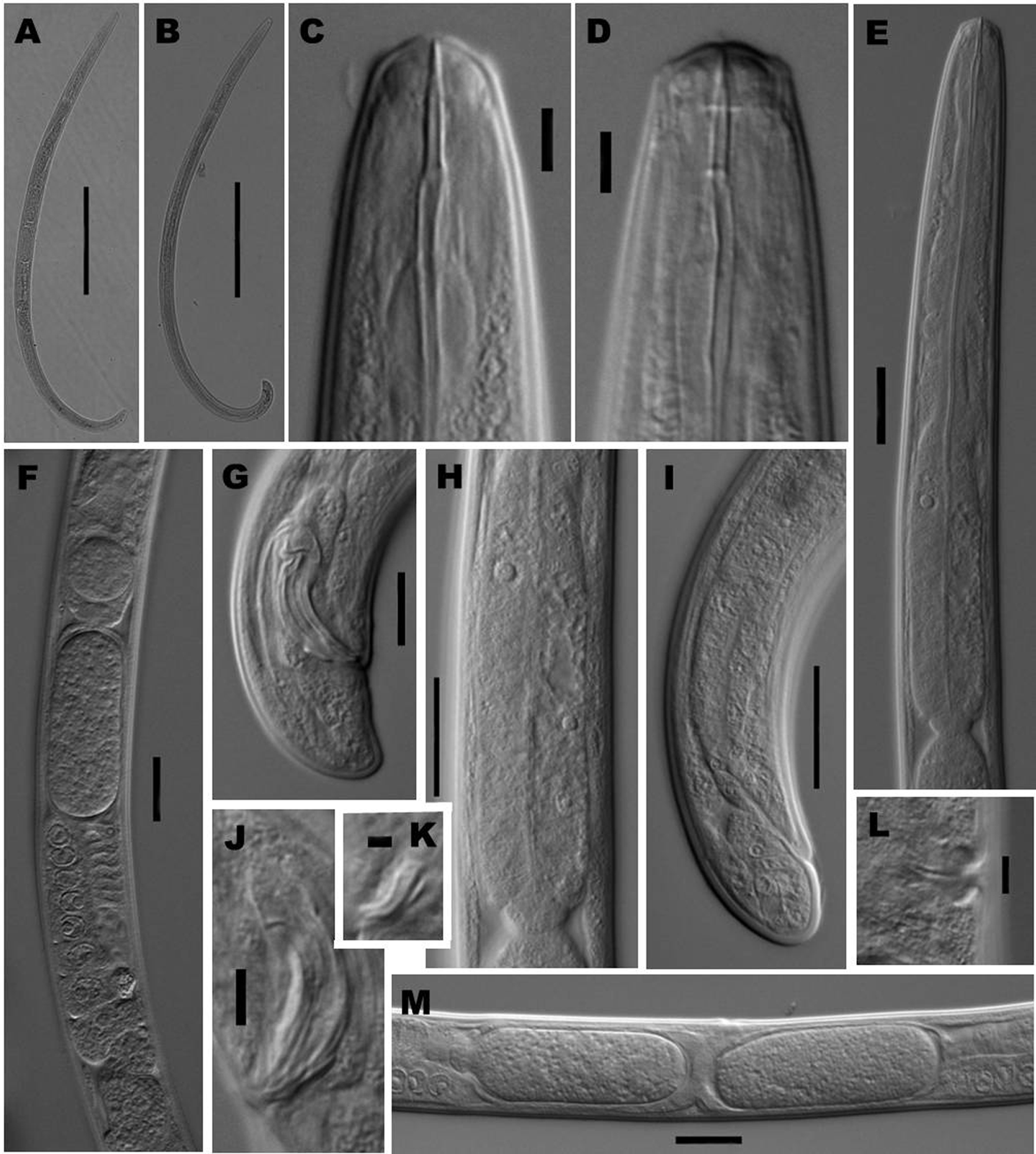
Nygolaimoides zarrinensis sp. n.
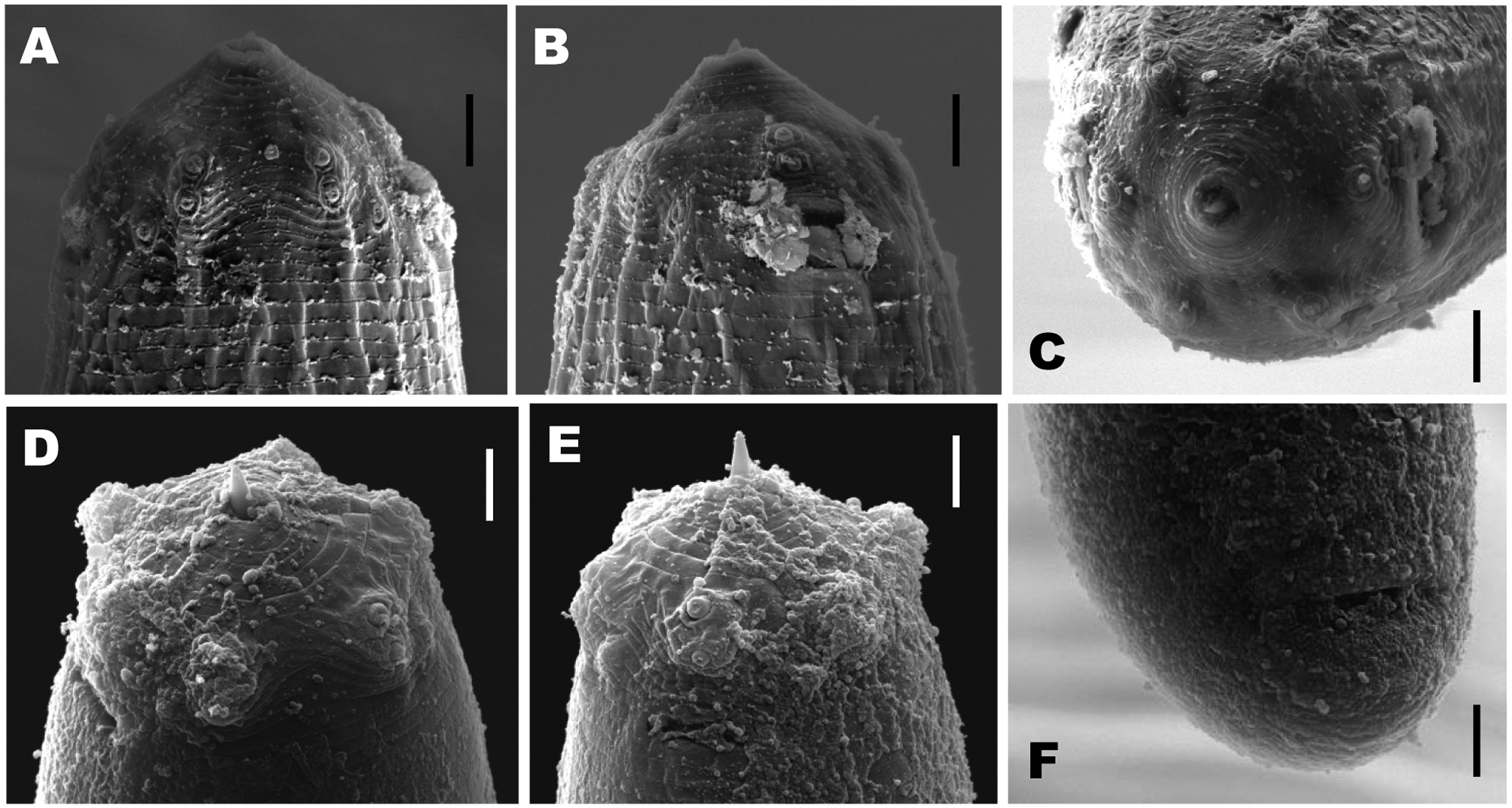
( Figs 1 View FIGURE 1 , 2 View FIGURE 2 & 3A–C View FIGURE 3. A – C )
Material examined. Fifteen females, ten males and eight juveniles, in very good condition.
Measurements. See Table 1.
Description. Adult: Moderately slender to slender (a = 29–35) nematodes of small size, 0.73–0.94 mm long. Body cylindrical, tapering towards both extremities but more so towards the anterior end as the tail is short and rounded. Habitus regularly curved ventrad upon fixation, C-shaped in female and J-shaped in male. Two-layered cuticle, 1.5–2.0 µm thick throughout most body. Body pores indistinct. Lip region continuous with the adjacent body, about two-fifths of body diameter at neck base, and with the perioral area distinctly protruding or elevated; lips (under SEM, but see remarks) totally fused; labial and cephalic papillae button-like, each surrounded by a ringlike, visibly elevated annulus, and with a large pore at the center; inner labial papillae situated at the margin of oral field, very close to outer labial papillae; oral field wide, with six or seven fine concentric incisures around the oral aperture. Amphid fovea with a large elliptical aperture about twice as wide as long and occupying ca one-third of lip region diameter. Cheilostom a truncate cone 5 µm long. Odontostyle typical of the genus, as long as lip region diameter, 6–7 times longer than broad, and with aperture occupying about two-fifths its total length. Guiding ring simple, distinct. Odontophore rod-like, lacking any differentiation, 1.5 times the odontostyle length. Pharynx consisting of a more slender but muscular anterior section gradually enlarging into the basal expansion, which is 4.6–4.8 times longer than wide, 3.1–3.2 times longer than body diameter at neck base, and occupies 42–43% of total neck length; gland nuclei very distinct in a few specimens, DN larger than S1N which are larger than S2N ( Fig. 2 View FIGURE 2 H), located as follows (n = 2): DO = 65, DN = 69–70, S1N1 = 83, S1N2 = 83–85, S2N = 90–91. Pharyngo-intestinal junction consisting of a short and rounded cardia surrounded by three moderately developed cardiac cells. Prerectum nearly twice and rectum about equal to the corresponding body diameter long. Tail short, conoid and tip rounded, ventrally straighter in males.
Female: Genital system didelphic-amphidelphic, with both branches very well developed, 203–339 µm long or 21–33% of total body length each. Ovaries large, 146–189 µm long, surpassing the oviduct-uterus junction. Oviduct 82–118 µm or 2.8–4.1 times the corresponding body diameter long, consisting of a slender, distal portion made of prismatic cells and an about twice longer than wide pars dilatata often containing ellipsoid sperm (5–6.5 ×1.9 µm) cells inside. Uterus a simple tube-like structure, 32–33 µm long or slightly longer (1.1 times) than body diameter, rarely containing eggs (66–70×23–26 µm). Vagina extending inwards to about one-third of body diameter, poorly differentiated but its pars distalis is easily seen. Vulva a short transverse slit.
Male: Genital system diorchic, with opposite testes. In addition to the adcloacal pair, there are two (very exceptionally one or three) ventromedian supplements out of the range of the spicules, the posterior ones 32–41 µm from the adcloacal pair, the anterior pair at 20–25 µm from the most posterior ones. Spicules dorylaimoid, about 5.0 times longer than wide and 1.4 times longer than body diameter at level of the cloacal aperture; curvature 134º; head visibly broader than blade, in some specimens apparently folded and irregular; ventral side lacking a distinct hump and hollow; median piece occupying one-half of maximum spicule width, reaching the posterior tip; posterior end 2 µm wide. Lateral guiding pieces visibly curved ventrad, about 6 µm long and four times longer than wide. Gubernaculum weakly curved, 3.5–4.5 µm long.
Diagnosis. The new species is characterized by its moderately slender to slender (a = 29–35), 0.73–0.94 mm long body, lip region continuous, 9.5–10.5 µm broad, odontostyle 9.5–11.0 µm long, neck 162–194 µm long, pharyngeal expansion 78–83 µm long, V = 46–50, female tail short and rounded conoid (11–15 µm, c = 63–84, c’ = 0.7–0.9), male tail rounded conoid (14–18 µm, c = 43–55, c’ = 0.9–1.1), spicules 21–25 µm long and with irregular head, and two (exceptionally one or three) ventromedian supplements bearing hiatus.
Relationships. The new species is easily distinguishable from their congeners in the combination of four features: small general size (body less than 1.0 mm long), anterior vulva (V = 46–50 vs V often more than 50), spicules with thickened head, and usually two (vs one) ventromedian supplements. See below the key to species for additional information.
Type locality and habitat. Iran, West Azarbaijan province, Miandoab County, Heidarabad region (GPS Coordinates N: 36˚56ˈ17 –E: 46˚09ˈ48.7 ̎), in soil and stump samples of an old Populus alba nursery.
Type material. Female holotype, ten female paratypes, six male paratypes and five juveniles deposited with Nematode Collection , Nematology Lab., Faculty of Agriculture , University of Tabriz, Iran . Two female and two male paratypes with Nematode Collection , University of Jaén, Spain .
Etymology. The species epithet refers to the Zarrinehrood River, close to Heidarabad region, where nematodes were collected from.
Remarks. The SEM pictures ( Fig. 3 View FIGURE 3. A – C ) are the first ones available for a representative of Nygolaimoides . In spite of their non-optimal quality, they confirm the pattern observed under LM, as the labial papillae form two circles of six papillae each situated close together, at the margin of the oral field. The amphid apertures are distinctly elliptical and large, differing from the usual shape observed in other dorylaims (having a much longer than wide transverse slit). It is also remarkable that the oral field is not differentiated and is conspicuously protruding (elevated) around the oral aperture.
No known copyright restrictions apply. See Agosti, D., Egloff, W., 2009. Taxonomic information exchange and copyright: the Plazi approach. BMC Research Notes 2009, 2:53 for further explanation.