
Itostenhelia golikovi ( Chislenko, 1978 ) Karanovic & Kim, 2014
|
publication ID |
https://doi.org/ 10.11646/zootaxa.3783.1.1 |
|
publication LSID |
lsid:zoobank.org:pub:E6155BDC-AEAE-475D-BC83-61B3B863344C |
|
DOI |
https://doi.org/10.5281/zenodo.5062436 |
|
persistent identifier |
https://treatment.plazi.org/id/6878D460-FFEE-FF9B-64D0-F9BB0135FED9 |
|
treatment provided by |
Felipe |
|
scientific name |
Itostenhelia golikovi ( Chislenko, 1978 ) |
| status |
comb. nov. |
Itostenhelia golikovi ( Chislenko, 1978) comb. nov.
( Figs. 43–47 View FIGURE 43 View FIGURE 44 View FIGURE 45 View FIGURE 46 View FIGURE 47 )
Type locality. Russia, Primorsky Krai, Posyet Bay, Minonosok inlet, benthic sands at 3–4 m depth, 42.609258°N 130.861661°E GoogleMaps .
Specimens examined. Two females dissected on one slide each (collection numbers NIBRIV0000232699 and NIBRIV0000232700), male dissected on one slide (collection number NIBRIV0000232701), one male and five females together on one SEM stub (collection number NIBRIV0000232702), 10 females (four ovigerous) and two copepodids together in ethanol (collection number NIBRIV0000232703), 10 females together in ethanol (collection number NIBRIV0000232704), four males and 40 females (10 ovigerous) together in ethanol (collection number NIBRIV0000232705), 10 females destroyed for DNA sequence (five amplifications successful, Codes 0734, 0832, 0631, 0330 & 0433), type locality, 06 May 2012, leg. J. Trebyakova.
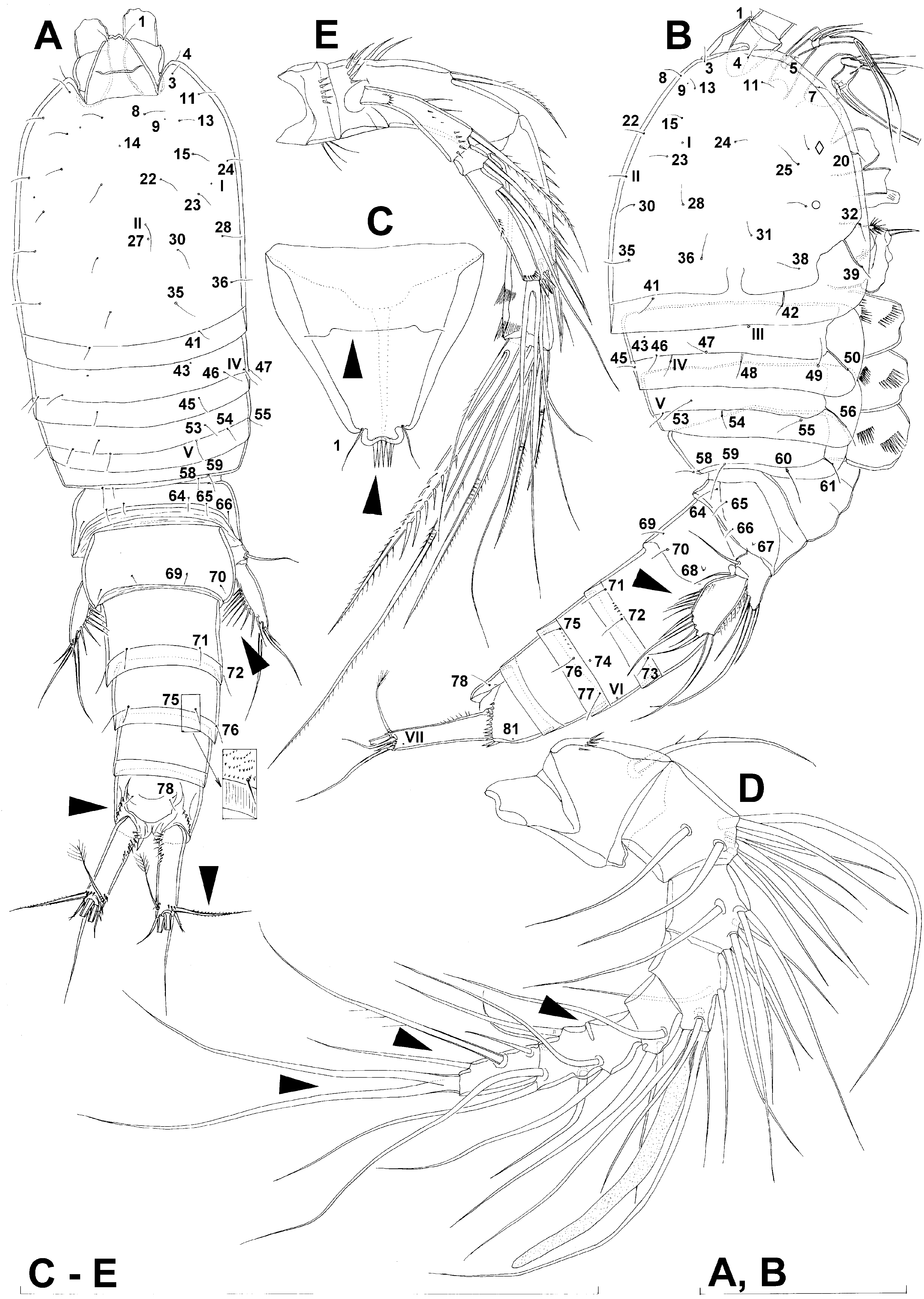
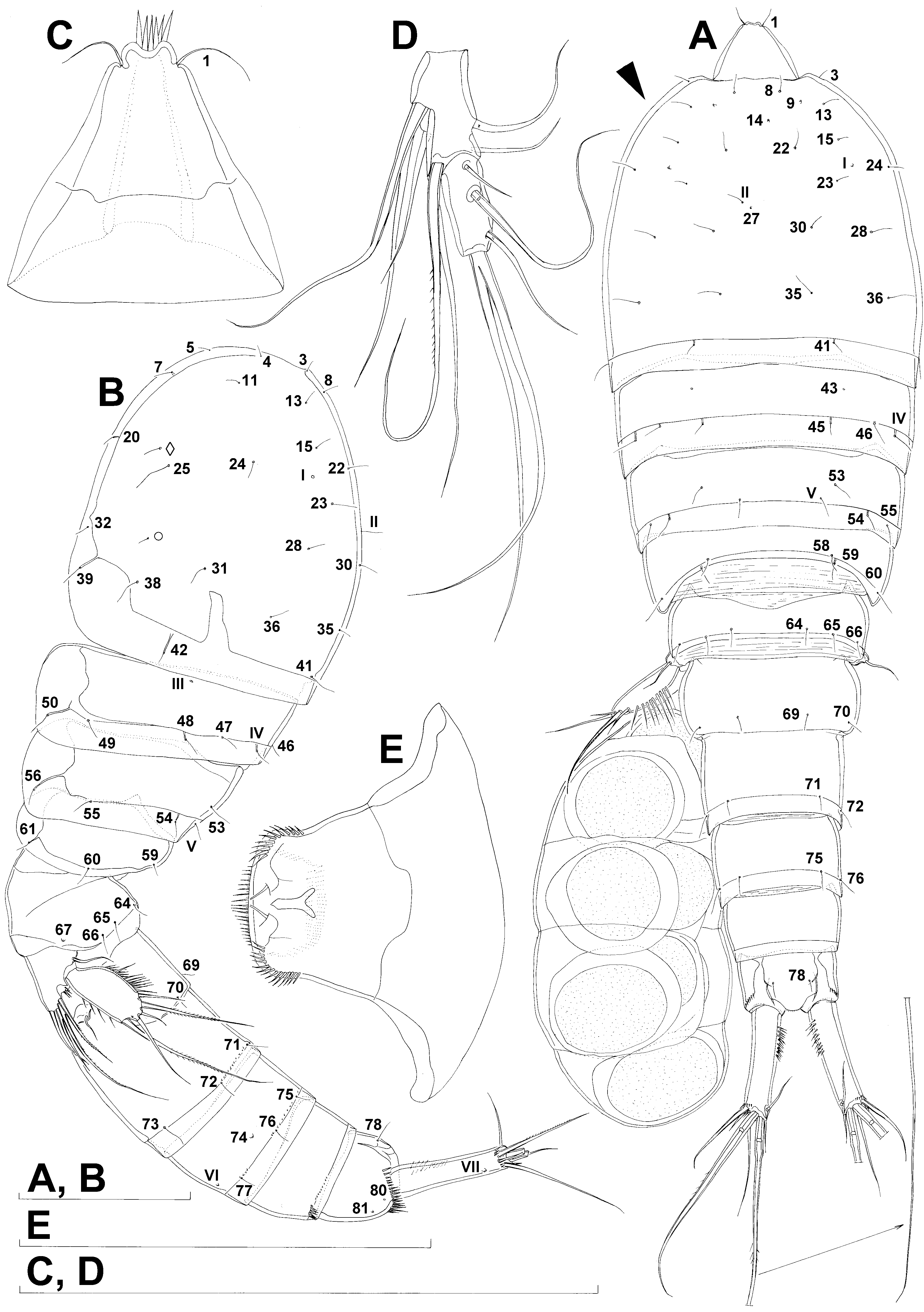
Redescription. Female (based on ten specimens). Body length from 545 to 648 µm. Body segmentation, nauplius eye, hyaline fringes, integument thickness, surface appearance of somites, and all somite ornamentation as in Itostenhelia polyhymnia sp. nov., including all sensilla and pores, and minute dorso-lateral rows of spinules on urosomites. However, colour of preserved specimens more translucent. Habitus ( Figs. 43A, B View FIGURE 43 , 46A View FIGURE 46 ) more robust than in Itostenhelia polyhymnia , with prosome/urosome length ratio about 1.1, body length/width ratio 3.4, and cephalothorax 1.8 times as wide as genital double-somite. Five specimens with paired egg-sacs, each containing between seven and 10 eggs.
Rostrum ( Fig. 43C View FIGURE 43 ) as in Itostenhelia polyhymnia , including apical bunch of spinules, dorsal suture, position of anterior pair of sensilla no. 1; only slightly smaller in comparison to cephalothorax.
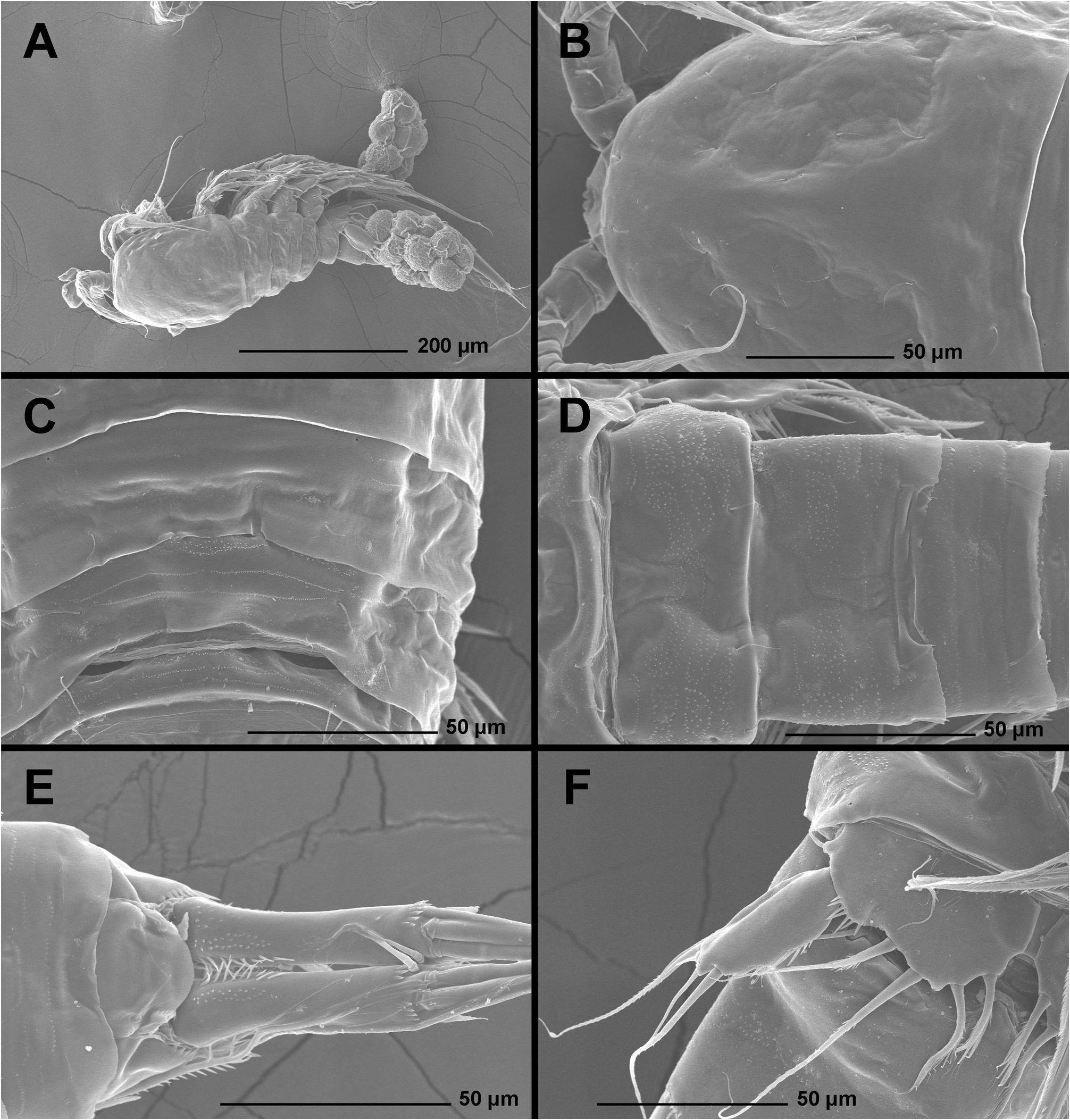
Cephalothorax ( Figs. 43A, B View FIGURE 43 , 46B View FIGURE 46 , 47C View FIGURE 47 ) about 0.9 times as long as wide; comprising 26% of total body length. Surface of cephalothoracic shield ornamented exactly as in Delavalia polyhymnia (all homologous pores and sensilla indicated with Arabic numerals, geometric shapes or Roman numerals in illustrations).
Pleuron of second pedigerous somite ( Figs. 43A, B View FIGURE 43 , 46C View FIGURE 46 , 47D View FIGURE 47 ), pleuron of third pedigerous somite ( Figs. 43A, B View FIGURE 43 , 46C View FIGURE 46 , 47D View FIGURE 47 ), and pleuron of fourth pedigerous somite ( Figs. 43A, B View FIGURE 43 , 46C View FIGURE 46 , 47D View FIGURE 47 ) ornamented exactly as in Itostenhelia polyhymnia .
First urosomite ( Figs. 34A, B View FIGURE 34 ) as in Itostenhelia polyhymnia , except slightly narrower both in dorsal and lateral view.
Genital double-somite ( Figs. 43A, B View FIGURE 43 , 44A View FIGURE 44 , 46D View FIGURE 46 ) as in Itostenhelia polyhymnia , except internal structure slightly narrower (arrowed in Fig. 44A View FIGURE 44 ).
Third urosomite ( Figs. 43A, B View FIGURE 43 , 44A View FIGURE 44 , 46D View FIGURE 46 ) and preanal somite ( Figs. 43A, B View FIGURE 43 , 44A View FIGURE 44 , 46E View FIGURE 46 ) as in Itostenhelia polyhymnia .
Anal somite ( Figs. 43A, B View FIGURE 43 , 44A View FIGURE 44 , 46E View FIGURE 46 ) also as in Itostenhelia polyhymnia , except lateral pore no. 80 present.
Caudal rami ( Figs. 43A, B View FIGURE 43 , 44A View FIGURE 44 , 46E View FIGURE 46 ) as in Itostenhelia polyhymnia , except additional field of small spinules present on anterior ventral surface close to inner margin, in addition to large inner spinules.
Antennula ( Figs. 43D View FIGURE 43 , 47C View FIGURE 47 ) as in Itostenhelia polyhymnia , except sixth and seventh segments completely fused.
Antenna, labrum ( Figs. 43E View FIGURE 43 , 47A View FIGURE 47 ), paragnaths ( Fig. 47A View FIGURE 47 ), mandibula, maxillula ( Fig. 47A View FIGURE 47 ), maxilla ( Fig. 47B View FIGURE 47 ), maxilliped ( Fig. 47B View FIGURE 47 ), first swimming leg, second swimming leg, third swimming leg ( Fig. 44B View FIGURE 44 ), and fourth swimming leg ( Fig. 44C View FIGURE 44 ) as in Itostenhelia polyhymnia .
Fifth leg ( Figs. 43A, B View FIGURE 43 , 44D View FIGURE 44 , 46F View FIGURE 46 ) as in Itostenhelia polyhymnia , except pore on basis missing and endopodal lobe slightly more convex. Length ratio of endopodal setae, starting from inner side, 1: 1.3: 2.9: 1.8. Length ratio of exopodal setae, starting from inner side, 1: 0.5: 0.15: 0.7: 0.3.
Sixth leg ( Figs. 44A, E View FIGURE 44 , 46F View FIGURE 46 ) as in Itostenhelia polyhymnia , except for additional spiniform process next to plumose seta.
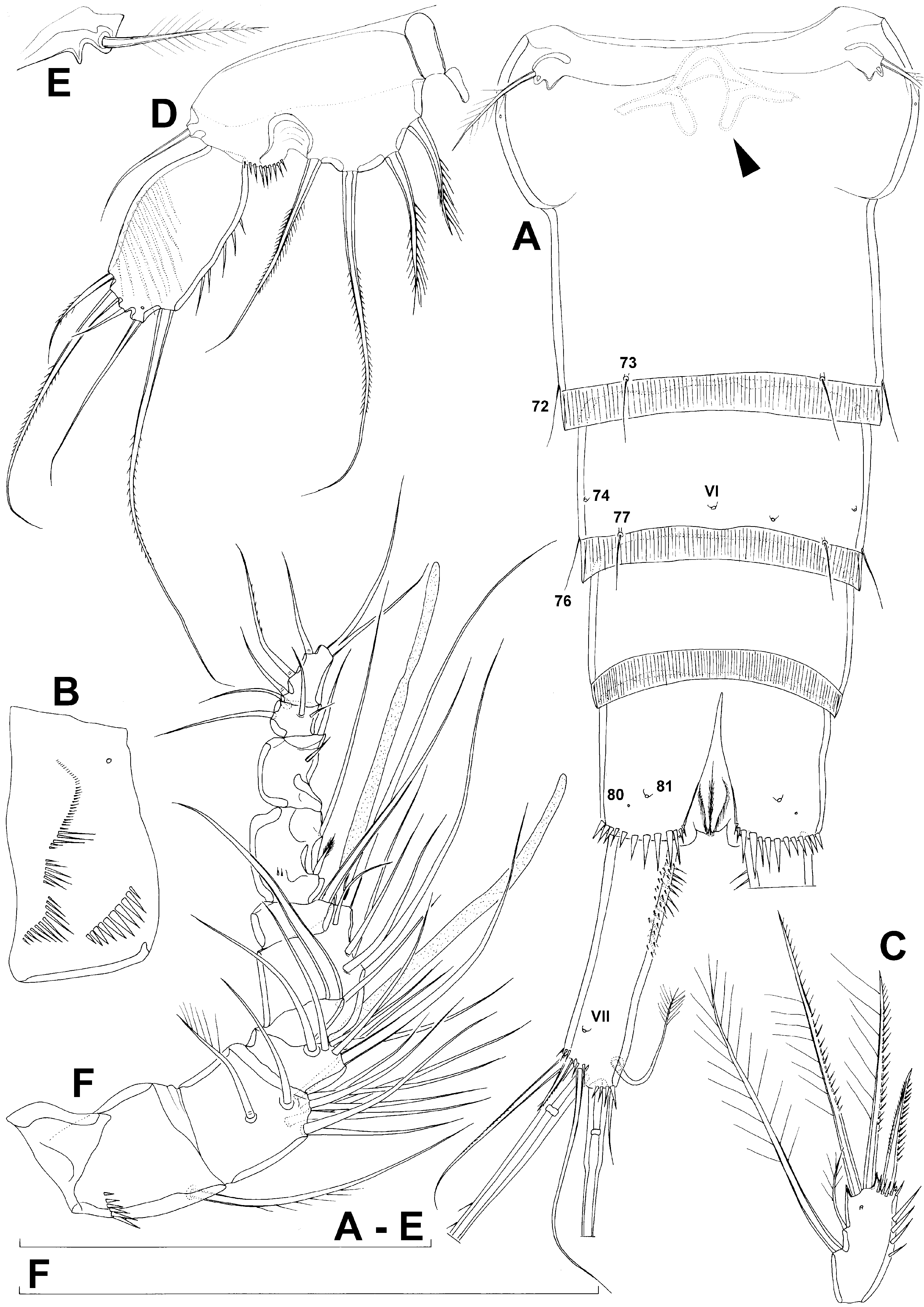
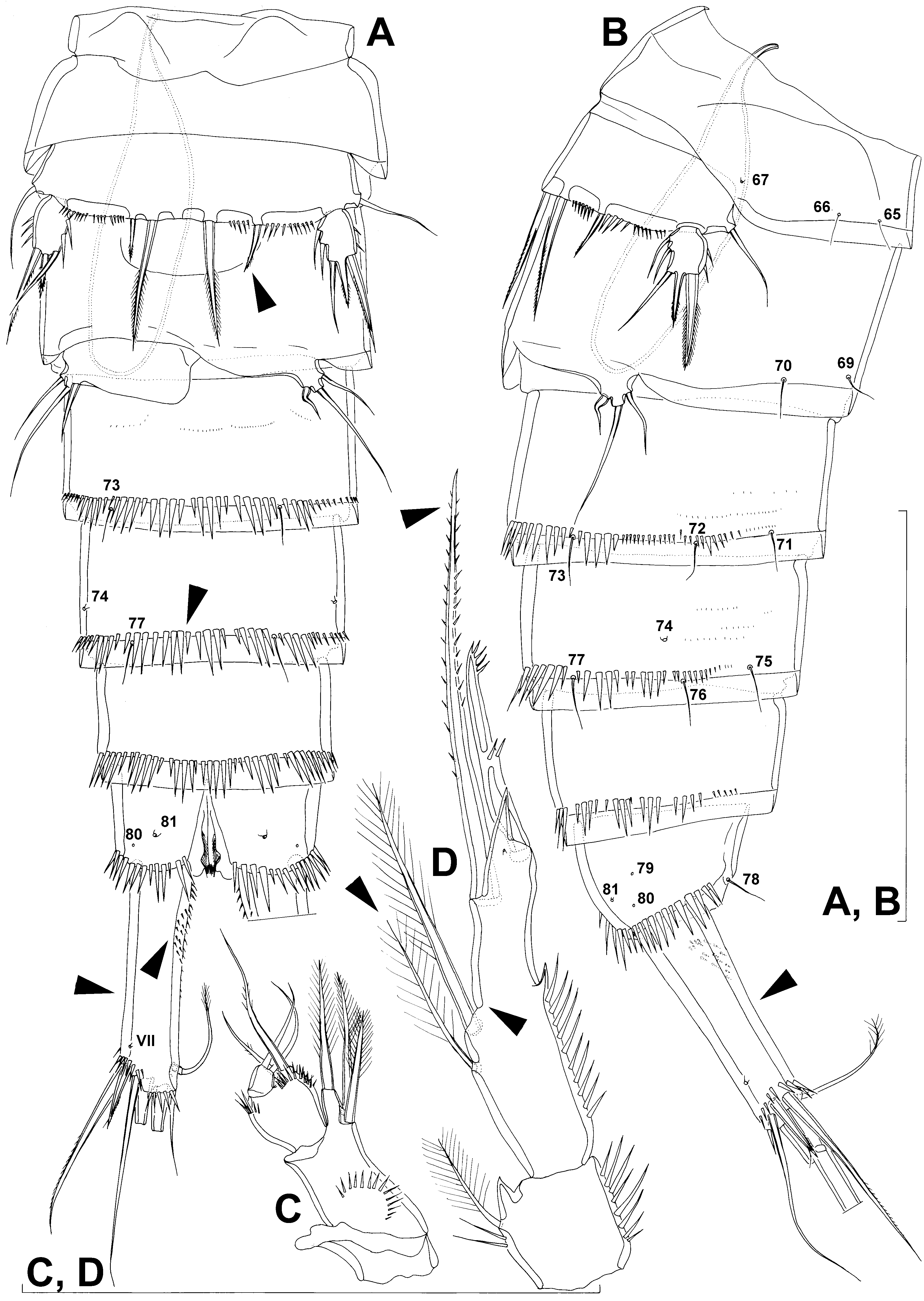
Male (based on three specimens). Body length from 548 to 604 µm. Habitus, colour, rostrum, shape and all ornamentation of cephalothorax, shape and ornamentation of second, third, and fourth pedigerous somites, ornamentation of first urosomite ( Figs. 45A, B View FIGURE 45 , 47E View FIGURE 47 ), caudal rami ( Figs. 45A, B View FIGURE 45 , 47F View FIGURE 47 ), antenna, labrum, paragnaths, mandibula, maxillula, maxilla, maxilliped ( Fig. 45C View FIGURE 45 ), first swimming leg, third swimming leg, and fourth swimming leg as in female.
Genital somite ( Figs. 45A, B View FIGURE 45 , 47E View FIGURE 47 ) as in Itostenhelia polyhymnia , except lateral pore no. # missing; relatively small spermatophore visible inside, positioned longitudinally on right side, about 3.5 times as long as wide, its neck reaching slightly beyond anterior margin of first urosomite.
Third urosomite ( Fig. 45A, B View FIGURE 45 ) as in Itostenhelia polyhymnia .
Fourth urosomite ( Fig. 45A, B View FIGURE 45 ) as in Itostenhelia polyhymnia , except ventral pair of pores no. VI missing (arrowed in Fig. 45A View FIGURE 45 ).
Preanal and anal somites ( Figs. 45A, B View FIGURE 45 , 47F View FIGURE 47 ) as in Itostenhelia polyhymnia .
Caudal rami ( Figs. 45A, B View FIGURE 45 , 47F View FIGURE 47 ) as in Itostenhelia polyhymnia , except slightly shorter (arrowed in Fig. 45A, B View FIGURE 45 ) and with additional patch of ventral spinules (arrowed in Fig. 45A View FIGURE 45 ), about four times as long as wide in ventral view, with two rows of relatively short inner spinules in anterior third.
Antennula ( Fig. 44F View FIGURE 44 ) as in Itostenhelia polyhymnia , except less clasped; general shape and segmentation as in Wellstenhelia calliope ; only ornamentation proximal row of spinules on first segment and two dorsal spinules on sixth segment; aesthetascs on third and fourth segments long and slender, no aesthetasc on ninth segment; setal formula 1.10.6+ae.8+ae.1.2.2.4.5.
Second swimming leg coxa, basis, and exopod as in female; endopod ( Fig. 45D View FIGURE 45 ) as in Itostenhelia polyhymnia , except inner spiniform process marking ancestral boundary between second and third segment missing (arrowed in Fig. 45D View FIGURE 45 ), proximal seta on ancestral second segment proportionately longer (arrowed in Fig. 45D View FIGURE 45 ), and inner spiniform element on ancestral third segment also proportionately longer (arrowed in Fig. 45D View FIGURE 45 ).
Fifth leg ( Figs. 45A, B View FIGURE 45 , 47E View FIGURE 47 ) as in Itostenhelia polyhymnia , except outer endopodal spine proportionately much longer (arrowed in Fig. 45A View FIGURE 45 ).
Sixth legs ( Figs. 45A, B View FIGURE 45 , 47E View FIGURE 47 ) as in Itostenhelia polyhymnia .
Variability. One male has the inner spine on left sixth leg curled, while the same element on right sixth leg is straight ( Fig. 45A, B View FIGURE 45 ).
Remarks. Chislenko (1978) reports a different armature formula of the swimming legs for this species than what we have found, with the minute inner distal setae missing on the third endopodal and exopodal segments of the fourth leg, as well as no inner seta on the first and second exopodal segments of the second leg. We examined all newly collected specimens from the type locality for these characters and found these setae always present. This implies that Chislenko (1978) either overlooked these setae (perhaps they were broken off on his specimen), or he was dealing with an aberrant specimen. We are inclined to favour the former, as Chislenko (1978) also provides a drawing of a maxilliped with only two setae on the basis, while all stenheliins have three setae here. Given the similarity of other features between our specimens and those studied by Chislenko (1978), the fact that our specimens were collected exactly from the type locality, and the apparent low diversity of stenheliins there, we believe that there is no chance that were are dealing with a different biological species.
Itostenhelia golikovi ( Chislenko, 1978) comb. nov. shows very few morphological differences from its Korean congener, Itostenhelia polyhymnia sp. nov., and they are all pointed out in the affinities of the latter species (see above) and also indicated with arrowheads in Figs. 43 View FIGURE 43 , 44 View FIGURE 44 , 45 View FIGURE 45 .

No known copyright restrictions apply. See Agosti, D., Egloff, W., 2009. Taxonomic information exchange and copyright: the Plazi approach. BMC Research Notes 2009, 2:53 for further explanation.
|
Kingdom |
|
|
Phylum |
|
|
Class |
|
|
Order |
|
|
Family |
|
|
Genus |