
Tretodictyum reiswigi, Boury-Esnault, Nicole, Vacelet, Jean, Dubois, Maude, Goujard, Adrien, Fourt, Maïa, Pérez, Thierry & Chevaldonné, Pierre, 2017
|
publication ID |
https://doi.org/ 10.11646/zootaxa.4236.1.6 |
|
publication LSID |
lsid:zoobank.org:pub:A2569FC8-0E88-416A-92A3-C61691E6FDFE |
|
DOI |
https://doi.org/10.5281/zenodo.6045573 |
|
persistent identifier |
https://treatment.plazi.org/id/697E87F1-FFD1-FF84-FF4E-AC8F9251FF02 |
|
treatment provided by |
Plazi |
|
scientific name |
Tretodictyum reiswigi |
| status |
sp. nov. |
Tretodictyum reiswigi n. sp. Boury-Esnault, Vacelet & Chevaldonné
Type-specimen. Sample VAL2-ACH-P15-ECH02. MNHN-HJV-04,
Type-locality. Valinco Canyon (Western Mediterranean) 41.662N / 8.795E, 397 m, 31/07/2014 GoogleMaps .
Other material (available in SME collection): Sample CS-ACH-P03- ECH 06 About ECH b, a small fragment encrusting a brachiopod shell, Cassidaigne canyon (W Mediterranean) 43.047N / 5.397E, 370 m, 28/10/2009 GoogleMaps . Sample VAL- ACH-P12- ECH 01 About ECH , small fragments, Valinco Canyon , Corsica, 41.663N / 8.795E, 444 m, 24/08/2010 GoogleMaps . Sample from CYLYCE cruise, dive 12 of Cyana submersible, fragments of two tubes, Ile Rousse Canyon, NW Corsica, 42.833N / 8.953E, 320 m, 25/05/1997, Slides from specimens of BALGIM Cruise, R / V Cryos, Alboran Sea, Station CP 135-168, 35.435N / GoogleMaps -4.301W and DR152–257, 35.935N/-5.569W, 390–560m, May–June 1984.
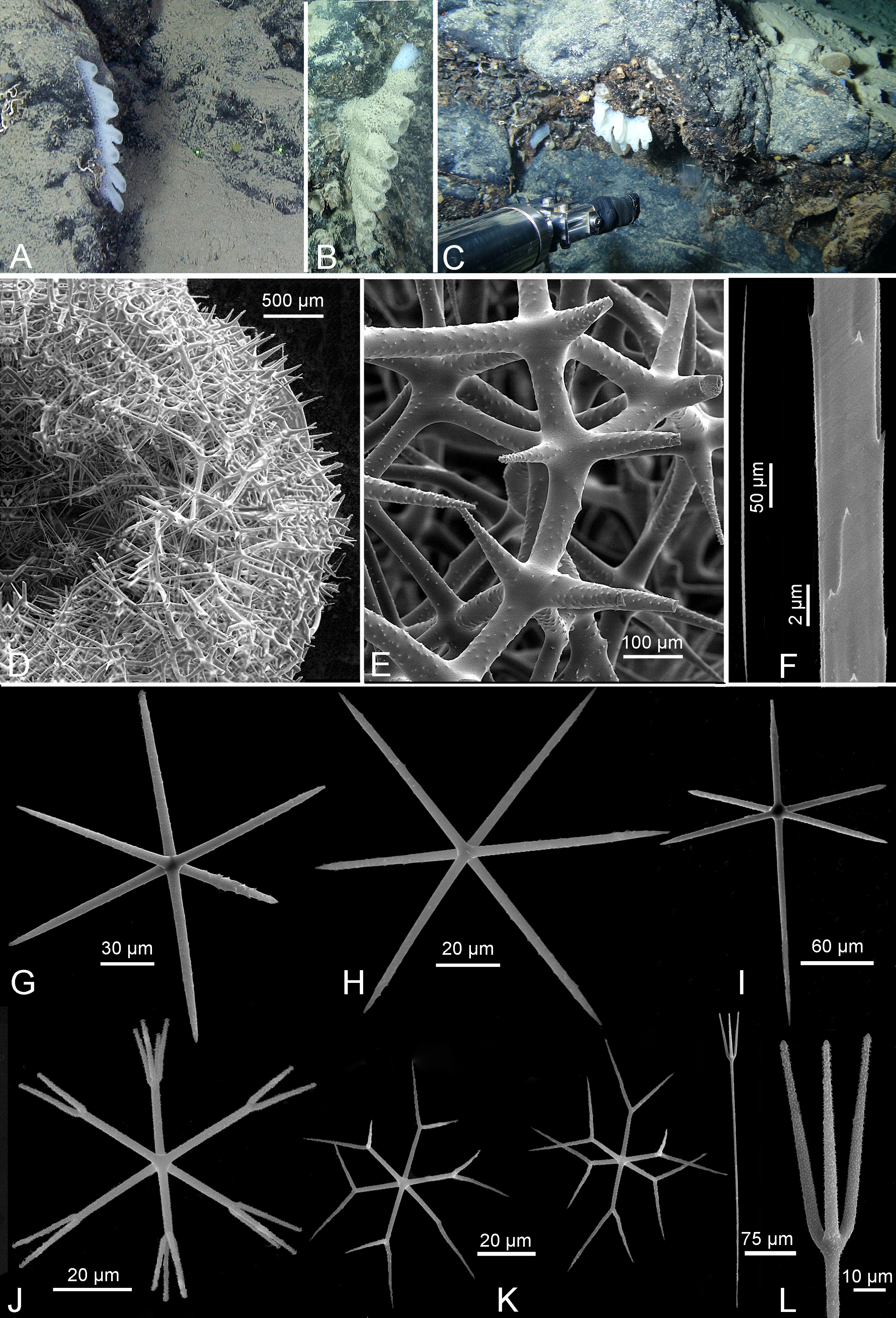
External morphology. The sponge is formed by several tubes more or less coalescent, expanding from a narrower base of attachment to the substrate. The tubes are most often arranged in a single or a double line ( Fig. 3 View FIGURE 3 ), with each line formed by up to 12 tubes. The maximum size estimated from ROV images is ca 15 x 7 x 5 cm, with tubes either entirely joined or free for up to 5 cm. The tubes are 1–2.2 cm in diameter with a central canal 0.55–0.65 cm in diameter. The wall of the tubes is ca 3 mm thick. The surface is pierced by numerous small depressions. The color is pure white in living or freshly dead specimens, grey in dead skeletons due to sediment deposits. Specimens with both living parts and dead skeletons have often been observed ( Fig. 3 View FIGURE 3 B). The consistency is hard, but the sponge is easily broken.
Numerous observations of sponges with very similar external characters have been made between 199 and 632 m depth from submersible and ROV dives, for examples Fig. 3 View FIGURE 3 A, 3B; Fig. 4 View FIGURE 4 B, 4C, 4D ( Table 3). Their identification to the same species is made with great confidence.
Skeleton. The main skeleton is a dictyonal framework of square meshes with 200–300 µm sides and beams 30–60 µm thick, with tubercles arranged in rows. Megascleres are dermal and parenchymal hexactins.
Strongyloscopules are present in dermal and atrial surfaces. Microscleres are oxyhexactins and two types of hexasters.

No known copyright restrictions apply. See Agosti, D., Egloff, W., 2009. Taxonomic information exchange and copyright: the Plazi approach. BMC Research Notes 2009, 2:53 for further explanation.
|
Kingdom |
|
|
Phylum |
|
|
Class |
|
|
Order |
|
|
Family |
|
|
Genus |