
Falcidens vasconiensis Salvini-Plawen, 1996
|
publication ID |
https://doi.org/ 10.1080/00222933.2014.958114 |
|
DOI |
https://doi.org/10.5281/zenodo.5196006 |
|
persistent identifier |
https://treatment.plazi.org/id/6B1787E6-7C75-AB56-6FB1-FF28DE14FE30 |
|
treatment provided by |
Felipe |
|
scientific name |
Falcidens vasconiensis Salvini-Plawen, 1996 |
| status |
|
Falcidens vasconiensis Salvini-Plawen, 1996 View in CoL
External morphology
Habitus
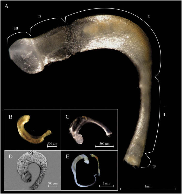
The specimens of Falcidens vasconiensis used in this study were 1.35–4.85 mm long and white or transparent in vivo, in some cases brownish due to the alimentary tract ( Figure 1 View Figure 1 ). After fixation in 70% ethanol, their colours are seldom altered. They have five body regions: anterium, neck, trunk, tail and tassel ( Figure 1A View Figure 1 ), differing by their morphology and the type of sclerites. The neck is delimited by two constrictions: one separating it from the anterium, the other one from the trunk. The specimens have a wide trunk and a tapering tail with very variable length, between a quarter and three quarters of the total length of the specimen ( Figure 1B–E View Figure 1 ). The pallial cavity bears two ctenidia.
Buccal shield
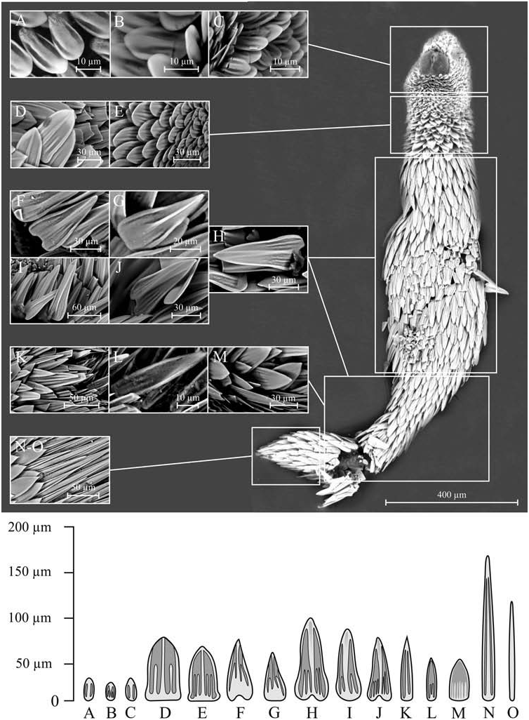
The buccal shield sometimes is difficult to distinguish in fixed specimens as the anterior part often contracts during fixation. In the specimens studied, the buccal shield ( Figure 2A, B View Figure 2 ) appears circular but really is horseshoe-shaped. It flanks the mouth opening laterally but is not closed preorally. The mouth is situated within a cleft formed by the flanks of the buccal shield; the cleft is three quarters as long as the shield diameter. The shield has a gland associated with it.
Sclerites
The scales are adpressed to the mantle and arranged in parallel to the longitudinal axis along the whole body ( Figure 2C View Figure 2 ), but occasionally they may be perpendicular to the mantle in the neck region ( Figure 2D View Figure 2 ). Each body region exhibits typical scales ( Figure 3 View Figure 3 ). In the anterior region, the scales are small (30–40 μm long × 10–15 μm wide), oval, flat, with two longitudinal grooves ( Figure 3A View Figure 3 ); these scales have two variants in the posterior part of the region: in the first variant, every groove has a small medial keel from the proximal end to the central area ( Figure 3B View Figure 3 ); in the second, the distal end of the scale is more pointed ( Figure 3C View Figure 3 ). The scales are larger in the neck (60–80 μm × 40–50 μm), triangular, flat, with a strengthened margin; they have a medial longitudinal keel narrowing from the proximal to the apical region and one or two shorter lateral keels from the base to the medial part of the scale ( Figure 3D, E View Figure 3 ). There are large scales in the trunk (70–100 μm × 25–50 μm), sagittiform, flat, with a more or less clear waist and a strengthened margin; they may occasionally exhibit a basal notch and very small wings on the base and they have a medial longitudinal keel, which is wider on the distal end, and one to three shorter lateral keels ( Figure 3F–J View Figure 3 ). Scales of the tail are similar to those described for the trunk, except for an additional three types of scales in their posterior part: lanceolate and flat scales (80–90 μm × 15–25 μm) with two longitudinal grooves and a medial keel ( Figure 3K View Figure 3 ), smaller, lanceolate and flat scales (50–65 μm × 10–20 μm) with a large central groove and two short striations extending from the base ( Figure 3L View Figure 3 ), and sagittiform, flat and wide scales (55–65 μm × 20–30 μm) with several short proximal striations ( Figure 3M View Figure 3 ). The tassel bears long acicular sclerites (100– 200 μm × 5–20 μm); the longest are slightly flattened and exhibit two proximal longitudinal grooves ( Figure 3N, O View Figure 3 ).
Internal anatomy
Digestive system
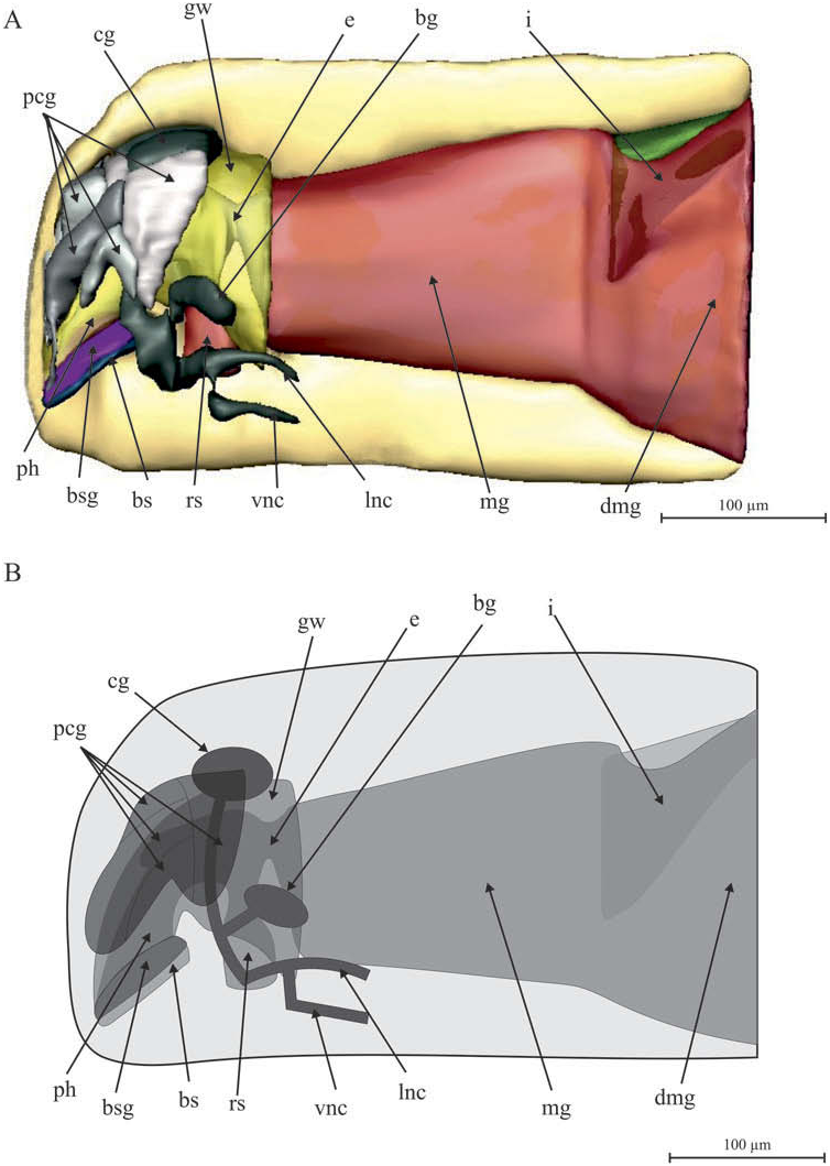
The mouth is in an anteroventral position, flanked by the buccal shield. The pharynx is short and the radular sac with radula is situated in its posterior end ( Figures 4 View Figure 4 , 5A, B View Figure 5 ). The radula ( Figure 5C, D View Figure 5 ) shows the typical structure of the genus, made up of a pair of sickle-shaped teeth with sclerotized tips and united at the base by a radular symphysis attached to the cone, two pairs of lateral supports of different sizes with sclerotized distal tips and two support muscles. It is lacking a central plate. The pharynx leads into a short oesophagus ( Figures 4 View Figure 4 , 5A, B View Figure 5 ). The pharynx walls and especially the oesophagus walls are thick and glandular ( Figure 5E, F View Figure 5 ). The oesophagus opens into a wide midgut that is divided posteriorly into a long and narrow intestine and a very wide and large midgut sac ( Figures 4 View Figure 4 , 5A, B View Figure 5 ). The posterior end of the midgut sac coincides with the area where the trunk merges with the tail. The intestine is located dorsally, however slightly displaced to the right in the specimen used for reconstruction; the anus is placed between the bases of the ctenidia in the pallial cavity.
Nervous system
The cerebral ganglion is located in a dorsoanterior position ( Figures 4 View Figure 4 , 6 View Figure 6 ); frontally to this, there is a cerebral complex formed by the union of three pairs of precerebral ganglia, which clearly separate as they extend towards the anterior part ( Figure 6 View Figure 6 ). These precerebral ganglia innervate the buccal shield, the gland associated with it, and the oral region. The unpaired lobe found in the posterior part of the brain of other species of the genus is lacking. The anterior part of the cerebral ganglion releases the pairs of ventral, lateral and buccal connectives ( Figure 6 View Figure 6 ), which originate close to each other and separate progressively. The buccal connectives end in the buccal ganglia, which are located at both sides of the radular sac. The ventral and lateral connectives continue along the ventral and lateral cords, which are connected by ventral commissures and lateroventral connectives. Both ventral and both lateral cords extend along the animal and unite in the posterior region, forming a ganglion and the suprarectal commissure; from here they innervate the pair of ctenidia and the dorsoterminal sense organ, which is located in a long groove dorsal to the pallial cavity.


No known copyright restrictions apply. See Agosti, D., Egloff, W., 2009. Taxonomic information exchange and copyright: the Plazi approach. BMC Research Notes 2009, 2:53 for further explanation.