
Synoicum stewartense ( Michaelsen, 1924 )
|
publication ID |
https://doi.org/ 10.1080/00222933.2014.896487 |
|
publication LSID |
lsid:zoobank.org:pub:5ADC2C9D-28AC-4348-8B4D-F262A43DEA66 |
|
persistent identifier |
https://treatment.plazi.org/id/6C5C87F5-FFE8-3B42-FE48-51A12781FDCE |
|
treatment provided by |
Felipe |
|
scientific name |
Synoicum stewartense ( Michaelsen, 1924 ) |
| status |
|
Synoicum stewartense ( Michaelsen, 1924) View in CoL
( Figures 8 View Figure 8 ; 7B View Figure 7 )
Macroclinum stewartense Michaelsen, 1924: p. 413 View in CoL –421, figs 26, 27, 28.
Synoicum stewartense: Millar 1982: p. 15 View in CoL , fig. 3.
Material examined
New records: Caswell Sound, Hansard Point (45° 00.57’S, 167° 08.93’E, vertical rock wall, coll. M. Page, 15 m, 3 February 2009, NIWA 49984 View Materials ), Wet Jacket Arm, Acheron Pinnacle (45° 40.577’S, 166° 43.954’E, rock wall, coll. M. Page, 22 m, 1 February 2009, NIWA 49964 View Materials , four colonies) GoogleMaps .
Description
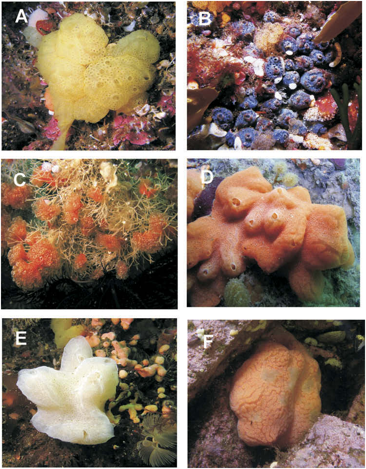
This species is cryptic, often found with heads of the colonies embedded among bryozoans and turfing red algae. The colonies are bright blue in life (PB 4/10) measuring 70–100 mm across and 20 mm thick, and are red when fixed in formalin. They are composed of numerous flat-topped, chalice-shaped heads that taper to a common basal mat. The heads are approximately 10 mm in diameter and 15 mm high. Each head generally has a circular system of 15–20 zooids around a central raised common cloacal aperture. Some colony heads may have two systems ( Figure 7B View Figure 7 ). Sand invests the basal test and sparsely invests the posterior half of the colony heads. The test is firm but gelatinous and the zooids heavily pigmented red (R 5/10) when fixed in formalin.
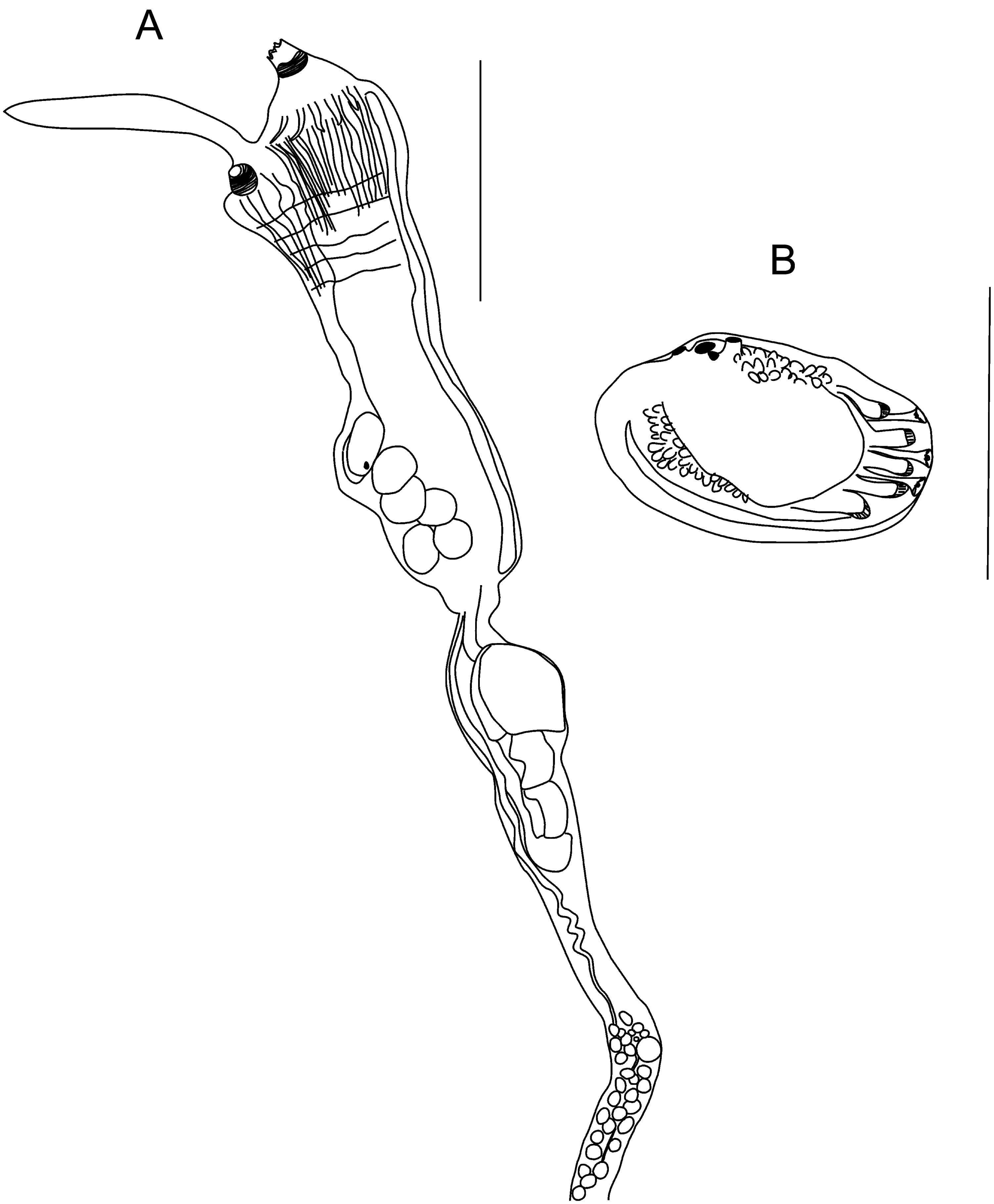
Zooids of this species are large, measuring approximately 13 mm total length. The thorax averages 4 mm, the abdomen 1.2 mm, and the post-abdomen 7 mm long. Six sharply pointed lobes surround a stout branchial aperture. The atrial aperture is produced into a short muscular siphon surmounted by a long narrow atrial languet with an occasionally bifid end ( Figure 8A View Figure 8 ). The branchial sac has 18–21 rows of stigmata with 18 stigmata per half row, and there are obvious transverse vessels with no papillae between rows of stigmata. Up to 14 longitudinal muscles are found on each side of the body wall of the thorax. These coalesce at the abdomen and form a band of muscle, which runs on the dorsal side of the stomach and down the abdomen to the distal end of the post-abdomen as one muscle band. There is a large finely areolated bell-shaped stomach with a constriction between the duodenum and the mid-intestine ( Figure 8A View Figure 8 ). The oesophagus is curved entering the stomach on its dorsal border. The post-abdomen is long and narrow with five ova located in the posterior third, anterior to approximately 30 oval testis follicles. The vas deferens runs a typical polyclinid course, running anteriorly from the left side of the post abdomen across the gut loop to the right of the stomach and up the right side of the thorax on the mesial side of the intestine. As many as four larvae were present in the posterior atrial cavity of the zooids. The larvae have an average trunk length of 0.7 mm. There are five pairs of lateral ampullae crowded on each side of three slender median adhesive papillae ( Figure 8B View Figure 8 ). Vesicles are located at the posterior end of the larva below the tail and on the dorsal surface.
Remarks
Michaelsen (1924) first described Synoicum stewartense from Stewart Island, noting the central position of the common cloacal opening surrounded by a low raised rim. This feature is evident from the in situ photograph of this species in Fiordland ( Figure 7B View Figure 7 ). In addition, the distinctive red/purple colour of the colonies and zooid tissue in our fixed specimens was also noted by Millar (1982) in colonies from the Chatham Rise. Michaelsen (1924) also described unique fine polygonal 30 µm ‘fielding’ on the stomach wall, of this species (see Michaelsen 1924; fig 28a). This character was not obvious in zooids from Fiordland. Other similar species from New Zealand, Synoicum apectetum Millar, 1982 and Synoicum arenaceum ( Michaelsen 1924) , are distinguished from S. stewartense by fewer rows of stigmata, a smooth non-areolated stomach and colonies with slender sand-coated sinuous stalks and either small pearshaped or flat-topped cormidia, respectively.
No known copyright restrictions apply. See Agosti, D., Egloff, W., 2009. Taxonomic information exchange and copyright: the Plazi approach. BMC Research Notes 2009, 2:53 for further explanation.
|
Kingdom |
|
|
Phylum |
|
|
Class |
|
|
Order |
|
|
Family |
|
|
Genus |
Synoicum stewartense ( Michaelsen, 1924 )
| Page, M. J., Willis, T. J. & Handley, S. J. 2014 |
Synoicum stewartense: Millar 1982 : p. 15
| Millar, RH 1982: 15 |
Macroclinum stewartense
| Michaelsen, W 1924: 413 |