
Paroecobius Lamoral, 1981
|
publication ID |
https://doi.org/ 10.11646/zootaxa.4527.1.3 |
|
publication LSID |
lsid:zoobank.org:pub:981018E0-2132-433D-878E-801C97C460C9 |
|
DOI |
https://doi.org/10.5281/zenodo.5946436 |
|
persistent identifier |
https://treatment.plazi.org/id/6D4287D2-FFE0-8F58-9D94-26CB8D1B00A9 |
|
treatment provided by |
Plazi |
|
scientific name |
Paroecobius Lamoral, 1981 |
| status |
|
Genus Paroecobius Lamoral, 1981 View in CoL
Paroecobius Lamoral, 1981: 508 View in CoL
Type species. Paroecobius wilmotae Lamoral, 1981 .
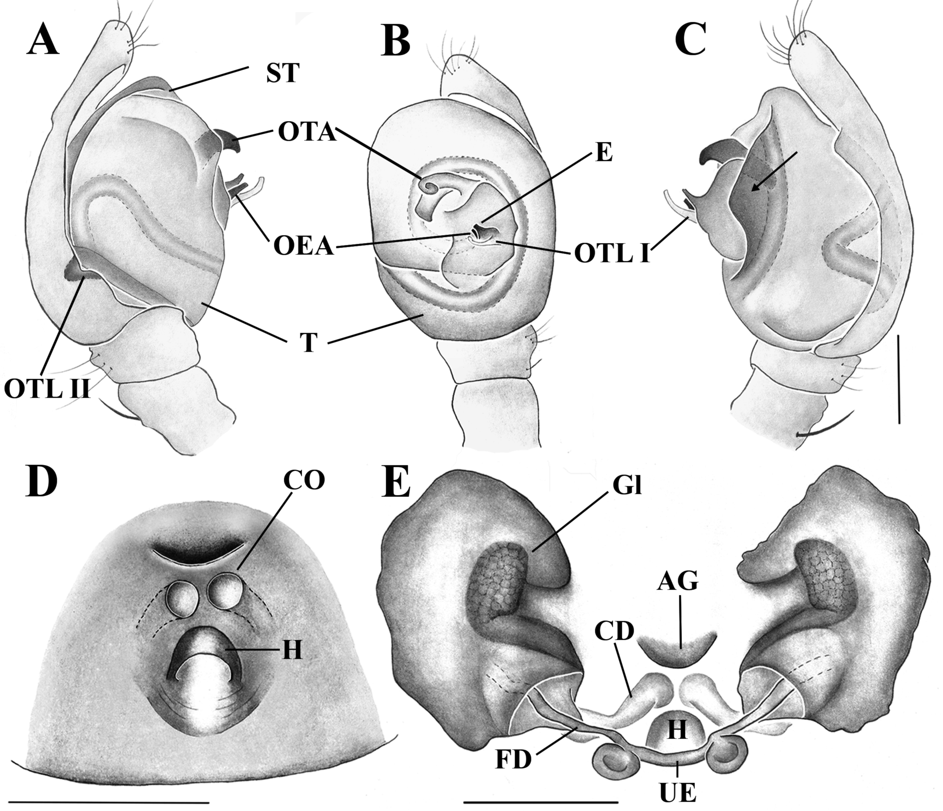
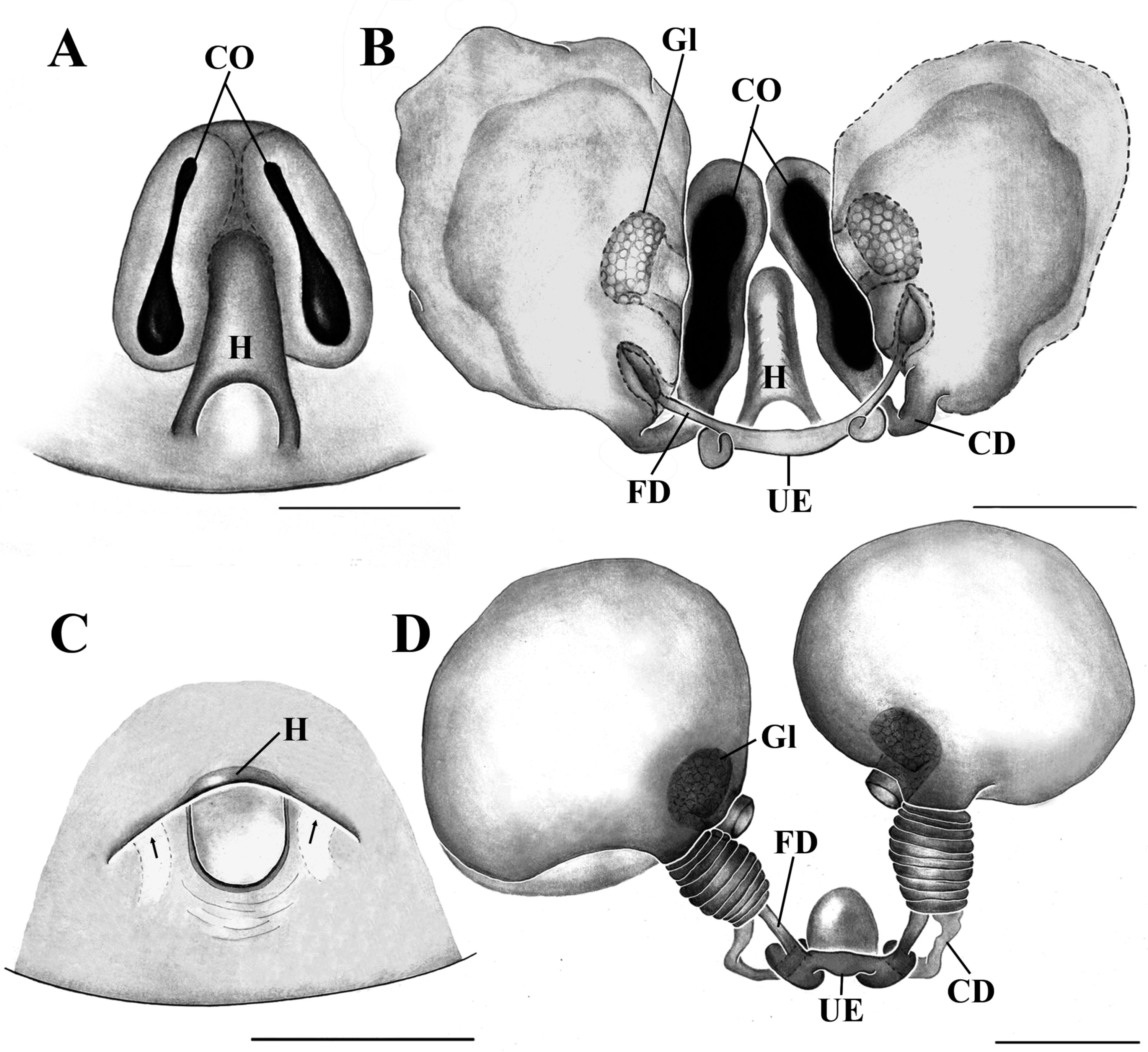
Emended genus diagnosis. Paroecobius can be distinguished from Oecobius and Platoecobius by the anterior median eyes dark and the largest, with a black ring on cuticular base, while all other eyes are opalescent ( Fig. 4 View FIGURE 4 C– D; Lamoral 1981: figs 2, 3). The calamistrum of females are in a single row, situated in proximal half of metatarsus IV, males do not have calamistrum. The female epigynum has a blind transverse invagination (hood, Figs 2 View FIGURE 2 D–E, 3A–D, 5E; Lamoral 1981: fig. 6). The spermathecae have a porous and globular glandular chamber inside, with a large external opening ( Figs 2E View FIGURE 2 , 3B, D View FIGURE 3 ; Lamoral 1981: fig. 7). The pedipalpus has a short subtegulum. The embolus, the tegular apophysis and the Oecobiidae tegular lobe I are reduced, located in a small area of the tegulum ( Fig. 2 View FIGURE 2 A–C; Wunderlich 1995b: figs 2–4).
Remarks. As mentioned in this study, Paroecobius seems to be a monophyletic group supported by sharing the epigynal hood, reduced embolus and tegular apophysis, both located in a small median area in the tegulum. The Oecobiidae tegular lobe I also seem to be reduced, which is consistent with the reduction of the embolus and all tegular apophysis in the genus.
We report an intriguing internal structure in the female spermathecae of two oecobiid genera. This structure was originally reported for Uroecobius ecribellatus Kullman & Zimmermann, 1976 as a muscular attachment surface, probably because of its wired surface as seen under SEM ( Kullmann & Zimmermann 1976: figs 12–14). A similar structure was illustrated in Paroecobius wilmotae ( Lamoral 1981: fig. 7) and is present in the three Paroecobius species described here and called the internal glandular chamber (Gl). It is currently impossible to ascertain the presence of this structure in P. nicolaii Wunderlich, 1995 because its internal female genitalia were not described or illustrated in detail (see Wunderlich 1995b). Although nothing is known about the function of this structure, its shape and the presence of numerous pores on its internal surface suggest that it could be a glandular chamber. The presence of this internal glandular chamber in the spermathecae suggest that Paroecobius could be phylogenetically close to Uroecobius .
Composition. Five species: Paroecobius wilmotae Lamoral, 1981 ; P. nicolaii Wunderlich, 1995 , and three species newly described below.
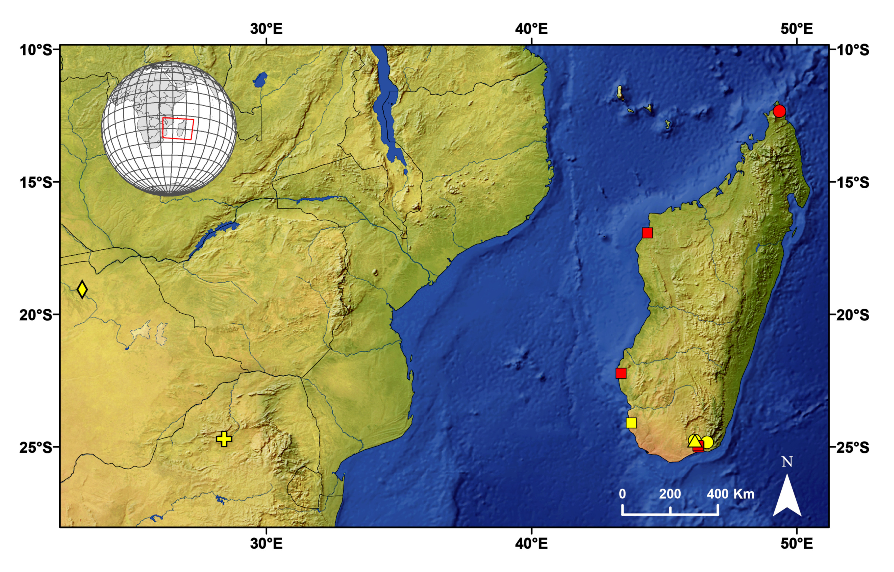
Distribution. Previously known only from the Afrotropical region, with two species from Botswana and South Africa, recorded here for the first time in Madagascar ( Fig. 6 View FIGURE 6 ).

No known copyright restrictions apply. See Agosti, D., Egloff, W., 2009. Taxonomic information exchange and copyright: the Plazi approach. BMC Research Notes 2009, 2:53 for further explanation.
|
Kingdom |
|
|
Phylum |
|
|
Class |
|
|
Order |
|
|
Family |
Paroecobius Lamoral, 1981
| Magalhães, Mayara D. F. & Santos, Adalberto J. 2018 |
Paroecobius
| Lamoral, B. H. 1981: 508 |