
Connelicita haiphongensis Wang et Zhang
|
publication ID |
https://doi.org/ 10.11646/zootaxa.4018.4.5 |
|
publication LSID |
lsid:zoobank.org:pub:0DEFFE93-514F-4329-8B7D-6A376776CCC1 |
|
DOI |
https://doi.org/10.5281/zenodo.6108151 |
|
persistent identifier |
https://treatment.plazi.org/id/6E2D8794-736B-A831-2CC1-FA73FD72FEB2 |
|
treatment provided by |
Plazi |
|
scientific name |
Connelicita haiphongensis Wang et Zhang |
| status |
sp. nov. |
Connelicita haiphongensis Wang et Zhang View in CoL , sp. nov.
( Figs 2–21 View FIGURES 2 – 7 View FIGURES 8 – 12 View FIGURES 13 – 21 )
Etymology. The Latin name refers to the locality of the species in Haiphong, Vietnam.
Diagnosis. The new species appears similar to C. backyensis Stroiński et Bourgoin , sp. nov. but differs in the head capsule shape and male genitalia conformation as indicated in the diagnosis of the type species— C. backyensis sp. nov.
Description. Length: male (including forewing) (N=1): 10.2 mm, forewing: 8.1 mm; female (including forewing) (N=2): 10.8–11.0 mm, forewing: 8.8–9.0 mm.
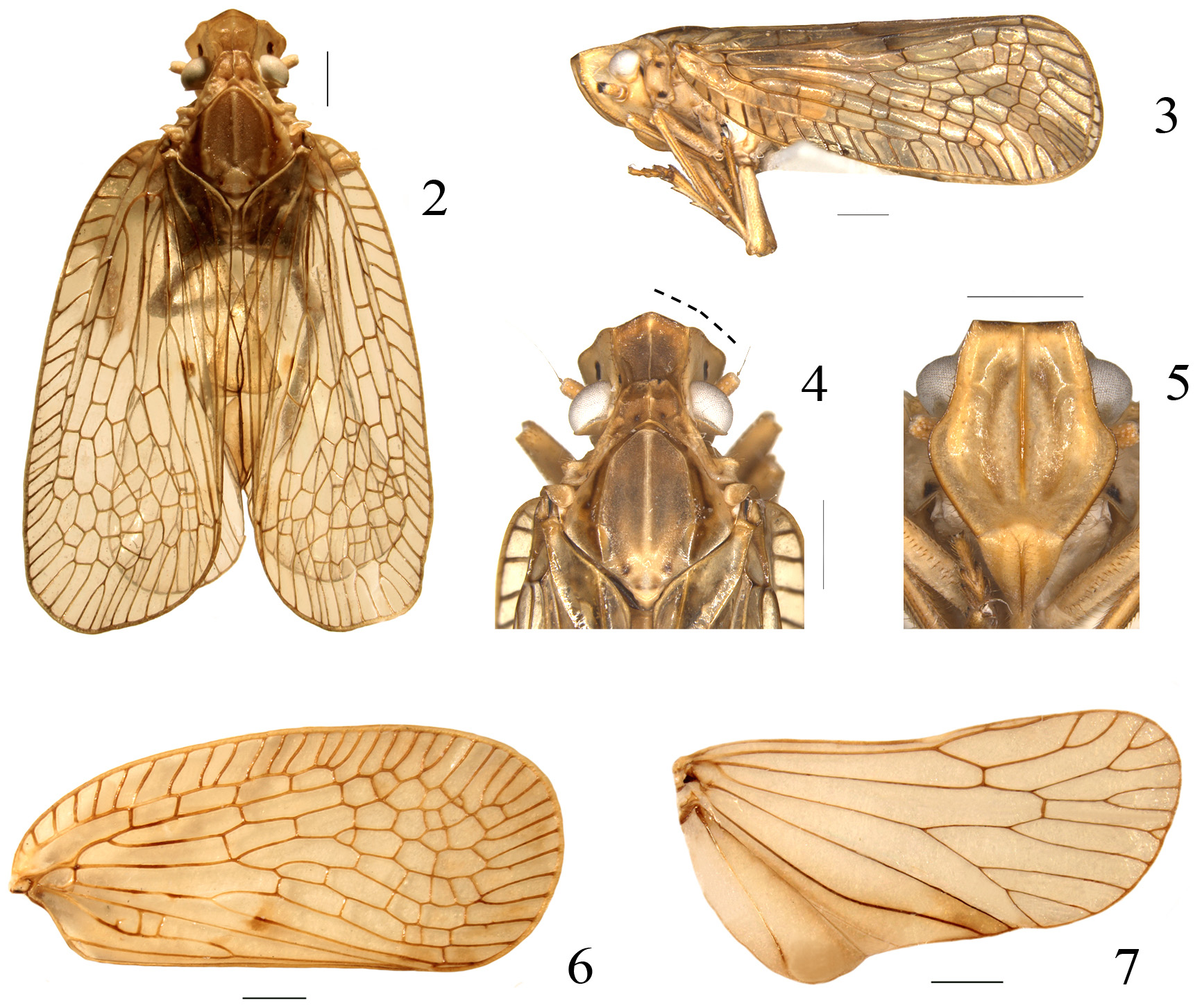
HEAD. Vertex 1.1 times longer in middle line than wide at base ( Fig. 4 View FIGURES 2 – 7 ). Frons 2.0 times longer in middle line than broad at upper margin, 1.1 times longer in middle line than widest part ( Fig. 5 View FIGURES 2 – 7 ).
THORAX. Pronotum 4.2 times wider at widest part than long in midline ( Fig. 4 View FIGURES 2 – 7 ). Mesonotum 0.9 times wider at widest part than long in midline ( Fig. 4 View FIGURES 2 – 7 ) and 1.6 times longer than cumulative length of the vertex and pronotum in midline ( Fig. 4 View FIGURES 2 – 7 ). Forewing 2.2 times longer than widest part ( Fig. 6 View FIGURES 2 – 7 ).
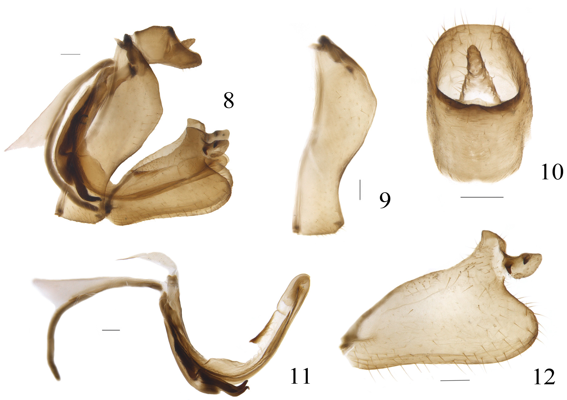
MALE TERMINALIA. Anal tube 1.4 times longer than width in dorsal view, with apical margin almost straight and lateral margin nearly parallel, epiproct short, located at upper half of anal tube ( Fig. 10 View FIGURES 8 – 12 ), ventral margin of anal tube slightly sinuate in lateral view ( Fig. 8 View FIGURES 8 – 12 ). Gonostylus with capitulum located at apical 1/4, posterior margin of gonostylus vertically in apical half, dorsal margin of capitulum slightly sloping posteriorly, additional lobe of capitulum with two spines separately in dorsal margin with larger one near base and small one near the middle, both directed downward ( Fig. 12 View FIGURES 8 – 12 ). Pygofer in lateral view with posterior margin deeply inclined posteriorly at upper 1/3 and slightly sinuate at lower 2/3 part ( Figs 8, 9 View FIGURES 8 – 12 ). Aedeagus smooth at apex ( Fig. 11 View FIGURES 8 – 12 ). Ventral process of periandrium hook-like ( Fig. 11 View FIGURES 8 – 12 ).
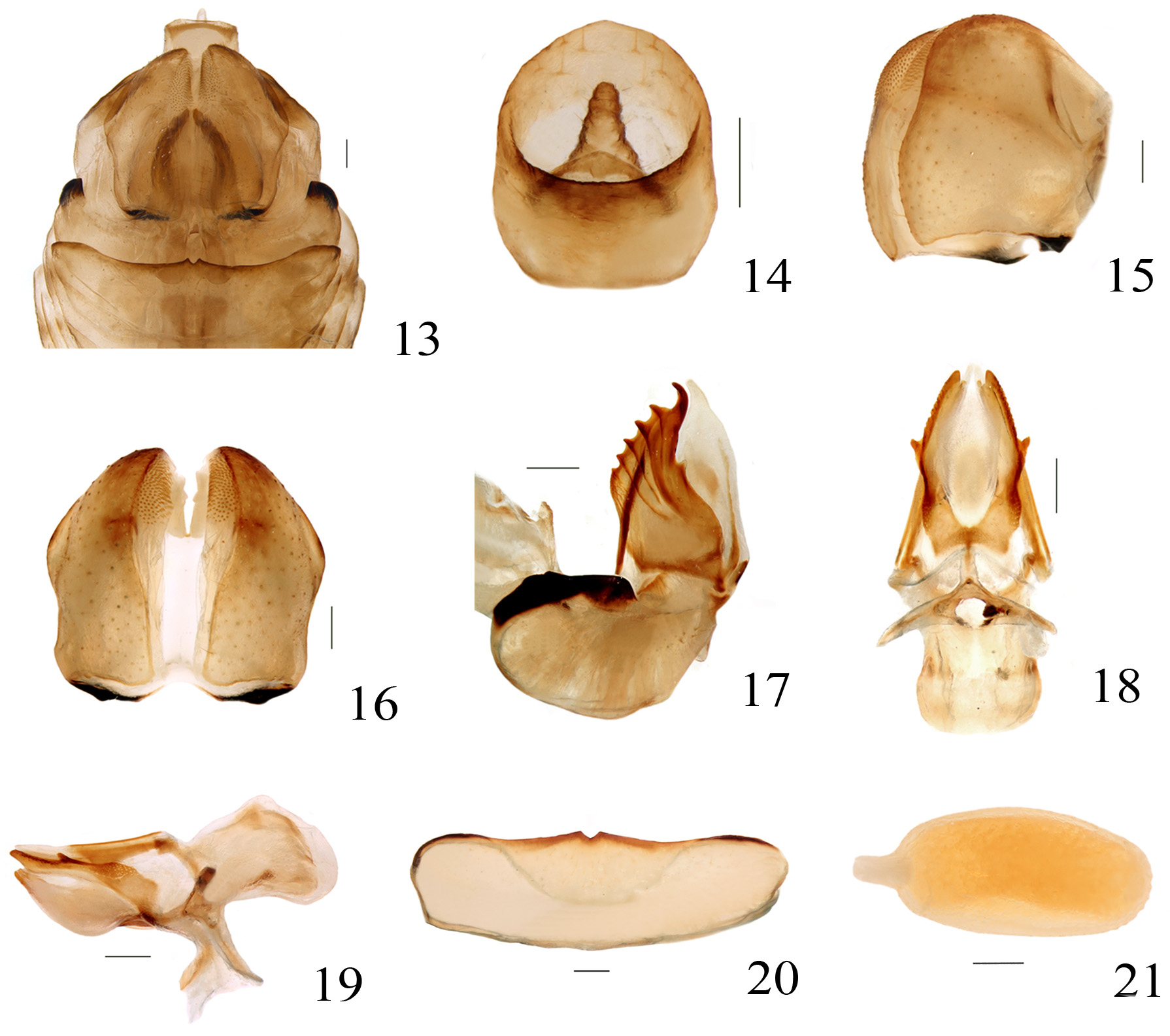
FEMALE TERMINALIA. Anal tube in dorsal view oval ( Fig. 14 View FIGURES 13 – 21 ). Hind margin of sternite VII nearly straight, shallowly incised medially ( Fig. 20 View FIGURES 13 – 21 ). Anterior connective lamina of gonapophysis VIII with six teeth at outer lateral margin and one tooth in inner lateral margin ( Fig. 17 View FIGURES 13 – 21 ). Endogonocoxal process developed and membranous, reaching the same level of the upper margin of gonapophysis VIII ( Fig. 17 View FIGURES 13 – 21 ). Gonocoxa VIII subquadrangular. Posterior connective lamina of gonapophysis IX membranous in medial part and sclerotized in outer part, with small tooth near middle each side ( Fig. 18 View FIGURES 13 – 21 ). Gonospiculum bridge large ( Figs 18, 19 View FIGURES 13 – 21 ). Gonoplacs rounded in lateral view ( Fig. 15 View FIGURES 13 – 21 ), fused at middle in apical half ( Fig. 16 View FIGURES 13 – 21 ), outer margin slightly broaden near middle, with numerous small teeth at inner side of apex ( Fig. 16 View FIGURES 13 – 21 ). Egg oval with long and narrow operculum ( Fig. 21 View FIGURES 13 – 21 ).
COLORATION. Vertex yellowish brown, with median carina pale yellow on disc, anterior and lateral carinae brown, lateral margins slightly widen from base to apex ( Figs 2, 4 View FIGURES 2 – 7 ). Compound eyes grey ( Figs 2, 4 View FIGURES 2 – 7 ). Ocelli red. Frons yellow, with median carina tawny, two brown curved vertical bands beside the median carina, anterior and lateral margins black ( Fig. 5 View FIGURES 2 – 7 ). Clypeus yellow, with median carina tawny ( Fig. 5 View FIGURES 2 – 7 ). Genae yellow, with black patch below antenna ( Figs 3, 4 View FIGURES 2 – 7 ). Pronotum yellowish brown, with median carina pale yellow and two small depressions beside median carina on disc ( Fig. 4 View FIGURES 2 – 7 ). Mesonotum yellowish brown, with median and lateral carinae pale yellow, two dark brown vertical bands along lateral carinae and two small brown spots near base ( Fig. 4 View FIGURES 2 – 7 ). Forewings with brown spot near middle of vein CuP and vein CuA2 ( Fig. 6 View FIGURES 2 – 7 ). Legs tawny, with apex of spines black.
Type materials. Holotype ♂, Vietnam: Cat Ba NP., Hai Phong Provinces, Vietnam Natural Forest, N 20º 48′ 258′′, E 107º 00′ 581′′, 132m, 2008. VII.17, Coll.: Zheng G., Sac. PD., Li SQ ( IZCAS). Paratypes: 2♀♀, same data as holotype. Specimens deposited in IZCAS.
Distribution. Vietnam: Hai Phong Province.
| IZCAS |
Institute of Zoology, Chinese Academy of Sciences |
No known copyright restrictions apply. See Agosti, D., Egloff, W., 2009. Taxonomic information exchange and copyright: the Plazi approach. BMC Research Notes 2009, 2:53 for further explanation.