
Aratricerca, Gustafsson & Bush, 2017
|
publication ID |
https://doi.org/ 10.11646/zootaxa.5104.4.2 |
|
publication LSID |
lsid:zoobank.org:pub:B2F30055-6675-4196-95E1-222DF756AE76 |
|
DOI |
https://doi.org/10.5281/zenodo.6335962 |
|
persistent identifier |
https://treatment.plazi.org/id/6E35BD52-A805-FF90-FF30-8F16B19EFD57 |
|
treatment provided by |
Plazi |
|
scientific name |
Aratricerca |
| status |
|
Key to the species of the genera Aratricerca , Timalinirmus , and Turdinirmoides
Note: We have not seen specimens of Turdinirmoides carpodaci and Turdinirmoides vasjukovae . These species were not completely described, and were illustrated with habitus photos, drawings of male genitalia and of frons. The male genitalia of T. vasjukovae are most similar to those of T. rozsai , whereas the male genitalia of T. carpodaci are not similar to those of any of the other species. We have placed Turdinirmoides carpodaci and T. vasjukovae in the key based on the characters given by Mey (2017), but redescriptions of both species are needed.
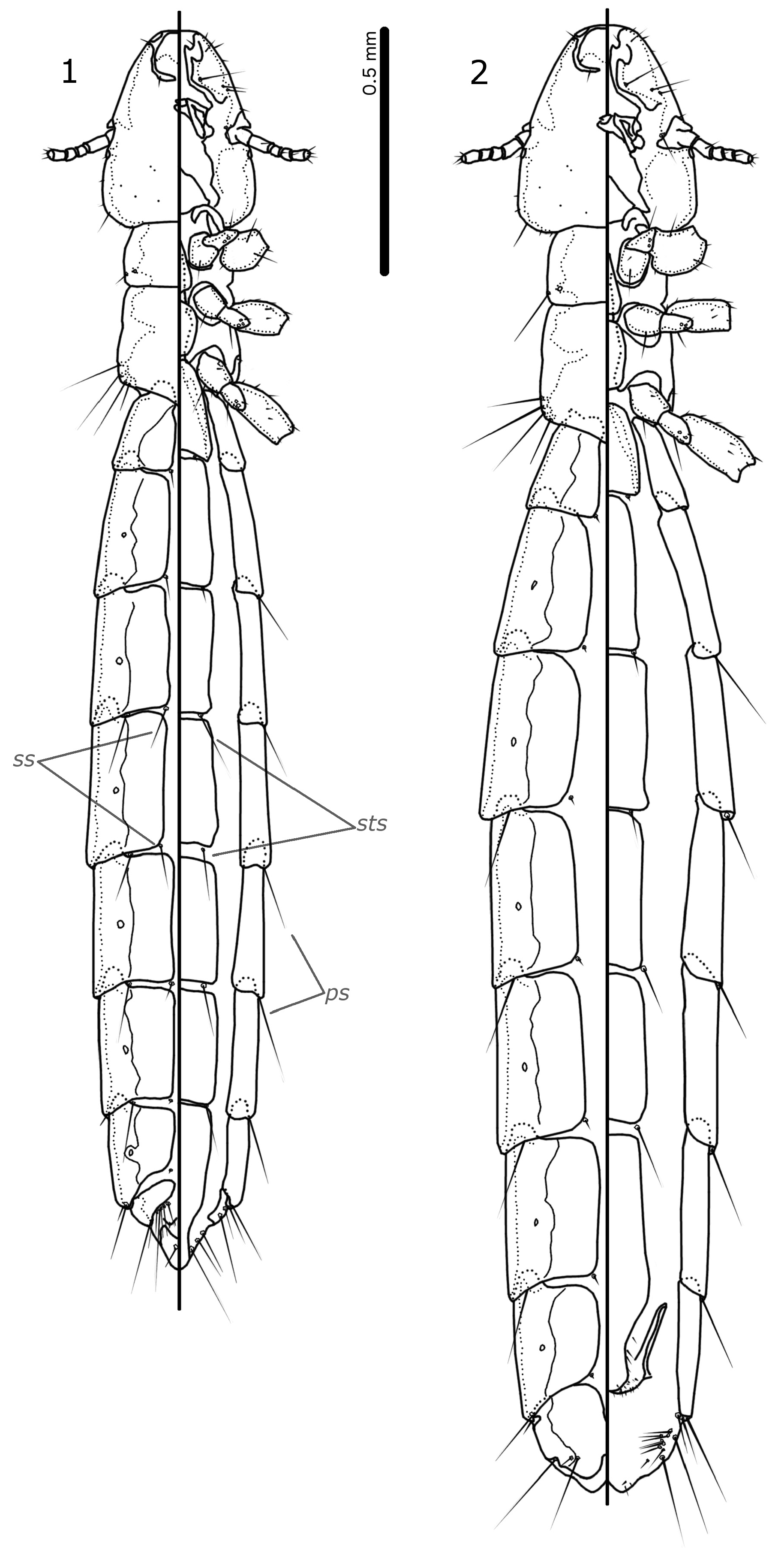
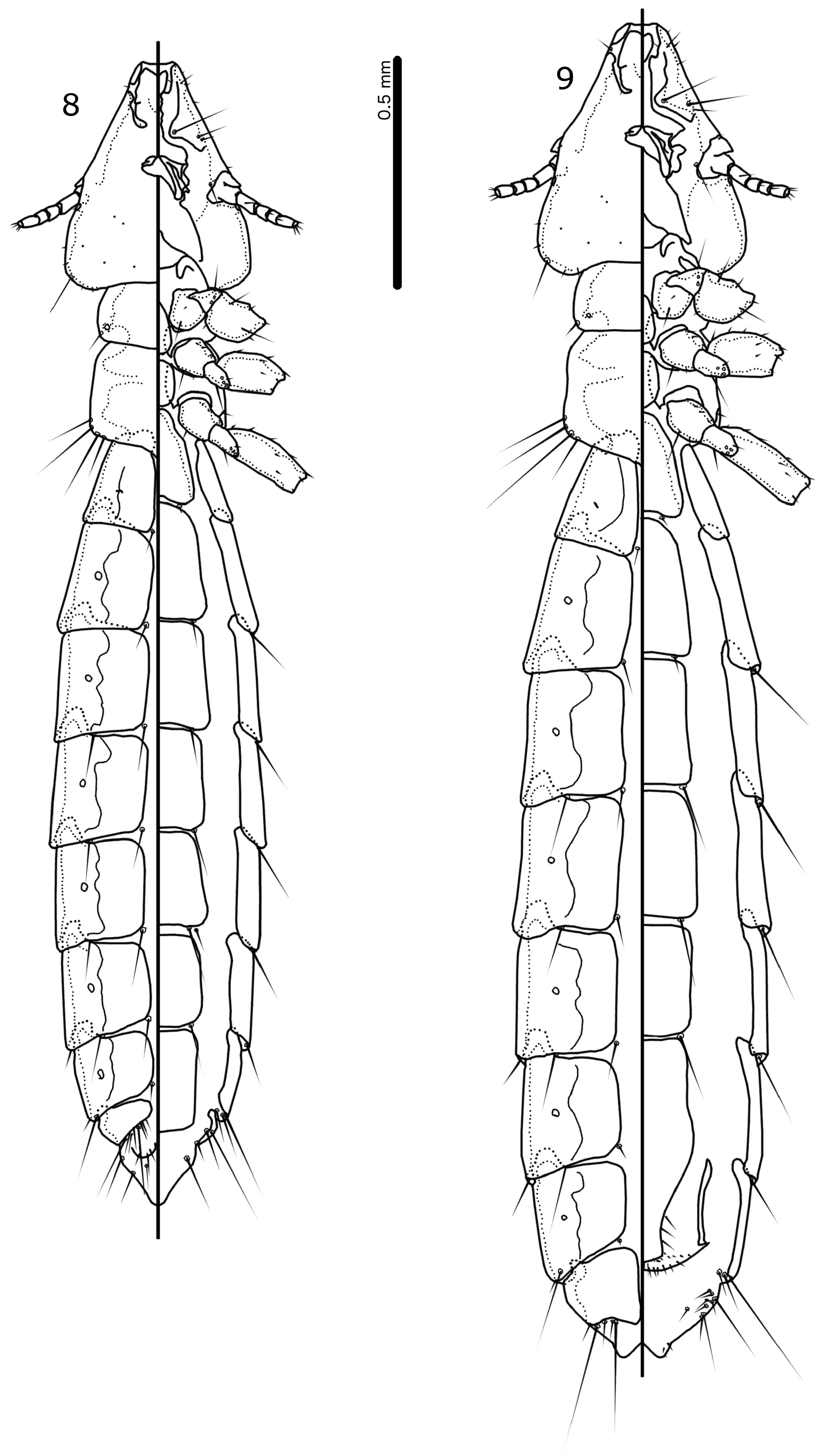
1. Lateral margins of pterothorax roughly parallel ( Fig. 1 View FIGURES 1–2 ); at least sternal plate II with antero-lateral thickening ( Fig. 1 View FIGURES 1–2 ); male abdominal segment XI extended into triangular tail ( Fig. 1 View FIGURES 1–2 )...................................................2.
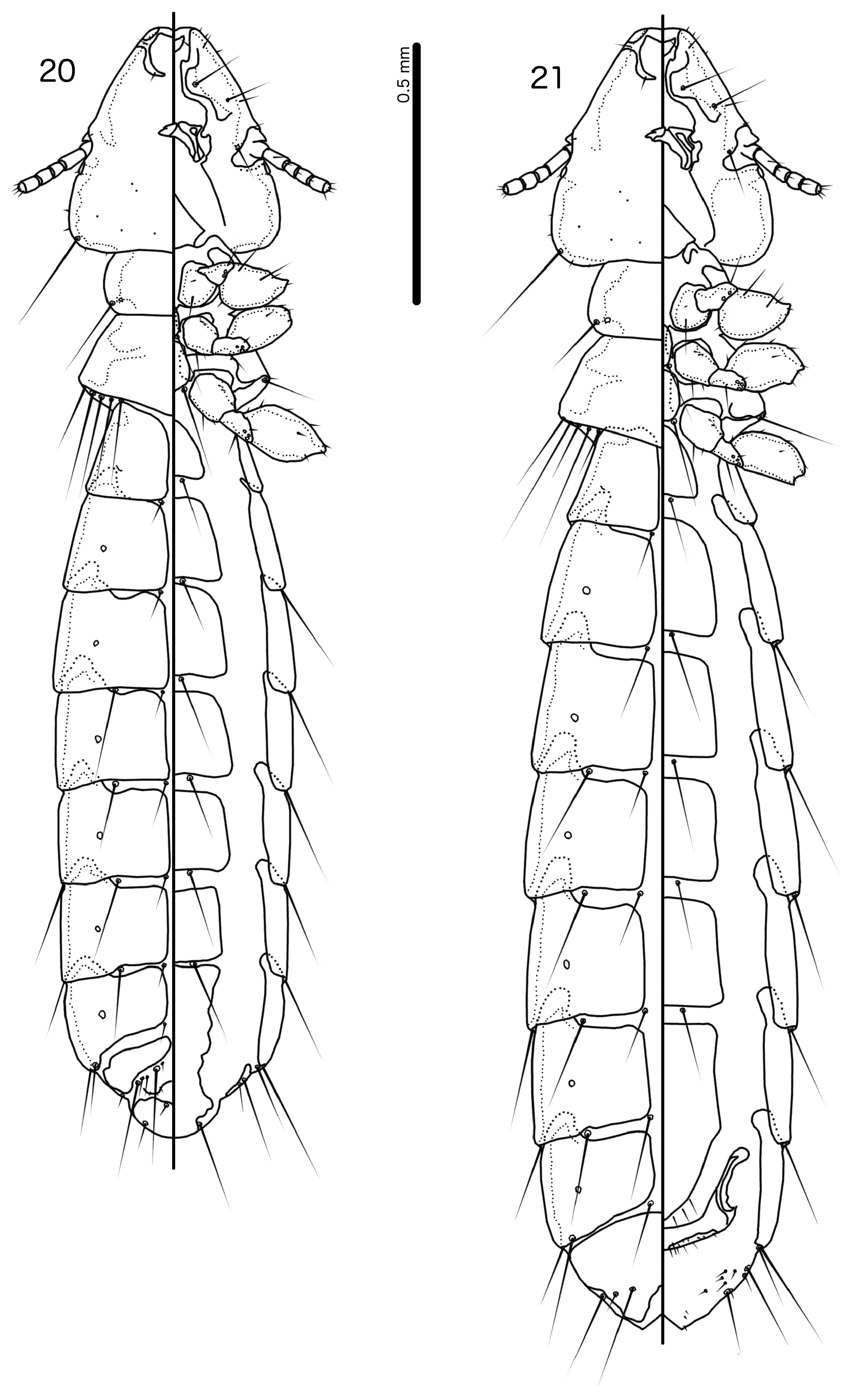
- Lateral margins of pterothorax divergent posteriorly ( Fig. 20 View FIGURES 20–21 ); no antero-lateral thickening of any sternal plate ( Fig. 20 View FIGURES 20–21 ); male abdominal segment XI not extended into triangular tail ( Fig. 20 View FIGURES 20–21 )............................................... 5.
2. Ventral anterior plate present ( Fig. 3 View FIGURES 3–7 ); parameres without marginal modifications ( Fig. 5 View FIGURES 3–7 ); proximal mesosome slender ( Fig. 6 View FIGURES 3–7 ); tail of male abdominal segment XI thinly sclerotised distally ( Fig. 1 View FIGURES 1–2 )............................................3.
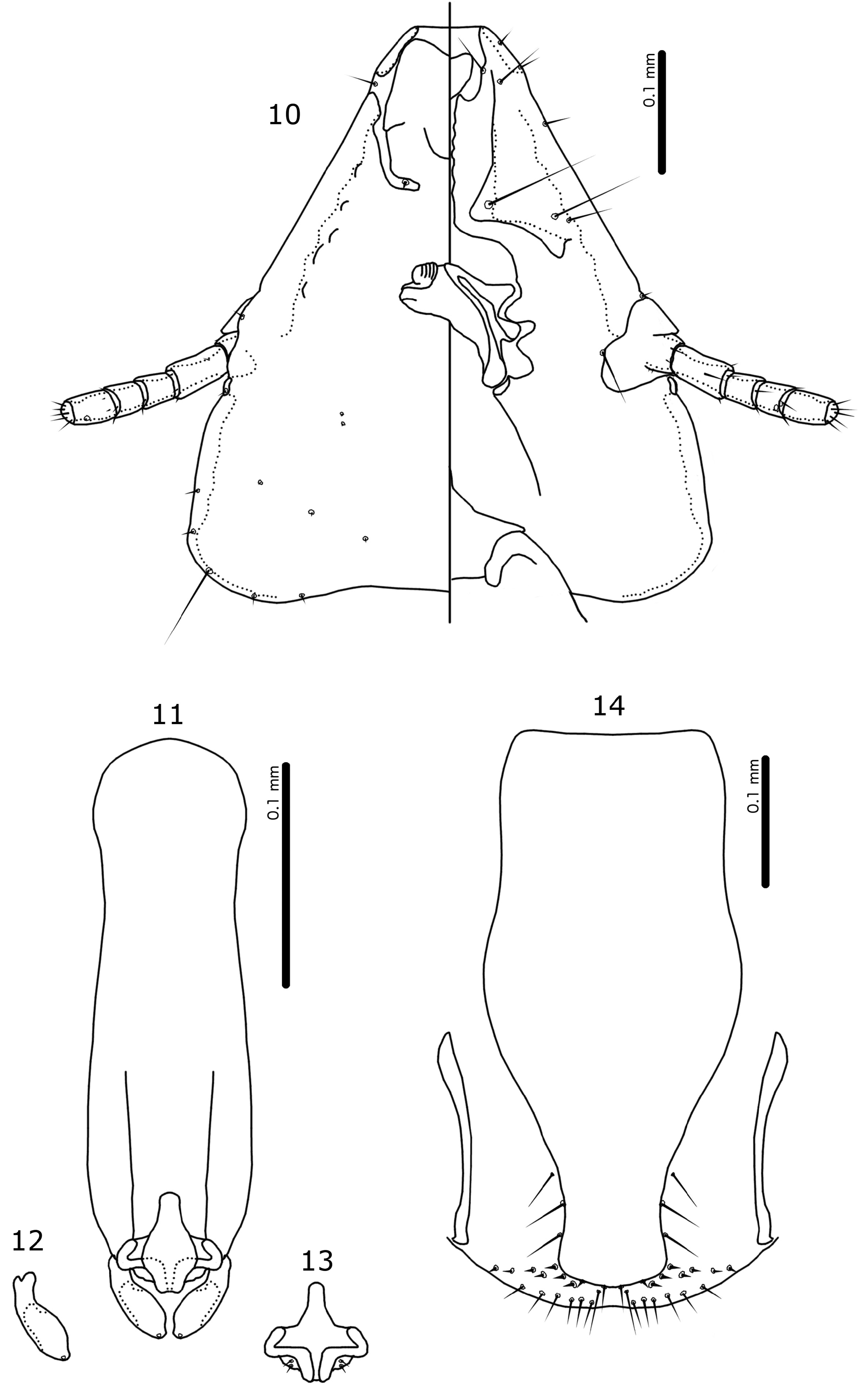
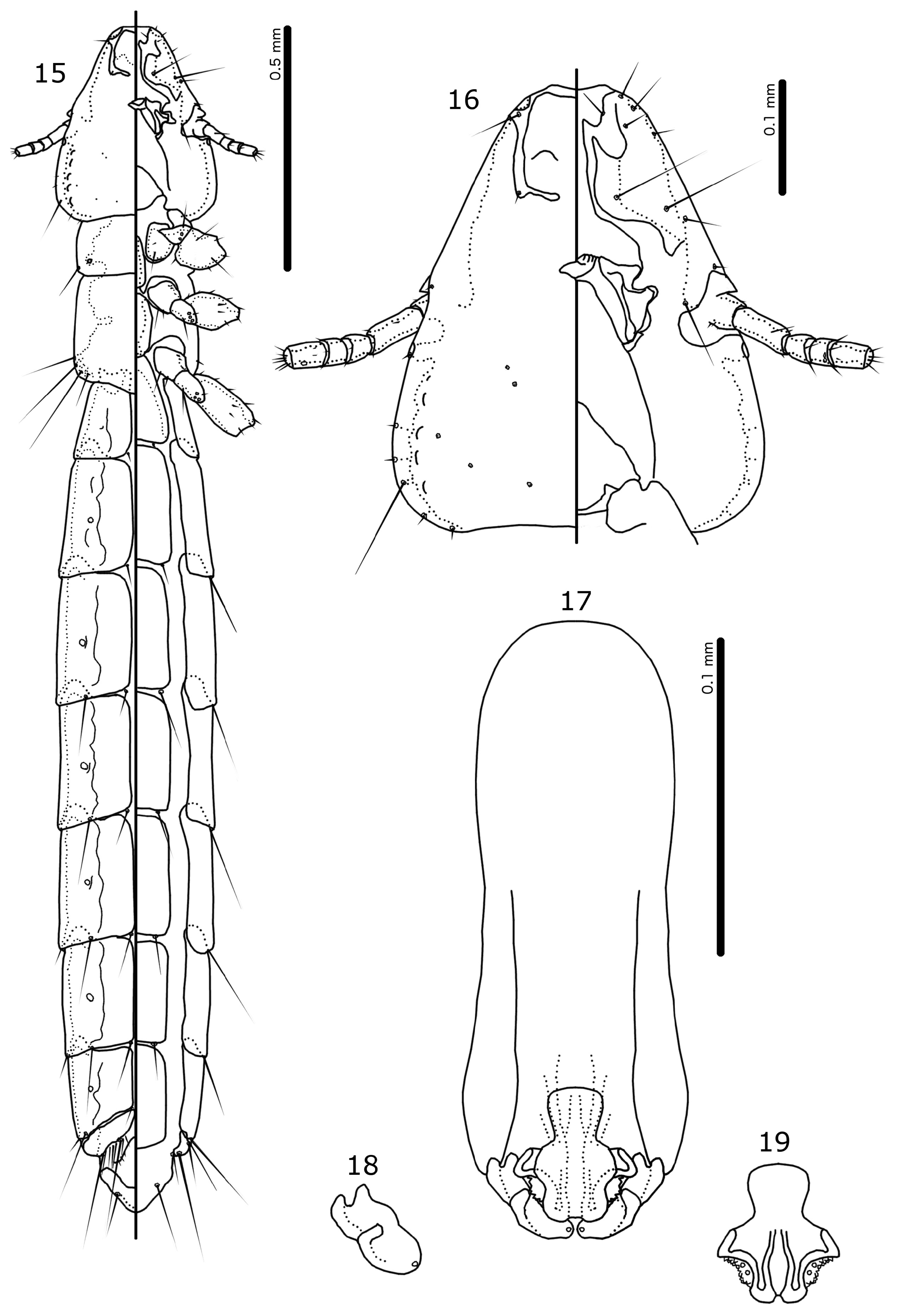
- Ventral anterior plate absent ( Fig. 10 View FIGURES 10–14 ); parameres with marginal modifications ( Figs 12 View FIGURES 10–14 , 18 View FIGURES 15–19 ); proximal mesosome broad ( Figs 13 View FIGURES 10–14 , 19 View FIGURES 15–19 ); tail of male abdominal segment XI with wide distal sclerotised plate ( Fig. 15 View FIGURES 15–19 )..............................4.
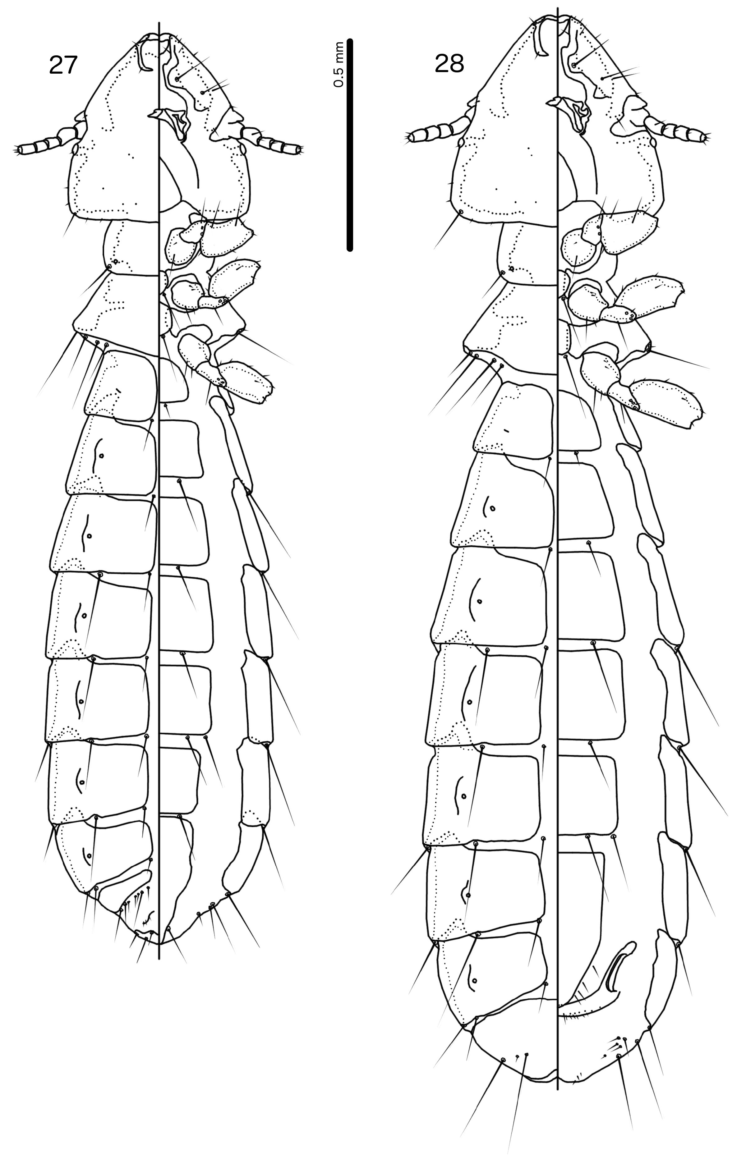
3. Male abdominal segment VI with 1 ps on each side ( Fig. 1 View FIGURES 1–2 ); proximal mesosome tapering slightly anteriorly ( Fig. 6 View FIGURES 3–7 ); distal margin of mesosome slight, smooth ( Fig. 6 View FIGURES 3–7 ); female sternite III without anterior thickening ( Fig. 2 View FIGURES 1–2 )....... Aratricerca macki
- Male abdominal segment VI with 2 ps on each side; proximal mesosome widening anteriorly; distal margin of mesosome bulging, fringed; female sternite III with anterior thickening........................................ Aratricerca cirithra
4. Frons convex ( Fig. 10 View FIGURES 10–14 ); lateral margins of preantennal area convex ( Fig. 10 View FIGURES 10–14 ); male tergopleurite VI with 1 ps on each side ( Fig. 8 View FIGURES 8–9 ); male proximal mesosome quadratic ( Fig. 13 View FIGURES 10–14 ); paramere with median, thumb-like modification ( Fig. 12 View FIGURES 10–14 ).................................................................................................... Aratricerca cerata
- Frons concave ( Fig. 16 View FIGURES 15–19 ); lateral margins of preantennal area straight ( Fig. 16 View FIGURES 15–19 ); male tergopleurite VI with 2 ps on each side ( Fig. 15 View FIGURES 15–19 ); male proximal mesosome rounded, widening anteriorly ( Fig. 19 View FIGURES 15–19 ); paramere with lateral, thumb-like modification ( Fig. 18 View FIGURES 15–19 )......................................................................... Aratricerca madagascariensis
5. Dorsal preantennal suture medianly continuous posterior to dorsal anterior plate...................................6.
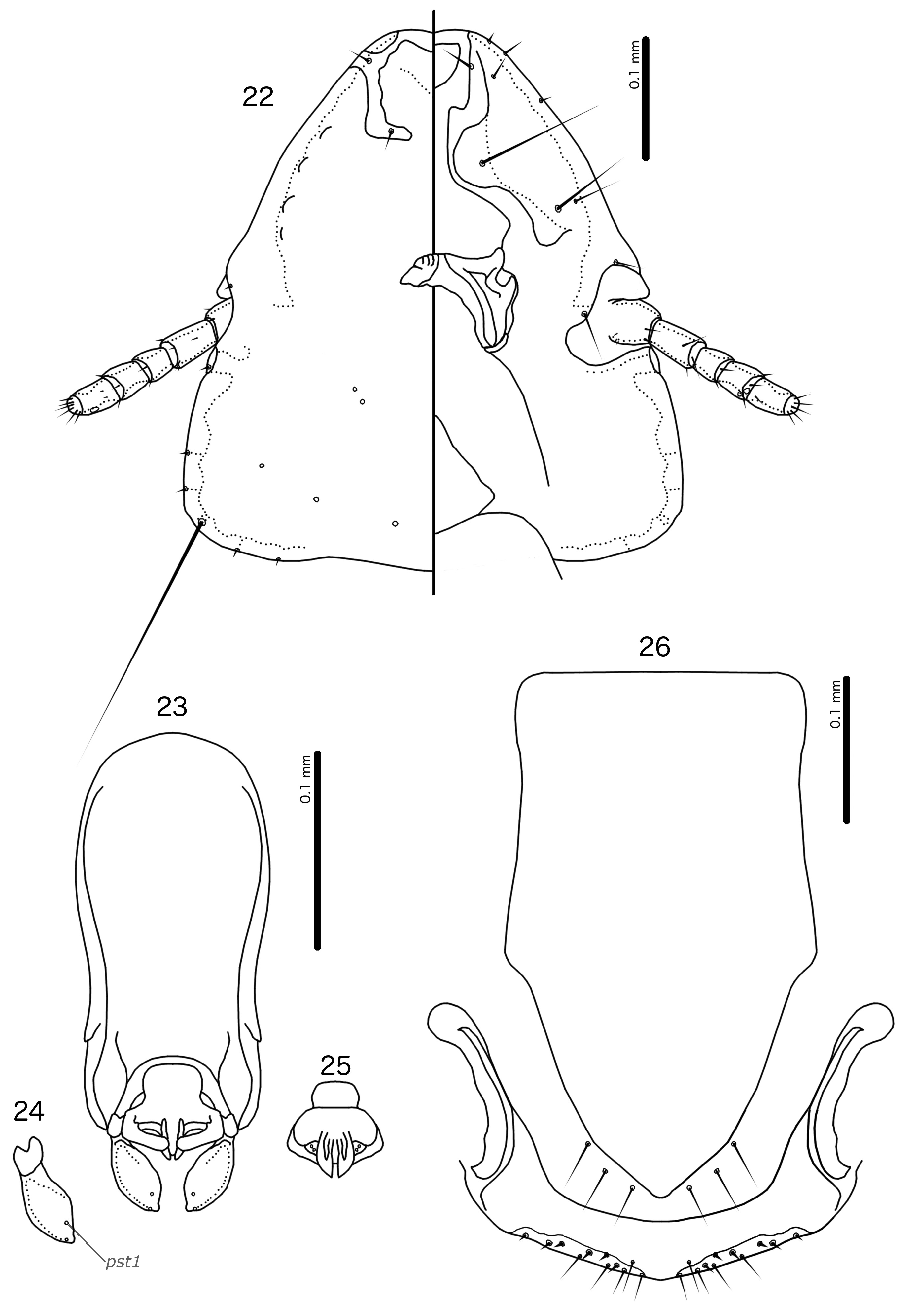
- Dorsal preantennal suture medianly discontinuous posterior to dorsal anterior plate ( Fig. 22 View FIGURES 22–26 )..........................7.
6. Female subgenital plate roughly triangular; proximal mesosome narrow...................... Turdinirmoides carpodaci
- Female subgenital plate roughly pentagonal; proximal mesosome broad..................... Turdinirmoides vasjukovae
7. Ventral anterior plate present ( Fig. 22 View FIGURES 22–26 ); male subgenital plate divided ( Fig. 20 View FIGURES 20–21 ); sts present on male abdominal segment VII ( Fig. 20 View FIGURES 20–21 )................................................................................................ 8.
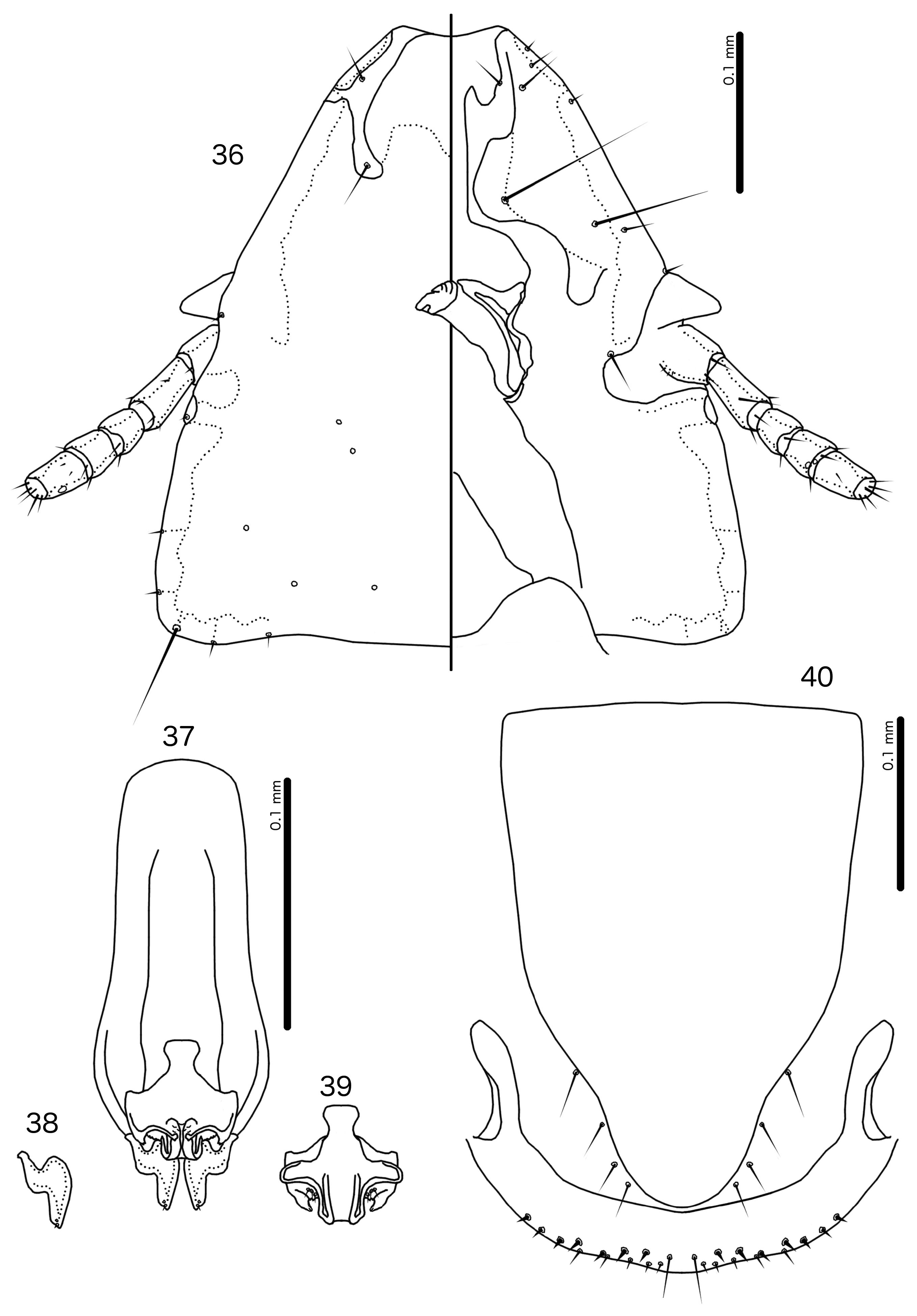
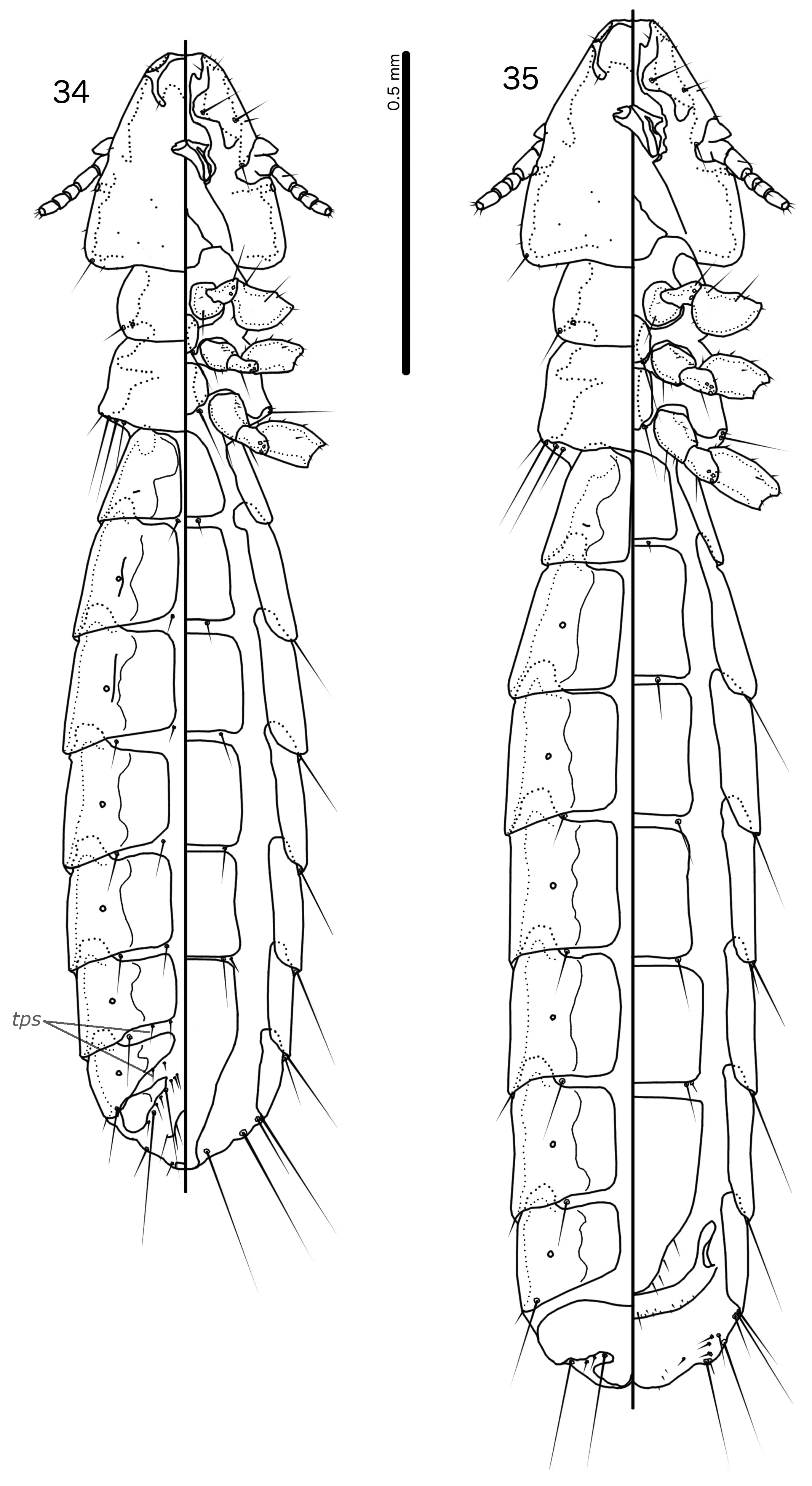
- Ventral anterior plate absent ( Fig. 36 View FIGURES 36–40 ); male subgenital plate not divided ( Fig. 34 View FIGURES 34–35 ); sts absent on male abdominal segment VII ( Fig. 34 View FIGURES 34–35 )...........................................................................................10.
8. Female subgenital plate triangular; tps present on male tergopleurites V–VIII; males with 1 ps on each side of abdominal segment VI; pst1 close to pst2 ......................................................... Turdinirmoides grandalae
- Female subgenital plate pentagonal ( Fig. 26 View FIGURES 22–26 ); tps absent on all male tergopleurites ( Fig. 20 View FIGURES 20–21 ); male with 2 ps on each side of abdominal segment VI ( Fig. 20 View FIGURES 20–21 ); pst1 not close to pst2 ( Fig. 24 View FIGURES 22–26 )............................................... 9.
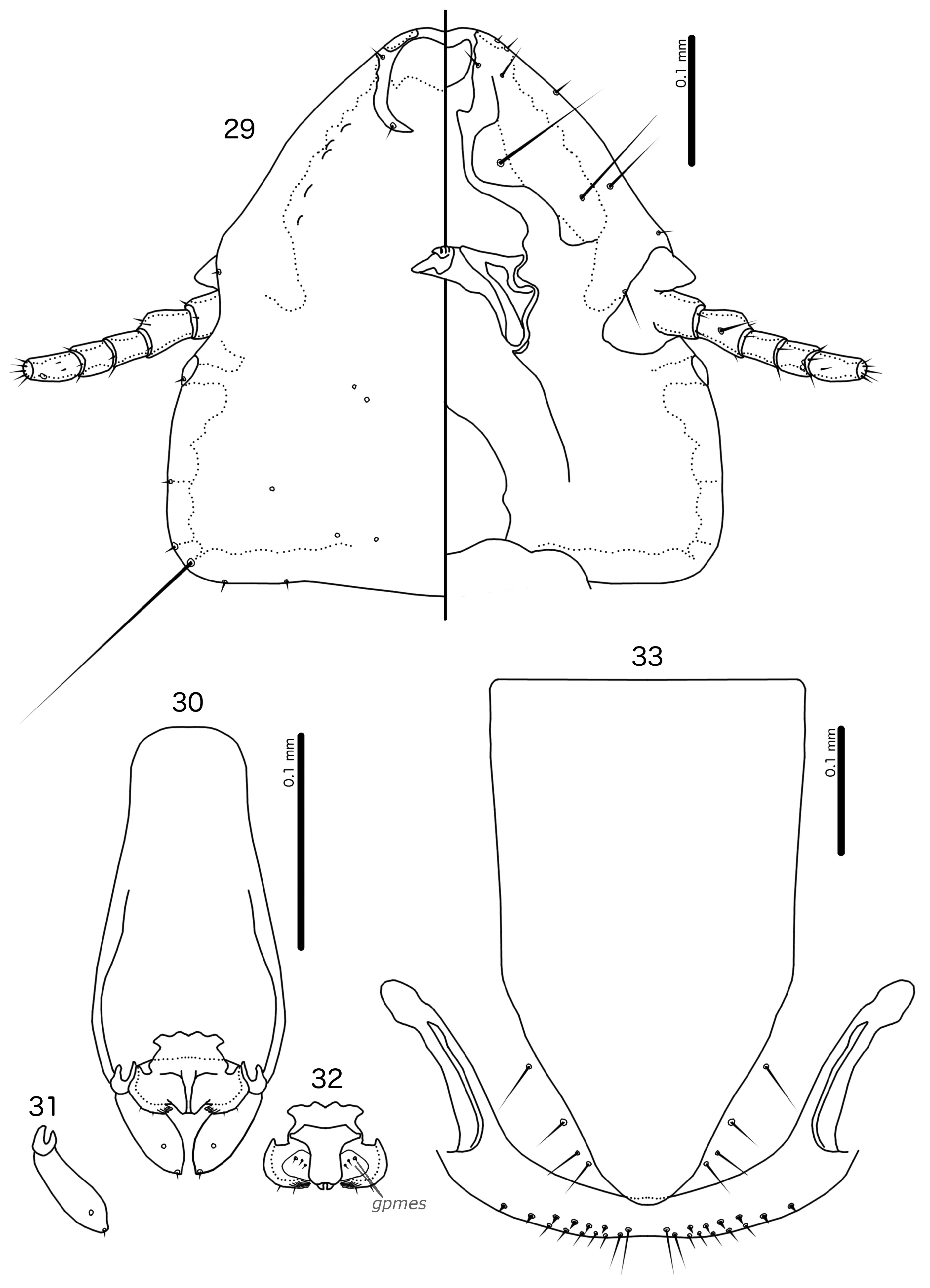
9. Female without ps on abdominal segment III ( Fig. 28 View FIGURES 27–28 ); proximal mesosome with irregular margins ( Fig. 32 View FIGURES 29–33 ); mesosomal lobes rounded with anterior hooks, distal fringe, and 2 lpmes microsetae on each side ( Fig. 32 View FIGURES 29–33 ); female subgenital plate overlapping with detached cross-piece ( Fig. 33 View FIGURES 29–33 ); detached cross-piece follows vulval margin for entire length ( Fig. 33 View FIGURES 29–33 ).................................................................................................. Turdinirmoides rozsai
- Female with 1 ps on each side of abdominal segment III ( Fig. 21 View FIGURES 20–21 ); proximal mesosome with smooth margins ( Fig. 25 View FIGURES 22–26 ); mesosomal lobes bluntly triangular, without hooks, fringes, or lpmes ( Fig. 25 View FIGURES 22–26 ); female subgenital plate not reaching detached crosspiece ( Fig. 26 View FIGURES 22–26 ); detached cross-piece reaches vulval margin only medianly and laterally ( Fig. 26 View FIGURES 22–26 )..... Turdinirmoides janigai
10. Male subgenital plate does not reach distal end of abdomen; female tergopleurite XI much reduced; proximal mesosome with pointed anterior margin................................................................ Timalinirmus hrabali
- Male subgenital plate reaches distal end of abdomen ( Fig. 34 View FIGURES 34–35 ); female tergopleurite XI large ( Fig. 35 View FIGURES 34–35 ); proximal mesosome with flat anterior margin ( Fig. 39 View FIGURES 36–40 )............................................................. Timalinirmus curvus

No known copyright restrictions apply. See Agosti, D., Egloff, W., 2009. Taxonomic information exchange and copyright: the Plazi approach. BMC Research Notes 2009, 2:53 for further explanation.
|
Kingdom |
|
|
Phylum |
|
|
Class |
|
|
Order |
|
|
Family |