
Pleurodema kriegi (Müller), Muller
|
publication ID |
https://doi.org/ 10.5281/zenodo.185006 |
|
DOI |
https://doi.org/10.5281/zenodo.5669074 |
|
persistent identifier |
https://treatment.plazi.org/id/6E5487E2-4C62-FFBD-F980-FD86FE2F76CF |
|
treatment provided by |
Plazi |
|
scientific name |
Pleurodema kriegi (Müller) |
| status |
|
( Fig. 2 View FIGURE 2 C, 2D)
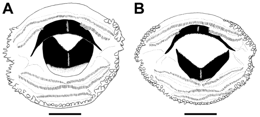
At stage 36 mean total length is 36.4 ± 1.3 mm, body is globose, depressed (mean BMH/BMW = 0.85 ± 0.04). The body length is about 40% of total length (mean BL/TL = 0.42 ± 0.01), body shape is oval in dorsal view with a constriction behind the cephalic region and the maximum width is placed at the posterior portion of the head, behind the eyes. Ventral contour is slightly convex in lateral view and dorsal contour is rounded, uniformly convex from the anterior edge of the oral disc to the eyes, and then slightly convex to the beginning of the dorsal fin. The snout is truncated, trapezoidal in dorsal view, and rounded in lateral view. The nostrils are rounded and placed in a depression; they are dorsolaterally located (mean EN/BWN = 0.33 ± 0.02), slightly closer to the eyes than to the tip of the snout (mean FN/ END = 1.03 ± 0.07), more visible dorsally than laterally. Their openings are directed dorsally and slightly to the front and present a slightly elevated marginal fleshy rim. The eyes are small (E/BWE = 0.17 ± 0.01) and dorsally located (mean IOD/BWE = 0.56 ± 0.03), dorsolaterally oriented, and not visible in ventral view. The pineal end organ is visible as a less pigmented spot between the anterior edges of the eyes. The spiracle is single, lateral, sinistral, short, posterodorsally directed, and placed in the second third of the body (mean RSD/BL = 0.61 ± 0.05). The spiracle inner wall is fused to the body wall except for the very distal end, which folds to delimit the spiracle opening. Spiracle opening is oval, placed below body midline, being its diameter smaller than the tube diameter; it is visible laterally and ventrally. The intestinal ansa was observed approximately at the center of the abdominal ventral surface or displaced to the left behind the spiracle opening. The vent tube is medial; it starts at midline but opens to the right in 60% of the examined specimens or to the left in the others, due to a variable folding of the ventral fin at its origin. A medium-sized saccular structure underlies the limb buds and encloses the vent tube. The tail is medium-sized (mean TAL/TL = 0.58 ± 0.01), and both fins are slightly shallower than body height (mean MTH/BMH = 0.94 ± 0.06). The dorsal fin originates on the body-tail junction and its free margin is regularly curved, convex, with the maximum height at the middle length of the tail. It is as high as the ventral fin (mean DFH/VFH = 1.0 ± 0.05), which originates from the saccular structure, and presents a smoothly convex free margin. The tail axis is straight, and the tail smoothly stretches towards the tip in it last half, ending bluntly rounded. The tail musculature does not reach the tail end, and myomeres are more visible in the proximal half. Neuromasts are very small and hardly visible, even with the help of magnification and after staining the tegument with methylene blue. The oral disc ( Fig. 3 View FIGURE 3 B) is anteroventral, medium-sized (mean OD/BMW = 0.41 ± 0.02), laterally emarginated, and has a large dorsal gap (mean DG/OD = 0.56 ± 0.04). Marginal papillae are arranged in a single alternated or double row, except in the ventral region in which the row is single for approximately the length of P3. Papillae are simple, small and longer than wide, sub-conical. Sub-marginal papillae are absent. Jaw sheaths very robust, finely serrated, heavily pigmented distally for about 2/3 to 1/2 their length. The free margin of the upper jaw sheath is widely arch shaped, whereas that of the lower jaw sheath is V-shaped. Labial tooth row formula is 2(2)/3(1). The gap in A2 is wide, almost one third the length of the row, and the anterior edge of the upper jaw sheath is placed within it. The gap in P1 is narrow. The length of P3 is about half the length of the other rows. The tadpole of P. k r i e g i belongs to the benthic ecomorphological guild (section II: A: 1) of McDiarmid and Altig (1999) as revised from Altig and Johnston (1989).
Variation: Four additional specimens from the same lot, stages 30–31, showed no remarkable variation. The vent tube opened to the left or to the right in equal numbers. One specimen at stage 30 showed a very small ventral gap in the marginal row of papillae. Tadpoles from lot MLP A. 4729 (stages 32–38) differed from those of MLP DB 5059 by having a more rounded snout in dorsal view and by lacking an evident pinealend organ. The vent tube opens to the right in seven specimens, while it opens to the left and medially in one specimen each. LTRF 2(2)/3(1) in eight specimens (four of them show non-medial small gaps in P2 and P3) and 2(2)/ 3 in one specimen at stage 34.
Measurements (in mm): Mean and Standard Deviation (range given in parentheses): TL = 36.4 ± 1.3 (35.4–38.2); BL = 15.1 ± 0.5 (14.6–15.8); TAL = 21.3 ± 1.1 (20.0–22.4); MTH = 8.0 ± 0.4 (7.6–8.5); TMH = 3.2 ± 0.1 (3.0–3.3); TMW = 2.8 ± 0.1 (2.6–2.9); IND = 1.2 ± 0.1 (1.2–1.3); IOD = 4.9 ± 0.1 (4.7–5.0); BMW = 10.0 ± 0.3 (9.6–10.5); BWN = 6.2 ± 0.2 (5.9–6.4); BWE = 8.7 ± 0.3 (8.2–9.1); BMH = 8.5 ± 0.5 (7.9–9.2); RSD = 9.2 ± 0.5 (8.6–9.7); FN = 1.8 ± 0.2 (1.5–2.0); END = 1.7 ± 0.2 (1.5–2.0); N = 0.3 ± 0.1 (0.2–0.3); E = 1.5 ± 0.1 (1.4–1.5); EN = 2.0 ± 0.1 (1.9–2.2); IO = 2.6 ± 0.1 (2.6–2.7); OD = 4.1 ± 0.1 (4.0–4.3); DG = 2.3 ± 0.1 (2.2–2.5); DFH = 1.7 ± 0.2 (1.4–1.9); VFH = 1.7 ± 0.2 (1.5–1.9).
Coloration in preservative: Body dark brown in dorsal view, with some less pigmented regions around the eyes. Perinasal regions heavily pigmented. In lateral view, body dark brown and abdomen almost black. Branchial region almost transparent. Gular region and saccular structure transparent. Caudal musculature is dark brown and fins are finely reticulated, except for the region of ventral fin insertion at caudal musculature. Caudal blood vessels dark.

No known copyright restrictions apply. See Agosti, D., Egloff, W., 2009. Taxonomic information exchange and copyright: the Plazi approach. BMC Research Notes 2009, 2:53 for further explanation.
|
Kingdom |
|
|
Phylum |
|
|
Class |
|
|
Order |
|
|
Family |
|
|
Genus |