
Mycotretus trifasciatus Guérin, 1956
|
publication ID |
https://doi.org/ 10.11646/zootaxa.4282.1.9 |
|
publication LSID |
lsid:zoobank.org:pub:C027C2B6-4D35-41B2-80FC-527D00C15A95 |
|
DOI |
https://doi.org/10.5281/zenodo.6041723 |
|
persistent identifier |
https://treatment.plazi.org/id/6F32B419-FFDD-FFBB-F3B5-F115FAAC864F |
|
treatment provided by |
Plazi |
|
scientific name |
Mycotretus trifasciatus Guérin, 1956 |
| status |
|
Mycotretus trifasciatus Guérin, 1956
Figs. 2 View FIGURE 2 , 3 View FIGURE 3 E–H, 4E–I, 6 and 7E.
Mycotretus trifasciatus Guérin 1956: 63 , fig. 26. Type locality: Brasil, Santa Catarina, Nova Teutonia; Alvarenga 1994: 37 [information on type series and distribution]; Campaner et al. 2008: 242 [list of Erotylidae View in CoL types deposited at MZUSP].
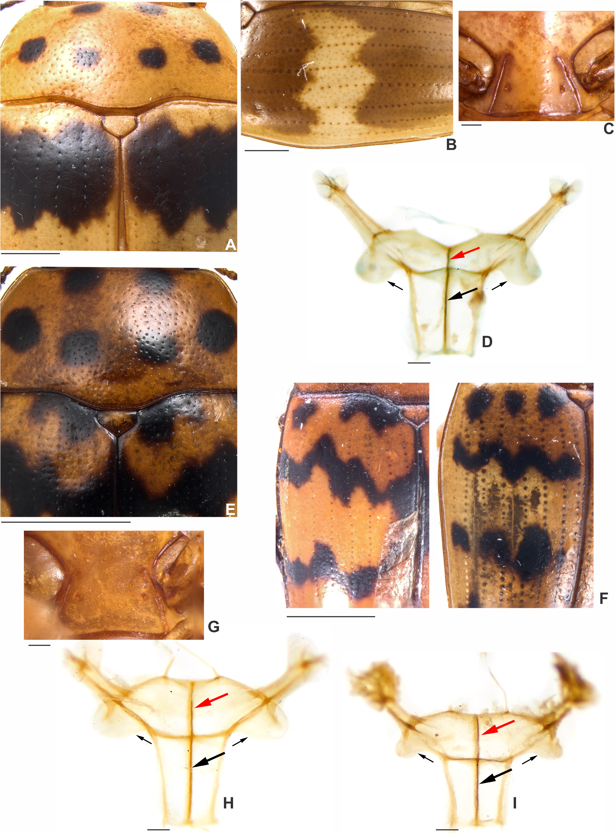
Adult diagnosis. There are two distinct color patterns in M. trifasciatus that seem to be geographic variations: (i) in the specimens like the holotype ( Figs. 2 View FIGURE 2 A–B), the pronotum has two circular black spots on the anterior portion and four circular free black spots on disc ( Figs. 2 View FIGURE 2 A, D, G, 4E). Each elytron has a bilobed scutellar black mark ( Fig. 2 View FIGURE 2 A, big white arrow) and a black spot close to the humerus (the spots reach the anterior elytral edge) ( Fig. 2 View FIGURE 2 A, small white arrow); each elytron has three transverse black bands, extending from the mesal suture to the outer edges ( Figs. 2 View FIGURE 2 A–B), each band being very sinuate and bearing a prominent peak at middle ( Fig. 2 View FIGURE 2 A, red arrow); along the mesal suture there is a black stripe which is narrowest between scutellar shield and first transverse band and between the second and third transverse bands, expanded at elytral apex; (ii) in specimens from Viçosa (MG) ( Figs. 2 View FIGURE 2 D–F and 2H) and Obidos (PA) ( Fig. 2 View FIGURE 2 G), the anteriormost spots in each elytron are separated into three spots (only the scutellar spots reach the anterior edge of elytra, Fig. 2 View FIGURE 2 D, white arrow); the second transverse marking is a fascia, formed by an outer elongate spot and an inner bilobed spot ( Fig. 2 View FIGURE 2 D); the third transverse marking is also a fascia ( Fig. 2 View FIGURE 2 D), formed by an outer elongate spot (close to the second transverse marking) and an inner bilobed spot (sometimes completely separated into two spots on each elytron), shorter than the previous fascia; the transverse markings are devoid of conspicuous peaks and do not reach the mesal suture; there is no longitudinal black stripe close to the mesal suture ( Fig. 2 View FIGURE 2 D) and no apical black spot ( Figs. 2 View FIGURE 2 D–E). Penial flagellum (in the internal sac) well-developed and sinuate ( Fig. 6 View FIGURE 6 A; length mean: 2.17 mm, n = 3, from the beginning of virga to the end of its head) with a median membranous portion bearing a comparatively broader sinuosity ( Fig. 6 View FIGURE 6 A, black arrow) and a sclerotization ( Fig. 6 View FIGURE 6 B, black arrow), the latter not observed in M. chilensis (compare to Fig. 5 View FIGURE 5 A).
Redescription. Length (in mm) = 3.55–6.24 (4.65 ± 0.51, n = 32). Body elongate, widest at the anterior onethird of elytra, TL/EW = 1.66–1.82 (1.73 ± 0.04), GD/EW = 0.59–0.73 (0.68 ± 0.03), glabrous and glossy, dorsal coloration yellowish to reddish-brown with black marks (bands and/or fasciae). Ventral coloration as the dorsal, without black marks; coxae, base and apex of femora, base of tibiae and apical portion of prosternal process blackish; head with a small circular black spot on frons (most prominent in individuals with the first color pattern); mentum plate bearing an almost black outline; antennae yellowish to reddish-brown with antennomeres VI to XI blackish. In individuals from Viçosa (MG) the ventral coloration (including legs) is homogenously yellowishbrown. Pronotum with six circular black spots, except for one malformed individual ( Fig. 2 View FIGURE 2 I).
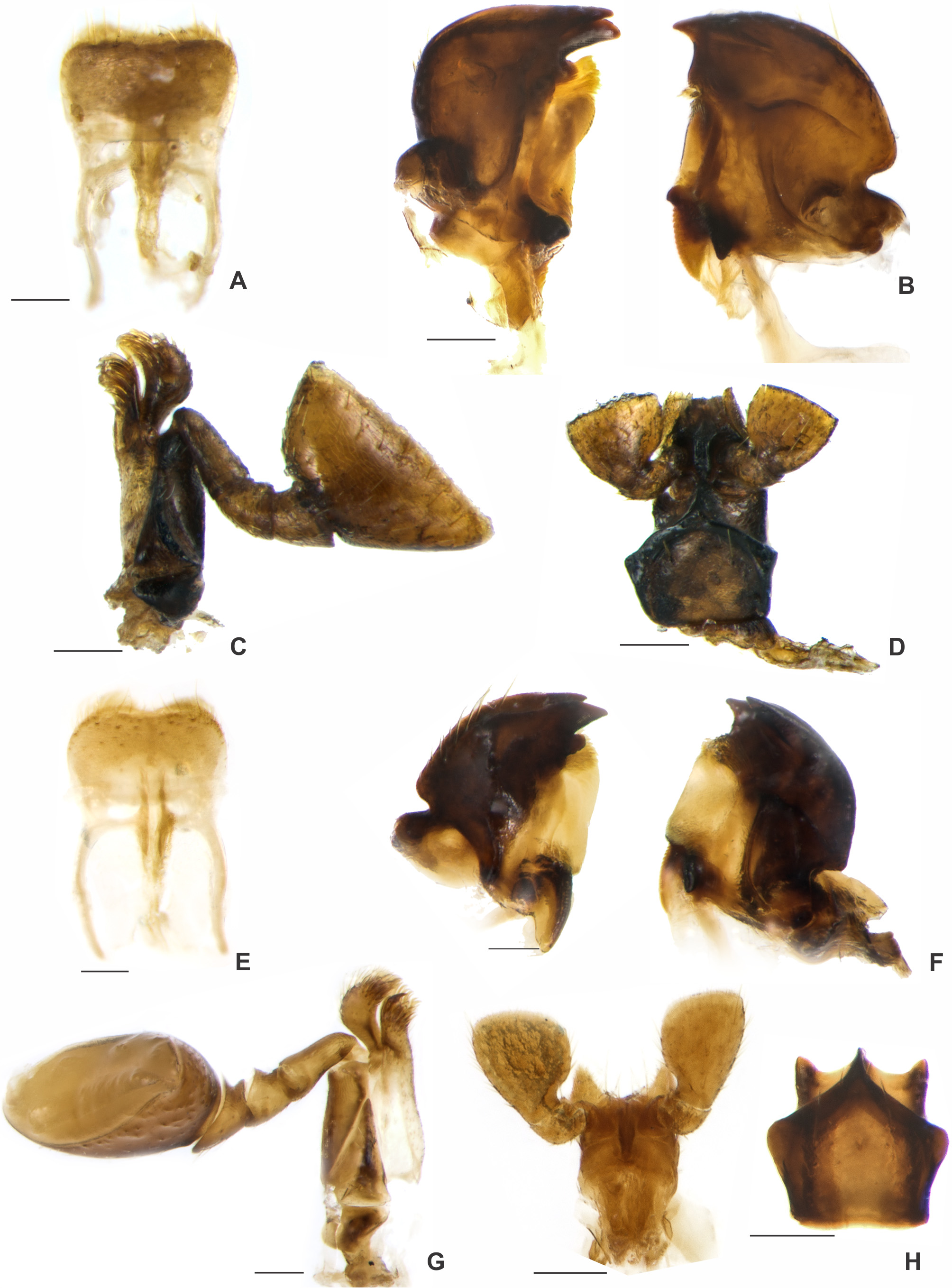
Head. Glabrous; punctation single, fine and sparse; frontoclypeal suture present but interrupted at middle. Clypeus. Shallowly and arcuately emarginated. Antennae. Right antenna measured in one individual: FL 0.56 mm, CL 0.32 mm, CL/FL 0.58; length of antennomeres (in mm), from antennomere one to eleven, as follows: 0.12, 0.07, 0.11, 0.08, 0.06, 0.05, 0.04, 0.07, 0.07, 0.12. Eyes. Glabrous (GW 0.30 mm), finely granulate. Mouthparts. ( Figs. 3 View FIGURE 3 E–H) Labrum free, sclerotized, pubescent, slightly emarginated at middle. Mandibles short and broad, distal portion with a distinct depression containing setae; apex with two teeth; mandibular base emarginated, with an additional outgrowth above mola; mola well developed, horseshoe-shaped, naked and distinctly costate; above mola there is a soft and pubescent prostheca with an additional tuft of setae. Maxillae with cardo subtriangular and stipes elongated; galea shorter but wider than lacinia, somewhat widened towards apex and there densely pubescent; lacinia much longer and narrower, apically covered with dense pubescence, with a highly sclerotized but barely visible hook; maxillary palpomere I about as long as II and III combined, II and III short, IV semicircular and approximately 3× as wide as long. Labium with apical palpomere of club-shaped; mentum pentagonal, strongly sclerotized, with depressions and setae at middle; paraglossae slightly sclerotized and pubescent; glossa strongly sclerotized.
Thorax. Pronotum. ( Fig. 4 View FIGURE 4 E) Subtrapezoidal with edges bordered and sides moderately arcuate, convergent anteriorly. PW/PL = 1.92–2.37 (2.11 ± 0.11), widest close to base in both sexes; shiny, punctation single and interspaces microreticulate; punctures separated by about 6 puncture-widths at disc and each puncture bearing a very short minute seta (barely visible even at a magnification of 110×); anterior edge slightly convex at middle and anterior angles sharp; color pattern: anterior portion bearing two circular black spots, disc with four circular free spots ( Figs. 2 View FIGURE 2 A and 2D). Scutellar shield. (BW 0.3 mm), subpentagonal, glabrous, bearing few punctures. Elytra. EL/EW = 1.25–1.43 (1.32 ± 0.05), EL/PL = 2.84–4.02 (3.21 ± 0.26); strongly margined basally, elongate, moderately convex, with six or seven longitudinal rows of punctures ( Fig. 4 View FIGURE 4 F, counted from the elytral suture to the outer edge of each elytron); punctures separated by about 4 puncture-widths; interspaces of rows microreticulate and with fine and sparse punctures, each bearing a minute seta (more discernible at the anterior one-third and barely visible even at magnification of 150×); elytral color patterns as in the diagnosis. Hind wings. Developed, apparently functional. Prosternum. Convex; anterior edge smooth and pubescent; notosternal sutures distinct and entire; procoxal cavities ovate; prosternal process abruptly expanded apically, shallowly emarginated at apex; procoxal lines shorter and nearly straight; on the prosternal process, there is a pair of secretory pores ( Fig. 4 View FIGURE 4 G; barely visible at a magnification of 130× and indistinguishable in a few individuals). Mesoventrite. Small, convex, mesocoxal lines straight to slightly arched; base sinuate, medially lobed. Metaventrite. Convex, glabrous, finely punctate, microreticulate between punctures; approximately 2.11× as long as mesoventrite; metacoxal lines conspicuous, approximately 0.69× as long as metaventrite; discrimen ( Figs. 2 View FIGURE 2 F, arrow) long, approximately 0.69× as long as metaventrite. Metendosternite well developed ( Figs. 4 View FIGURE 4 H and 4I), sclerotized and convex; central sclerotization of the anterior processes ( Figs. 4 View FIGURE 4 H and 4I, red arrow) approximately 0.39× as long as central sclerotization of the stalk ( Figs. 4 View FIGURE 4 H and 4I, black arrow); laminae with the inner outline without abrupt inflexion ( Fig. 4 View FIGURE 4 H and 4I, small black arrows). Legs. Pro- and mesocoxae almost globular, metacoxae transverse, cigaretteshaped. Femora elongated, smooth, without spines or other outgrowths. Tibiae long, somewhat widened apically. Apex of tibiae with a crown of wide flat setulae, two spurs on meso- and metatibiae and one reduced spur on protibia; tarsi densely pubescent.
Abdomen. Elongated; with coarse, shallow punctation; interspaces of punctures granulate; vestiture of sparse, slender setae. Coxal lines conspicuous and not continuous around coxae (approximately 0.92× the length of the first abdominal ventrite). Length of the ventrites one to five as follows (in mm, from base to apex of each ventrite at the longitudinal midline): 0.68, 0.34, 0.28, 0.26, 0.39. Male terminalia. ( Figs. 6 View FIGURE 6 A–D) Penis elongate, slightly curved ( Fig. 6 View FIGURE 6 A, pen); basal portion with a prominent and sclerotized projection linked to the apophyses; internal sac with a well-developed flagellum ( Fig. 6 View FIGURE 6 A, fla), sinuate (length mean: 2.17 mm, n = 3, from the beginning of virga to the end of head) with median portion accompanied by a membranous enlargement with a broad sinuosity ( Fig. 6 View FIGURE 6 A, arrow; length mean: 0.46 mm, n = 3) and a sclerotization not present in M. chilensis ( Fig. 6 View FIGURE 6 B, arrow). Apophyses ( Fig. 6 View FIGURE 6 A, apo) approximately 1.5× longer than penis (n = 3). Tegmen strongly sclerotized, with a membranous portion on dorsum; parameres reduced and strongly sclerotized, with densely pubescent outgrowths, dilated and pointed at apex ( Fig. 6 View FIGURE 6 C, arrow). Tergite IX and segment X well sclerotized; anteroventral edge of the segment IX with a truncate subgenital plate ( Fig. 6 View FIGURE 6 D, arrow). Female abdominal terminalia. ( Figs. 6 View FIGURE 6 E–I) Gonostyli ( Fig. 6 View FIGURE 6 E, black arrows) and gonocoxites ( Fig. 6 View FIGURE 6 F, red arrows) strongly sclerotized. Spermatheca ovate ( Figs. 6 View FIGURE 6 G, black arrow and 6H), very membranous in the dissected individuals from Viçosa (MG) ( Fig. 6 View FIGURE 6 G) to strongly sclerotized in dissected individuals from the remaining localities ( Fig. 6 View FIGURE 6 H); bursa copulatrix developed ( Fig. 6 View FIGURE 6 G, red arrow). Tergite VIII sclerotized and sternite VIII with a conspicuous median strut ( Fig. 6 View FIGURE 6 I, black arrow).
Holotype ( MZUSP). ( Figs. 2 View FIGURE 2 A–C) “ TIPO [red label, printed] \ N. Teutonia., S. Catarina. [printed], 12.948 [handwritten] \ Coll. J. Guérin., S. Paulo., Brasil. [printed], 18407 [handwritten] \ 194, Brasilien, Nova Teutonia, 27° 11 B, 52° 23’ L, Fritz Plaumann, 3500 m [printed] \ Mycotretus trifasciatus J. Guer [handwritten], J. Guerin det. [printed] 1954 [handwritten]”.
Other examined specimens. 1 male ( MZUSP, dissected) “COTIPO [red label, printed] \ N. Teutonia., S. Catarina. [printed], 12.949 [handwritten] \ Coll. J. Guérin., S. Paulo., Brasil. [printed], 18490 [handwritten] \ 194, Brasilien, Nova Teutonia, 27° 11 B, 52° 23’ L, Fritz Plaumann, 3500 m [printed] \ Mycotretus trifasciatus J. Guer [handwritten], J. Guerin det. [printed] 1954 [handwritten]” ; 1 male ( MZUSP, dissected) “COTIPO [red label, printed] \ Serro Azul, Rio Gr. do sul., S. Catarina.,8.939 [handwritten] \ Coll. J. Guérin., S. Paulo., Brasil. [printed], 18870 [handwritten] \ Serro Azul [printed], 8.39 [handwritten] \ Mycotretus trifasciatus J. Guer [handwritten], J. Guerin det. [printed] 1954 [handwritten]” ; 1 specimen ( MZUSP) “COTIPO [red label, printed] \ Cantareira. S. Paulo [printed], 12.929 [handwritten] \ Coll. J. Guérin., S. Paulo., Brasil. [printed], 17373 [handwritten] \ Mycotretus trifasciatus J. Guer [handwritten], J. Guerin det. [printed] 1954 [handwritten]” ; 1 specimen ( MZUSP) “COTIPO [pink label, handwritten] \ Morumbi [handwritten], Jabaquara., S. Paulo. [printed], 11.942 [handwritten] \ Coll. J. Guérin., S. Paulo., Brasil. [printed], 18164 [handwritten] \ MORUMBI, Sao Paulo – Capital, Dr. Nick. [printed], 29.11.42. [handwritten] \ Mycotretus trifasciatus J. Guer [handwritten], J. Guerin det. [printed] 1954 [handwritten]” ; 1 specimen ( MZUSP) “ Brasilien, Nova Teutonia, Santa Catharina [printed], 2.1936 [handwritten], B. Pohl [printed] \ Mycotretus trifasciatus J. Guer [handwritten], J. Guerin det. [printed] 1954 [handwritten]” ; 1 specimen ( MZUSP) “ BRASIL, S. Bento do Sul, Sta. Catarina, Dirings [printed] \ Mycotretus trifasciatus J. Guer [handwritten]” ; 1 female ( MZUSP, dissected) “ BRASIL, Rio Vermelho , Sta. Catarina, Dirings [printed], MAR 1952 [date printed on label back]” ; 1 male ( MZUSP, dissected) “ BRASIL, Rio Vermelho , Sta. Catarina, Dirings [printed], MAR 1952 [date printed on label back]” ; 1 specimen female ( MZUSP, dissected) “ BRASIL, Canta Galo ( Obidos ), PARÁ, Dirings [printed], JAN 1957 [date printed on label back]” ; 1 male (MCNZ, dissected) “Canela, RS, 06/I/1984, M.Hoffmann [handwritten] \ Col. MCN, 238429 [printed]”; 1 specimen (MCNZ) “Tapes, RS, (Faz. São Miguel), 17.XII.2003, Equipe Probio col. \ Col. MCN, 224774 [printed]”; 1 specimen ( CAMB) “ Brasil: MG, Viçosa, “ Mata do Paraíso ”, 08.xii.2014, leg. Pecci-Maddalena, Í.S.C & Lopes-Andrade, C. [printed] \ Bicho 9 Fungo 9 [printed] \ ex Mycena sp.” ; 1 specimen ( CERPE) “ Brasil: MG, Viçosa, “ Mata do Paraíso ”, 08.xii.2014, leg. Pecci-Maddalena, Í.S.C & Lopes-Andrade, C. [printed] \ Bicho 9 Fungo 9 [printed] \ ex Mycena sp.” ; 1 specimen ( CEMT) “ Brasil: MG, Viçosa, “ Mata do Paraíso ”, 08.xii.2014, leg. Pecci-Maddalena, Í.S.C & Lopes- Andrade, C. [printed] \ Bicho 9 Fungo 9 [printed] \ ex Mycena sp.” ; 13 specimens ( CELC, including two females and one male dissected) “ Brasil: MG, Viçosa, “ Mata do Paraíso ”, 08.xii.2014, leg. Pecci-Maddalena, Í.S.C & Lopes-Andrade, C. [printed] \ Bicho 9 Fungo 9 [printed] \ ex Mycena sp.” ; 3 specimens ( CELC, including one dissected male) “ Brasil: MG, Viçosa, “ Mata do Paraíso ”, 02.xii.2014, leg. Pecci-Maddalena, Í.S.C. [printed]\ Fungo 1 Bicho 1 \ ex Lentinus brumalis [printed]” ; 4 specimens ( CELC) “ Brasil: MG, Viçosa, “ Mata do Paraíso ”, 03.xi.2014; leg. A. Orsetti, S. Aloquio, I. Maddalena, A. Komonen & C. Lopes-Andrade [printed] \ Trilha do Pesquisador [printed], Polyporus brumalis Polyporaceae [handwritten]” ; 1 specimen ( CELC) “ Brasil: MG, Viçosa, “ Mata do Paraíso ”, 05.xi.2014; leg. A. Orsetti, S. Aloquio, I. Maddalena, A. Komonen & C. Lopes-Andrade [printed] \ Trilha do Pesquisador [printed], Polyporus brumalis Polyporaceae [handwritten]” ; 1 specimen female ( CELC, dissected) “ Brasil: MG, Viçosa, “ Mata da Biologia ”, 03.v.2014, leg. Lopes-Andrade et al. \ Fungo 2. Polyporus tricoloma [SIC]” ; 1 specimen ( CELC) “ Brasil: MG, Viçosa, Mata do Paraíso , 13.ii.2015, armadilha intercept. de voo, leg. S. Aloquio, A. Orsetti, C. Lopes-Andrade & M. Bento [printed]” ; 1 specimen ( RBINS) " Coll. R.I.Sc.N.B., Brazil: Mendes, Le Moult Vendit [printed] \ Mycotretus spp. [handwritten], det. P.E.Skelley [printed]".
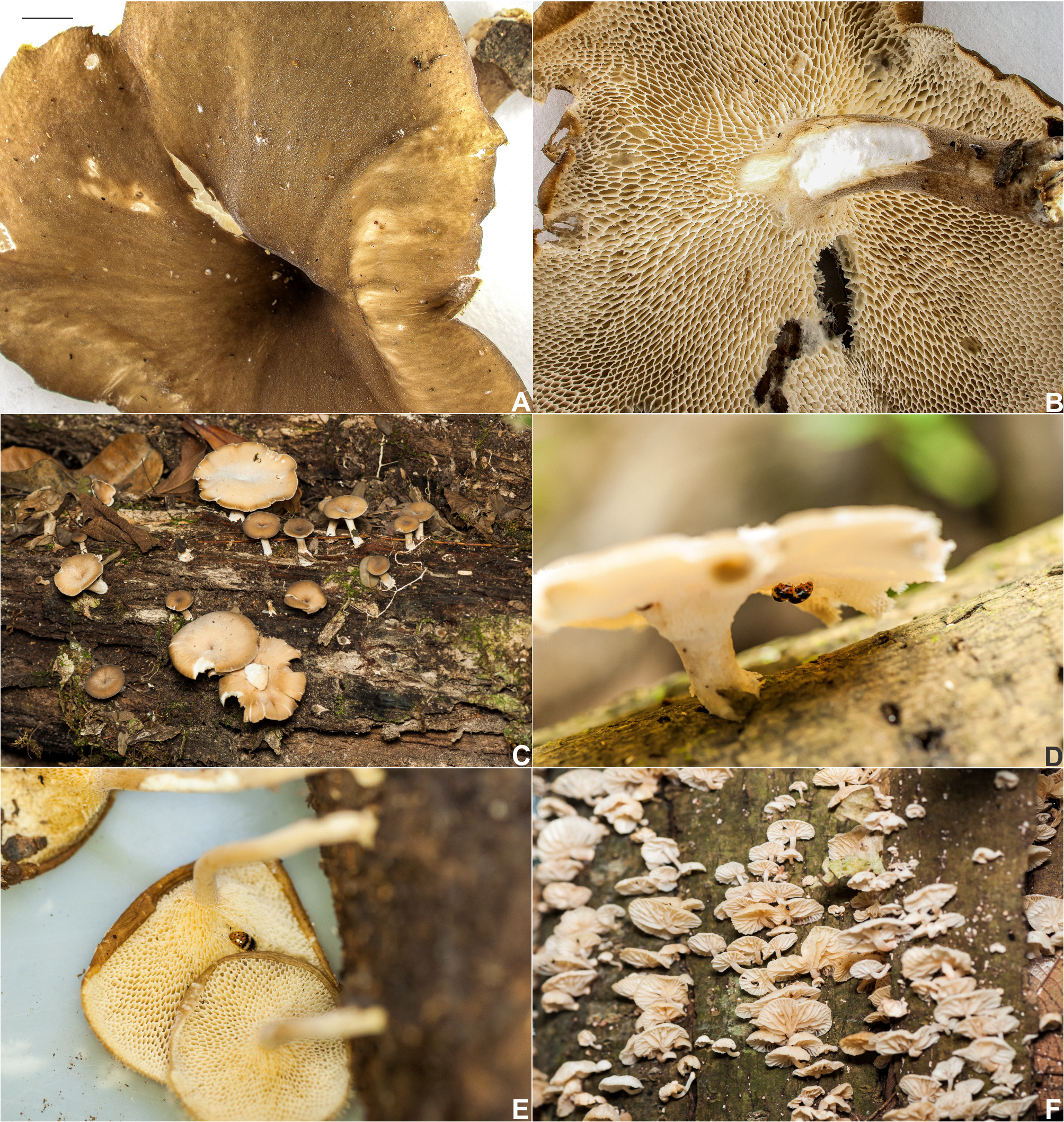
Ecology. Mycotretus trifasciatus has been collected in basidiomes of Polyporaceae (Polyporales) : Lentinus brumalis (Pers.) Zmitr. ( Fig. 7 View FIGURE 7 A–C), Lentinus tricholoma (Mont.) Zmitr. ( Fig. 7 View FIGURE 7 E) and in Mycenaceae (Agaricales) : Mycena sp. ( Fig. 7 View FIGURE 7 F). Specimens collected by us were found in two forest remnants in Viçosa (state of Minas Gerais, Southeast Brazil) in the beginning of summer, except for one collection on 0 3 May 2014.
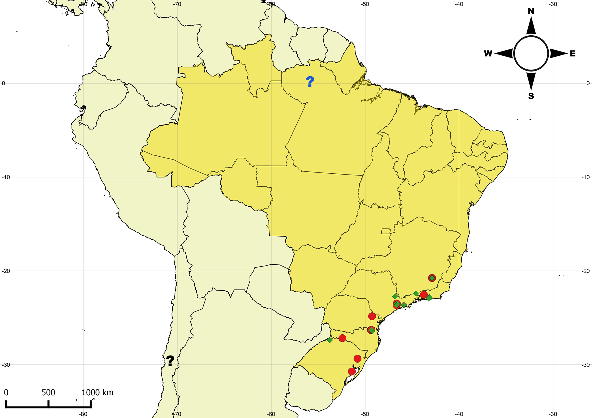
Distribution. Southern and southeastern Brazil, with one doubtful record in Obidos (in the state of Pará, North Brazil) ( Fig. 8 View FIGURE 8 , question mark blue).
Remarks. Specimens from Viçosa have free spots on elytral disc instead of bands or fasciae. The longitudinal elytral striae also vary between specimens, the punctures being coarser in part of the individuals of a single locality. In the latter case a seventh stria can be easily distinguished after the sixth in each elytron. In most cases, the coloration between the first and second transverse marking and between the third transverse marking and the end of the elytra is remarkably yellowish-brown ( Figs. 2 View FIGURE 2 A and 2G, black arrows). In live specimens we observed that this coloration can be whitish ( Fig. 2 View FIGURE 2 H). As observed by Guérin (1956) in the original description, in one paratype the anterior sides of pronotum are deformed and there are only five circular pronotal spots instead of six ( Fig. 2 View FIGURE 2 I).


No known copyright restrictions apply. See Agosti, D., Egloff, W., 2009. Taxonomic information exchange and copyright: the Plazi approach. BMC Research Notes 2009, 2:53 for further explanation.
|
Kingdom |
|
|
Phylum |
|
|
Class |
|
|
Order |
|
|
Family |
|
|
Genus |
Mycotretus trifasciatus Guérin, 1956
| Pecci-Maddalena, Italo Salvatore De Castro & Lopes-Andrade, Cristiano 2017 |
Mycotretus trifasciatus Guérin 1956 : 63
| Campaner 2008: 242 |
| Alvarenga 1994: 37 |
| Guerin 1956: 63 |