
Metacrangon haona, Komai & Ahyong, 2010
|
publication ID |
https://doi.org/ 10.1080/00222933.2010.520823 |
|
persistent identifier |
https://treatment.plazi.org/id/710787D3-6753-0F74-5BC2-FDC3FB80BEC3 |
|
treatment provided by |
Felipe |
|
scientific name |
Metacrangon haona |
| status |
sp. nov. |
Metacrangon haona View in CoL sp. nov.
( Figures 1–3 View Figure 1 View Figure 2 View Figure 3 )
Material examined
Holotype. Pukaki Rise, Campbell Plateau, 49 ◦ 21.50 ′ S, 171 ◦ 53.00 ′ E, 353 m, stn D0210, 26 January 1964, male (CL 6.8 mm), NIWA 6257 View Materials . GoogleMaps
Description
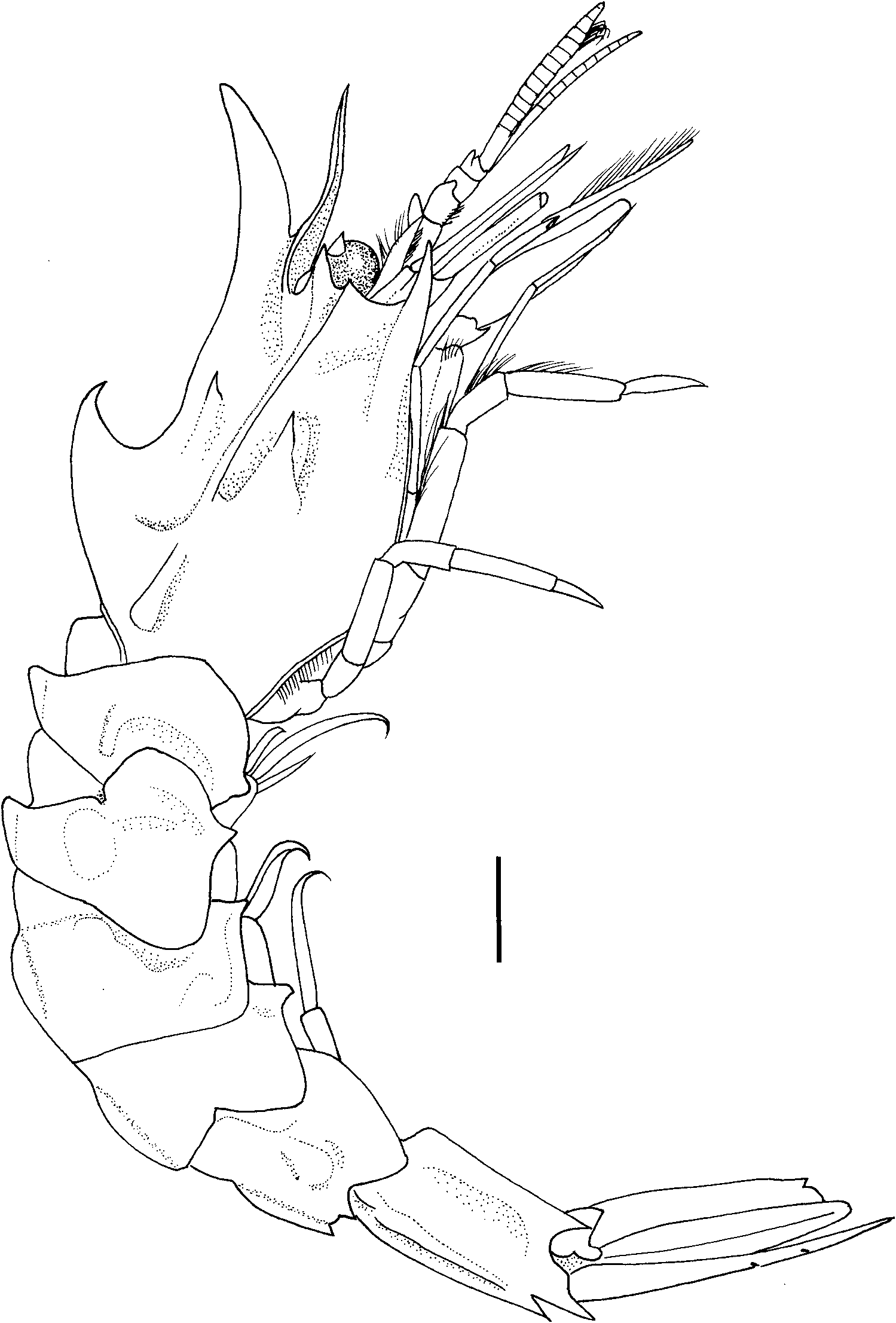
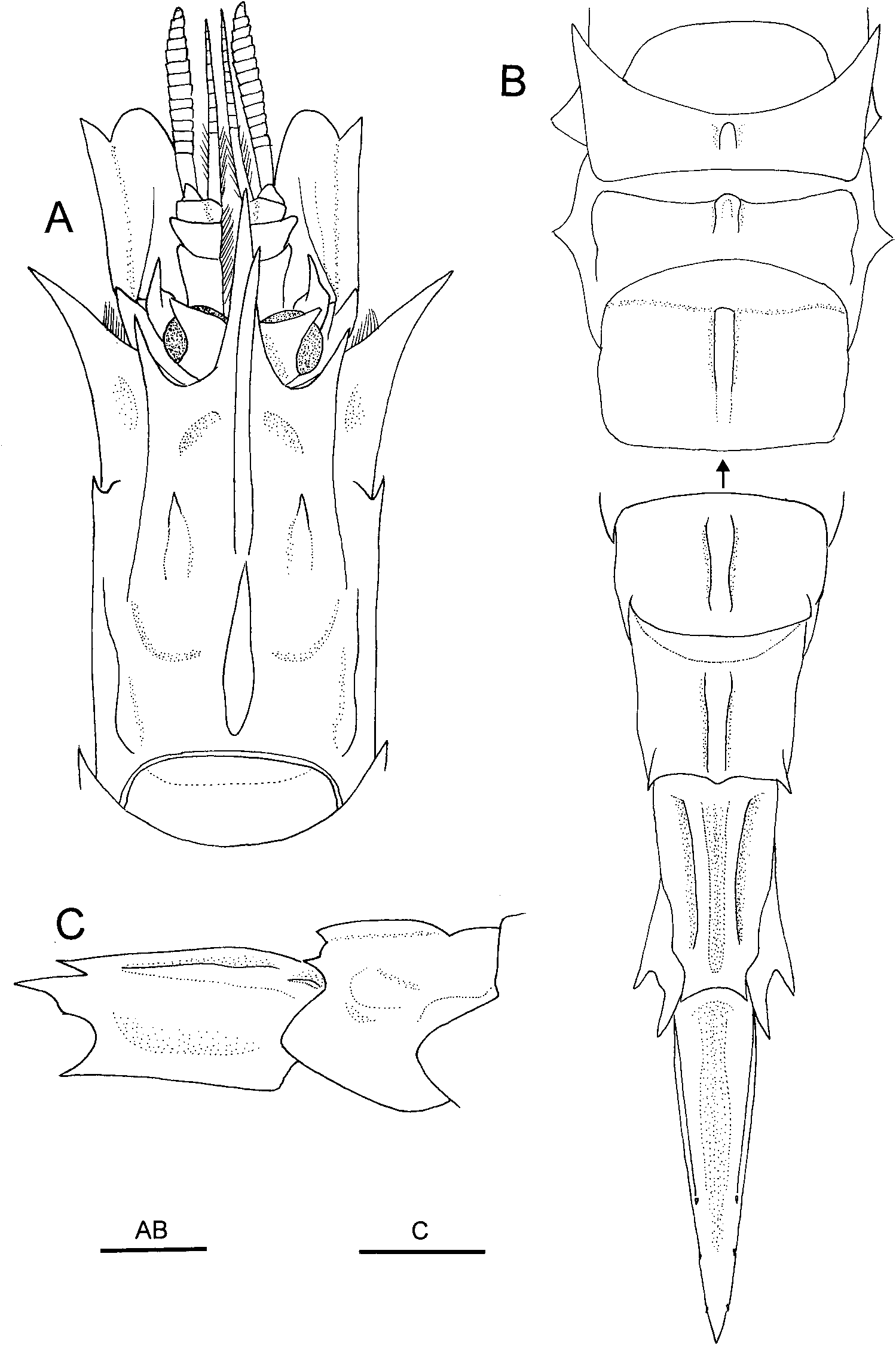
Body ( Figures 1 View Figure 1 , 2A,B View Figure 2 ) moderately robust. Rostrum ( Figures 1 View Figure 1 , 2A View Figure 2 ) dorsoventrally flattened, directed slightly dorsally, unusually elongate (0.55 times as long as carapace), reaching distal margin of antennular peduncle, sinuously curved in lateral view; tip acuminate in dorsal view; dorsal surface nearly flat; lateral margin merging into orbital margin; midventral carina low, blunt, present only in proximal half. Carapace ( Figures 1 View Figure 1 , 2A View Figure 2 ) not widened posteriorly, distinctly longer than wide postorbitally; surface covered with very short setae; dorsal midline with two very prominent, laterally compressed teeth; anterior tooth arcuate, strongly upturned (angle against horizontal plane of carapace about 50 ◦), arising distinctly anterior to rostral base, overlapping rostrum, tip slightly falling short of rostral tip; posterior (cardiac) tooth slightly smaller than anterior tooth, hooked, arising at 0.60 of CL; submedian tooth moderately small; hepatic tooth moderately small, followed by epibranchial ridge; antennal tooth moderately small, directed forward in lateral view and somewhat laterally in dorsal view, acuminate; orbital cleft absent; anterolateral margin between antennal and branchiostegal teeth with tiny denticle inferior to base of antennal tooth; branchiostegal tooth slender, recurved, directed laterally in dorsal view and slightly dorsally in lateral view, over-reaching level of dorsodistal margin of antennal basicerite; pterygostomial tooth small, not visible in lateral view; postorbital carina clearly delimited, accompanied by suture; epibranchial carina distinct.
Abdomen ( Figures 1 View Figure 1 , 2B View Figure 2 ) moderately sculptured; anterior five somites each with distinct, crested mid-dorsal carina, none of mid-dorsal carina on anterior four somites not extending to posterodorsal margin of each somite; mid-dorsal carina on first and second somites produced anterodorsally. First pleuron with blunt, but well-defined tooth at posteroventral angle; second pleuron with triangular tooth at middle of ventral margin; first and second pleura flared laterally, so ventral teeth visible in dorsal view; third and fourth pleura each with distinct triangular tooth at anteroventral angle. Fifth somite ( Figure 2C View Figure 2 ) with obsolete lateral ridge; dorsal margin nearly straight in lateral view; posterodorsal margin armed with small tooth on either side of median projection; pleuron rounded posteroventrally. Sixth somite 1.7 times longer than wide; submedian carinae distinct, slightly curving, not reaching posterodorsal margin; dorsolateral carina distinct, sinuous posteriorly; posterodorsal margin strongly produced, bi-toothed; pleuron strongly flared laterally, posteroventral tooth strong; posterolateral process strong, somewhat curved laterally, terminating in sharp tooth ( Figure 2C View Figure 2 ). Telson ( Figure 2B View Figure 2 ) with three pairs of dorsolateral spines, anteriormost pair located at posterior 0.4; several pairs of plumose setae distal to third pair of lateral spines.
Thoracic sternum widened posteriorly; fifth thoracic sternite with relatively broad, forwardly directed median tooth; sixth to eighth sternites with prominent median keels, those on sixth and seventh sternites each with sharp point anteriorly, that of eighth sternite sharply pointed dentiform. First to fourth abdominal sternites each with prominent median tooth, fifth sternite with short median ridge. Sixth sternite wide, shallowly depressed.
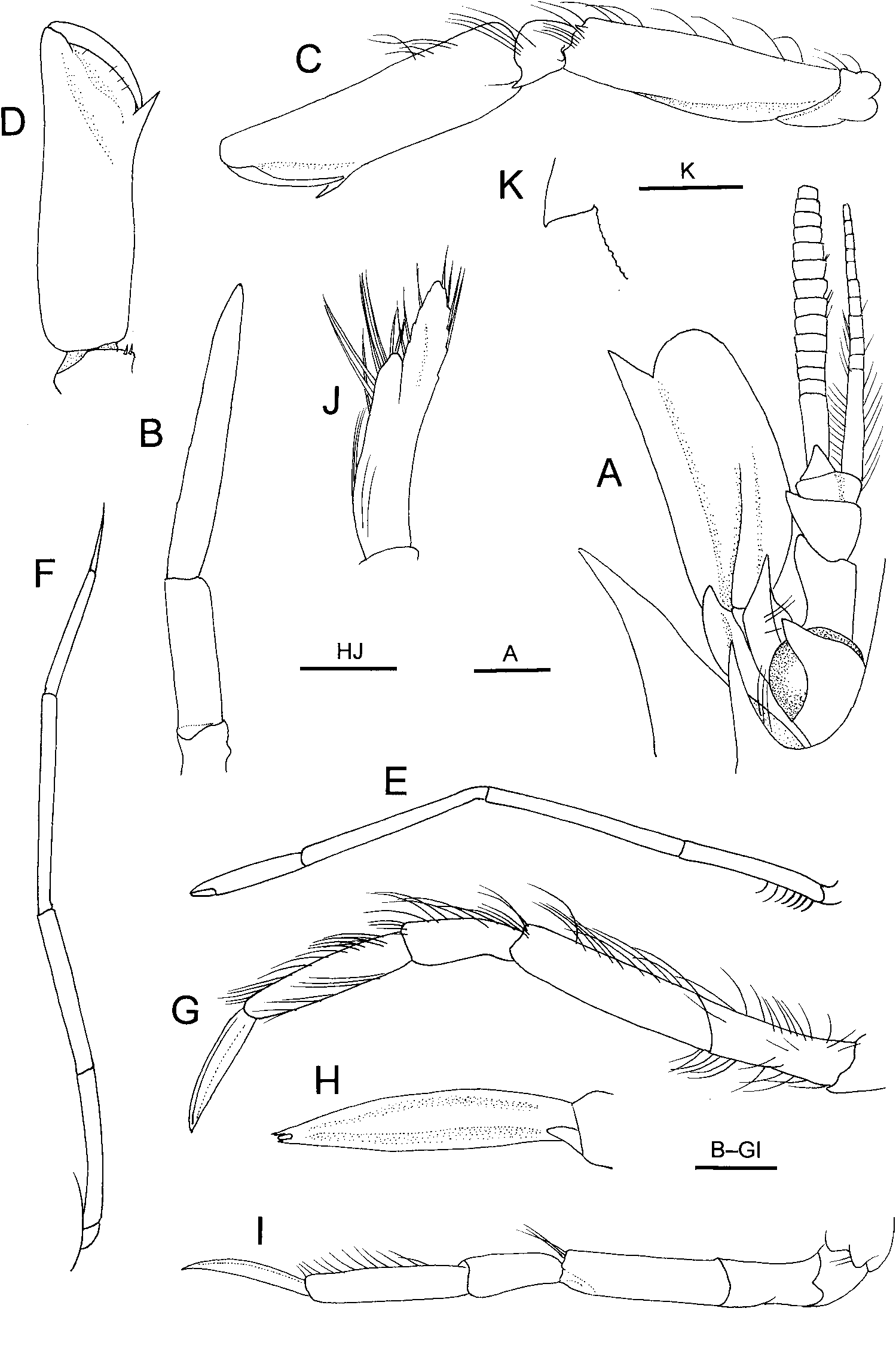
Eye ( Figures 2A View Figure 2 , 3A View Figure 3 ) as wide as long; cornea as wide as eyestalk, light greyish brown in preservative, corneal width about 0.20 of CL; eyestalk with prominent, acutely pointed dorsal tubercle over-reaching distal corneal margin.
Antennular peduncle ( Figures 2A View Figure 2 , 3A View Figure 3 ) reaching midlength of antennal scale. First segment with prominent, blunt distolateral process curving anteriorly, distomesial margin unarmed; stylocerite terminating in acute tooth, slightly falling short of tip of distolateral process of first segment, lateral margin obtusely angular. Second segment much wider than long, widened distally, with prominent, blunt distolateral process. Third segment wider than long. Outer flagellum over-reaching distal margin of lamella of antennal scale by 0.6 length, consisting of 14 or 15 articles.
Antennal basicerite ( Figures 2A View Figure 2 , 3A View Figure 3 ) stout, with produced, acutely pointed dorsodistal lateral angle, ventrolateral tooth reaching beyond dorsodistal lateral angle. Antennal scale 0.53 times as long as carapace and 2.3 times longer than wide; lateral margin nearly straight; distolateral tooth moderately wide, just reaching rounded distal margin of lamella.
Third maxilliped relatively slender, over-reaching antennal scale by half length of ultimate segment; ultimate segment ( Figure 3B View Figure 3 ) gradually tapering distally, about 7.5 times longer than wide; penultimate segment about 3.3 times longer than wide ( Figure 3B View Figure 3 ); antepenultimate segment unarmed on ventral surface.
First pereopod ( Figure 3C,D View Figure 3 ) reaching distal margin of antennal scale; palm 3.3 times longer than wide, not widened proximally or distally, lateral and mesial margins slightly sinuous; thumb relatively slender; carpus with small ventrolateral tooth; merus with one small dorsodistal tooth, ventral margin sinuous, crested. Second pereopod ( Figure 3E View Figure 3 ) reaching nearly to midlength of antennal scale; dactylus about 0.3 times as long as palm; length ratio of chela to merus 1: 1.7: 1.8. Third pereopod ( Figure 3F View Figure 3 ) nearly reaching distal margin of antennal scale by tip of dactylus; length ratio of dactylus to ischium 1: 1.9: 3.2: 2.3: 2.2. Fourth pereopod ( Figure 3G View Figure 3 ) moderately slender, slightly over-reaching distal margin of antennal scale by dactylus; dactylus ( Figure 3H View Figure 3 ) narrowly spatulate, about 0.7 times as long as propodus, margins naked; dactylus–propodus articulation about 60 ◦; propodus about 4.5 times longer than wide. Fifth pereopod ( Figure 3I View Figure 3 ) slightly shorter than fourth pereopod, slightly over-reaching base of branchiostegal tooth; dactylus subspatulate, about 0.70 times as long as propodus.
Second pleopod with appendix masculina reaching distal 0.30 of endopod, bearing about 10 spiniform setae ( Figure 3J View Figure 3 ). Uropodal exopod with nearly straight lateral margin, posterolateral tooth acute ( Figure 3K View Figure 3 ); no spinule mesial to posterolateral tooth.
Colouration in life
Not known.
Size
The only known specimen is a mature male, CL 6.8 mm.
Distribution
Known only from Pukaki Rise, Campbell Plateau, at depth of 353 m.
Etymology
From the Maori word “ haona ”, meaning horn, in reference to the unusually elongate rostrum and prominent, horn-like anterior mid-dorsal tooth of the carapace.
No known copyright restrictions apply. See Agosti, D., Egloff, W., 2009. Taxonomic information exchange and copyright: the Plazi approach. BMC Research Notes 2009, 2:53 for further explanation.
|
Kingdom |
|
|
Phylum |
|
|
Class |
|
|
Order |
|
|
Family |
|
|
Genus |