
Paramicrosphaeropsis amygdalus M.Mehrabi-Koushki, S.Artand, K.D.Hyde & Jayaward., 2022
|
publication ID |
https://doi.org/ 10.5252/cryptogamie-mycologie2022v43a7 |
|
DOI |
https://doi.org/10.5281/zenodo.7815362 |
|
persistent identifier |
https://treatment.plazi.org/id/71328784-9E6A-CB77-FEAC-2BA3FED0E9E4 |
|
treatment provided by |
Felipe |
|
scientific name |
Paramicrosphaeropsis amygdalus M.Mehrabi-Koushki, S.Artand, K.D.Hyde & Jayaward. |
| status |
sp. nov. |
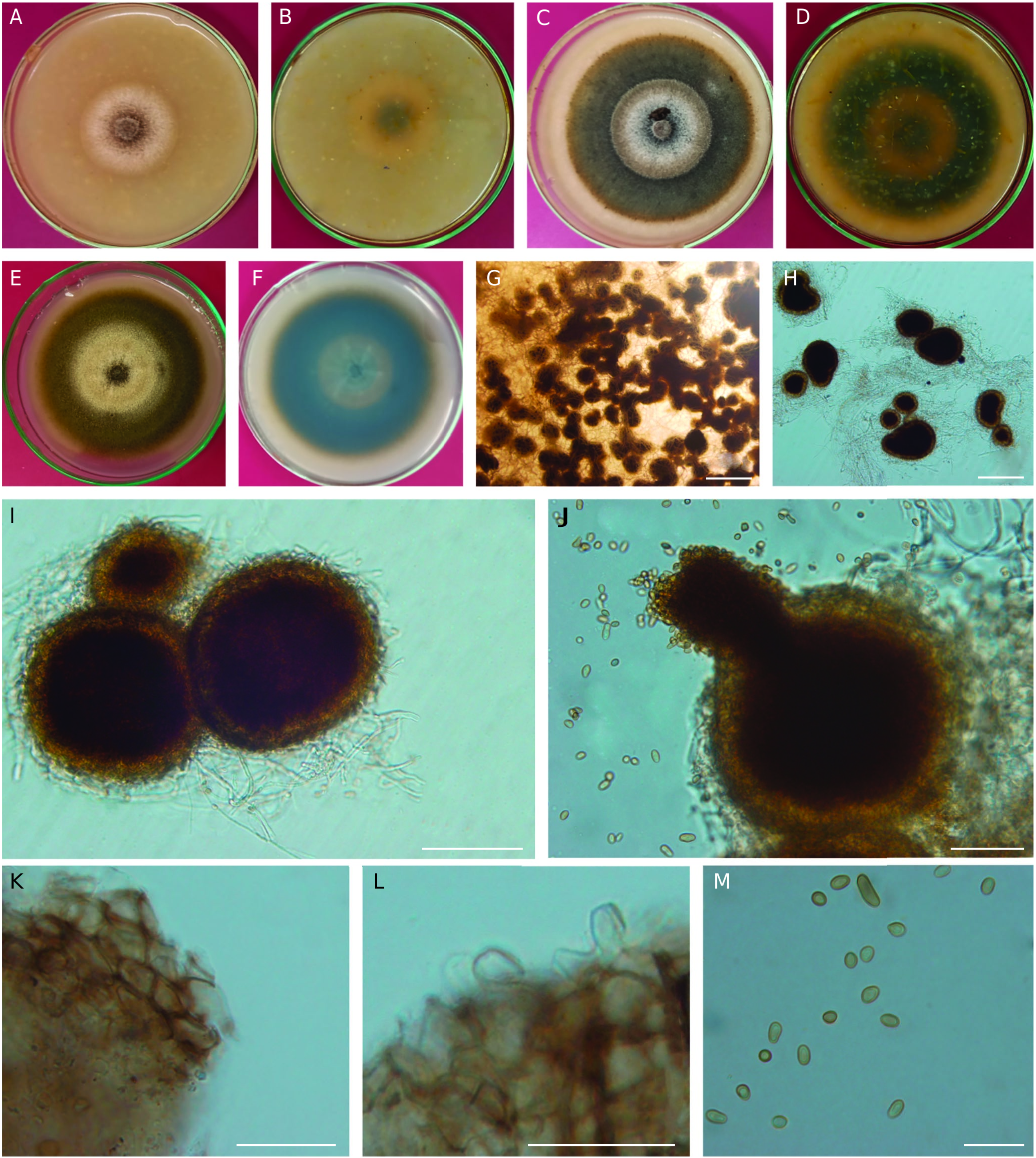
Paramicrosphaeropsis amygdalus M.Mehrabi-Koushki, S.Artand, K.D.Hyde & Jayaward. , sp. nov.
( Fig. 2 View FIG )
HOLOTYPE. — Iran. Khuzestan Province, Dezful, Shahiyoon (forest mountains of Segeryoo), from stem canker of Amygdalus scoparia, I.2019, S. Artand (holo-, IRAN [18146F]; ex-type living culture, IRAN [4451C] = SCUA-Ar-KS3-3).
ADDITIONAL SPECIMENS EXAMINED. — Iran. Khuzestan Province, Dezful, Sardasht (forest mountains of Saland-Kooh), from stem canker of Amygdalus scoparia, III.2021, S. Artand ( IRAN [4452C] = SCUA-Ar-SB2A); from leaf spot of an unknown tree, III.2021, S. Artand (SCUA-Ar-S6A).
ETYMOLOGY. — The name refers to the host genus Amygdalus L. from which it was isolated.
MYCOBANK. — MB 841492.
DESCRIPTION
Asexual morphology
Pycnidia scattered and irregular, solitary or aggregated, superficial on the medium or in aerial mycelium, globose to subglobose, sometime with a short neck, covered with hyphal outgrowths, usually inconspicuous ostiole, sometime with a conspicuous ostiole, slightly papillate or non-papillate, brown to dark brown with a paler wall, (107.2-)159.6-186.6(-268) × (102.2-)144.1-166.4(-241.2) µm, (x ± SD = 173.7 ± 6.7 × 155.6 ± 5.7 µm, n = 50). Pycnidial wall pseudoparenchymatous, composed of isodiametric to elongated cells, 3-6 layers, pale brown to brown, outer layers darker. Conidiogenous cells phialidic, hyaline, smooth-walled, discrete, ampulliform or doliiform. Conidia mostly ovoid to ellipsoidal but also allantoid, or irregular in shape, pale brown to brown, straight or curved, smooth- and thin-walled, guttulate, aseptate, (3.7-)5.9-6.9(-10.5) × (3.2-)4.0-4.3(-5.8) µm, (x ± SD = 6.3 ± 0.3 × 4.2 ± 0.1 µm, n = 50). Chlamydospores and swelling cells not observed.
Sexual morphology
Not observed.
| IRAN |
Iranian Research Institute of Plant Protection |
No known copyright restrictions apply. See Agosti, D., Egloff, W., 2009. Taxonomic information exchange and copyright: the Plazi approach. BMC Research Notes 2009, 2:53 for further explanation.