
Oculudentavis naga, Bolet & Stanley & Daza & Arias & Cernanský & Vidal-García & Bauer & Bevitt & Peretti & Evans, 2021
|
publication ID |
https://doi.org/ 10.1016/j.cub.2021.05.040 |
|
DOI |
https://doi.org/10.5281/zenodo.5013967 |
|
persistent identifier |
https://treatment.plazi.org/id/714E87AE-6E11-FFB4-9E66-FC95FC8FF875 |
|
treatment provided by |
Tatiana |
|
scientific name |
Oculudentavis naga |
| status |
sp. nov. |
Oculudentavis naga new species
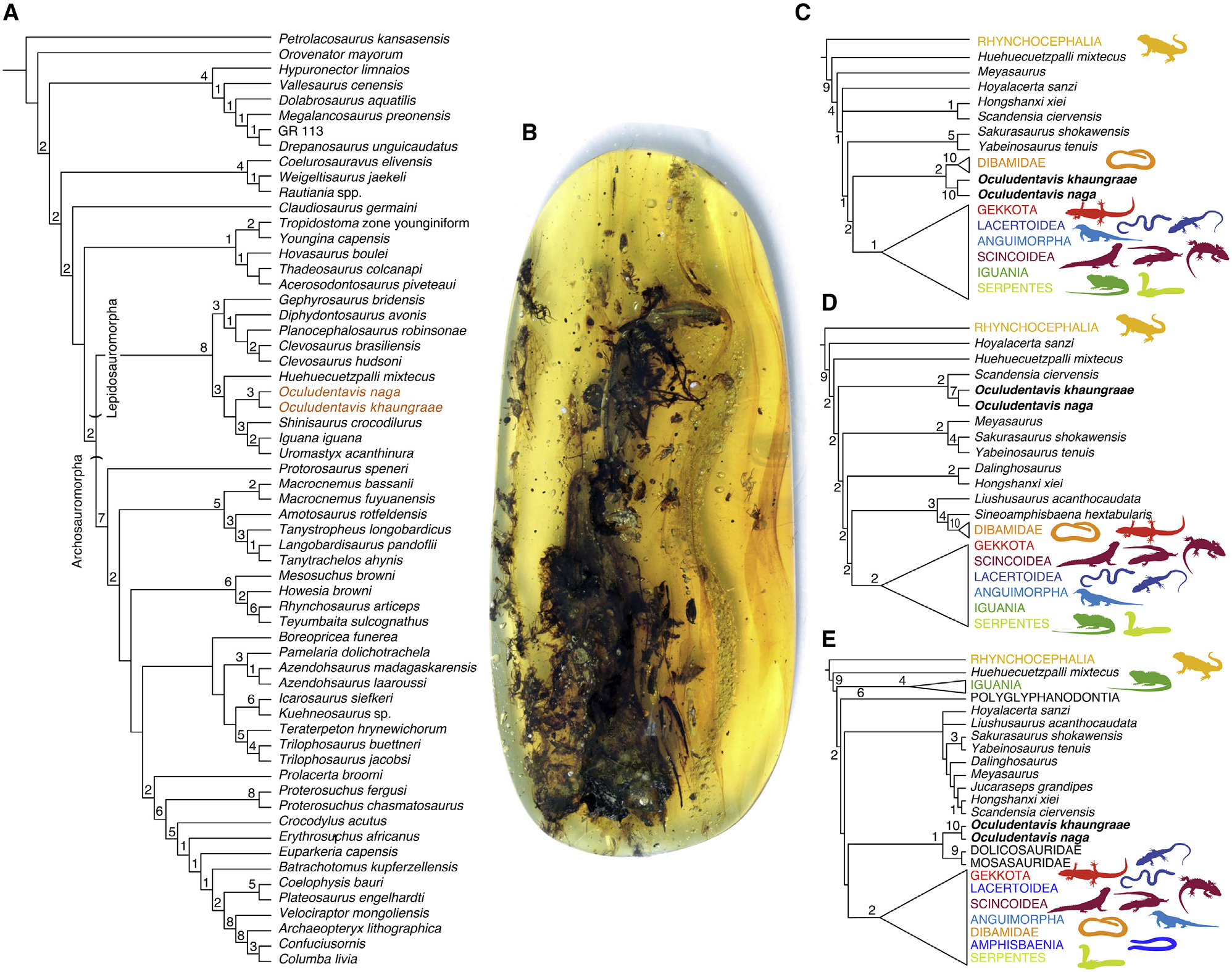
Holotype Peretti Museum Foundation, GRS-Ref-28627 , a skull and anterior postcranial skeleton ( Figures 1 View Figure 1 , 2A–2J View Figure 2 , 3A, 3C View Figure 3 , and S 1 View Figure 1 ). Three-dimensional model of new specimen available at https:// tinyurl.com/Oculudentavis-L-10420.
Type locality
The holotype specimen of Oculudentavis naga ( GRS-Ref-28627 ) and the holotype of O. khaungraae (HPG-15-3) were recovered from the same mine ( Aung Bar mine , 26 O 09 0 N, 96 O 34 0 E) .
Etymology
Combination of Oculudentavis (oculus = eye, dentes = teeth, and avis = bird) 1 and Naga, thename of oneof the many ethnic tribes living in the Burmese amber mines area. The Naga are mentioned in historical chronicles for their prominent role in amber trading. Divided into many sub-groups scattered across the hills and jungle of India (in Nagaland and other states) and in the Tiger valley region of Burma (where amber deposits are found), the Naga tribes are also reputed for their rich and fascinating culture.
Diagnosis
The holotype of O. naga (skull length = 14.2 mm) is somewhat smaller than that of O. khaungraae (skull length = 17.3 mm). Oculudentavis naga differs from O. khaungraae in having a jugal process of the maxilla that reaches caudally to less than 25% of orbit length; in having a long squamosal process of the postorbital; in having a relatively smaller braincase, with short, distally expanded basipterygoid processes (versus longer, unexpanded processes); and in having anterior palatal rami of pterygoids parallel, diverging posteriorly just behind the fossa columellae, interpterygoid vacuity nearly rectangular (versus divergent pterygoids, heart-shaped vacuity), rostral part of premaxilla shorter and proportionally wider than that of O. khaungraae , and less conspicuous platform on the dorsolabial surface of the posterior third of the dentary.
Notes
There are also differences between the two specimens in the robusticity of the postorbital (greater in O. khaungraae ); the height of the premaxillary crest (greater in O. naga ); the extent of the nasal emargination of the frontal (greater in O. naga ); the presence of a large anterior palatine fenestra ( O. naga ); the length and height of the coronoid process (larger and taller in O. naga ); the shape of the quadrate conch (more angular in O. khaungraae ); and in the overall shape of the rostrum (more pointed in O. khaungraae ) and postorbital skull (more vaulted in O. khaungraae ). Oculudentavis naga also displays a very large palatal fenestra between the vomers and palatines. This region is poorly preserved in the holotype of O. khaungraae , and the presence or absence of the fenestra cannot be determined. However, it is possible that at least some of these differences between the two specimens are due to a combination of individual variation, taphonomical deformation (also rendering some elements difficult to segment precisely), and perhaps sexual dimorphism (comparing a male of one species with a female of another could exaggerate interspecific differences like the premaxillary crest height). With only a single specimen of each species, individual variation is impossible to assess.Also note that the skull of O. khaungraae was reported as measuring 14 mm in length, 1 whereas our own measurement of the specimen gives a length of 17.3 mm.
Description
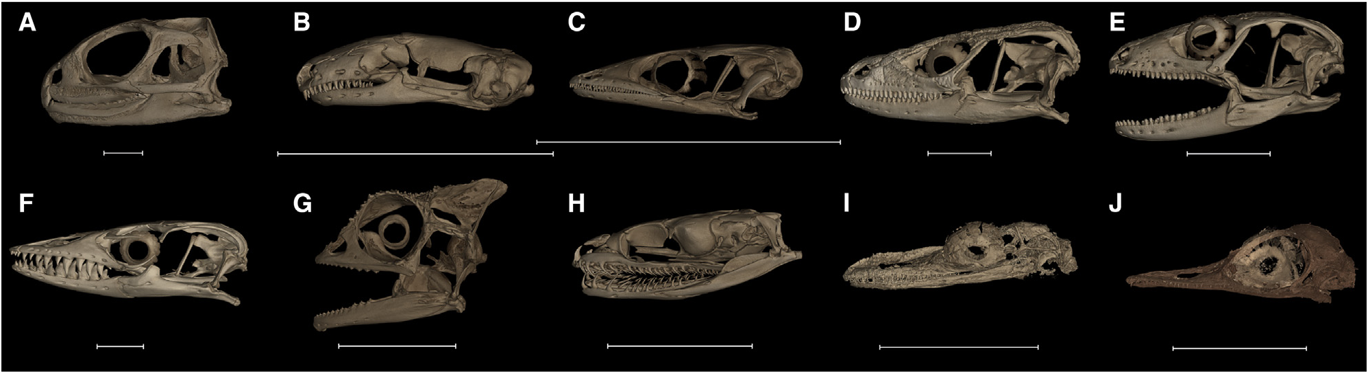
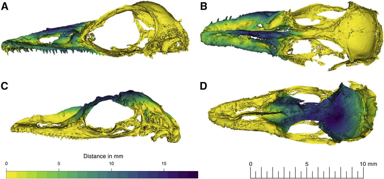
In its bird-like shape (vaulted cranium and tapering rostrum), the skull of Oculudentavis khaungraae appears strikingly different from that of any known lizard ( Figure 4 View Figure 4 ). The bird-like appearance is less striking in O. naga , which has a less compressed rostrum ( Figure 2 View Figure 2 ).
Despite thecompression of the rostrum,the two speciesshare many characters that distinguish them from other lizards. The nares are bounded by the premaxilla anterodorsally, the maxilla posteroventrally, and by the nasals posteriorly. The location of the nares is also the same, being placed at mid-length of the antorbital region and in having an elongated oval shape. The orbit is more intact in O. khaungraae , being nearly circular. In both species, the longest axis of the orbit is about 1/3 the total length of the skull and the orbit is complete and separated from the temporal fenestrae by a complete postorbital bar. The parietal supratemporal processes are aligned with the long and slender vertical supratemporals and fail to meet the squamosals ventrally. There is a complete circumorbital series in both specimens—jugal (ventrally), lacrimal (anteriorly), prefrontal (anterodorsally), frontal (dorsal), postfrontal (posterodorsally), and postorbital (posteriorly).
Premaxilla ( Figures 2, S2A–S2B, S 2I, and S2J View Figure 2 ). The upper jaw comprises an unpaired median premaxilla with slender, pointed teeth (9 in O. khaungraae and ~ 10 in O. naga ; count refers to one side of the element). The more anterior premaxillary teeth appear recurved in O. khaungraae , but the equivalent teeth in O. naga are partially obscured. Both species have a long crest along the premaxillary nasal process (the crest was considered taphonomic in O. khaungraae ), 1 which continues onto the nasals. The palatal shelf is broad and flat and has two narrow palatal processes that bound a large premaxilla-vomer fenestra in O. naga , which has a less deformed palate. The palatal processes of the premaxilla are also visible in O. khaungraae , but not the intervening fenestra (see below).
Maxilla ( Figures 2, S2C–S2E, and S2K–S2M View Figure 2 ). The maxilla of both species has a low, medially curved facial process, a long rostral component, and a suborbital ramus that does not reach the posterior margin of the orbit—extending up to one-quarter of the orbit length in O. naga and to about mid-orbit in O. khaungraae . It is excluded from the orbital rim by the jugal. The maxillary teeth are conical and pointed. There are 24 to 25 tooth maxillary loci in O. naga and 27–29 in O. khaungraae . The maxilla has two horizontal facets: one to support the prefrontal and another for the jugal.
Nasal ( Figures 2, S2F, and S2N View Figure 2 ). The paired nasals form a rhomboid plate, and combined with the maxilla, they define a long tapering rostrum with retracted narial openings. The nasals are paired, but they exhibit partial fusion along the crest and remain separated posterior to the crest.
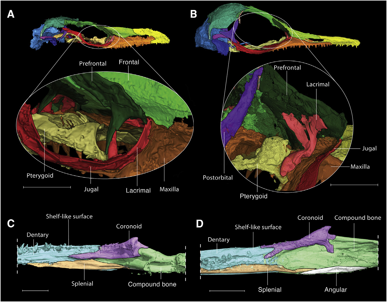
Prefrontal ( Figure 2 View Figure 2 ).The prefrontalscomprisea flatanterodorsal plate and a weakly concave orbital plate, contacting the ringshaped lacrimal ventrally. The anterodorsal plate seems less developed in O. khaungraae than O. naga . The lateral edge of the anterodorsal plate projects as a short angular ( O. naga ) or ridgelike ( O. khaungraae ) shelf that overhangs the lacrimal and maxilla. This shelf is autapomorphic among lizards, with a ridge, crest, or boss in this position variably present (e.g., some iguanians, including chameleons; some Phrynosoma ; and some Anolis ).
Lacrimal ( Figures 3A and 3B View Figure 3 ). The lacrimal of both species is unique among lizards and is one of several distinctive features that demonstrates their close relationship. It forms a ring, completely enclosing a large lacrimal foramen.
Jugal ( Figures 2, S2G, S2H, S2O, and S2P View Figure 2 ). In both species, the jugal forms a dorsomedially expanded flange that provides ventral support to the large eye. The orientation of the jugal is unusual for squamates, being dorsomedially inclined. The postorbital process of the jugal is short (distorted on the right side of O. naga ).
Frontal ( Figures 2 View Figure 2 , S 3A, and S3E View Figure 3 ). In both species, the unpaired median frontal has weak sub-olfactory processes and a deep V-shaped anterior emargination that receives the nasals. The frontal is overlapped extensively by the nasals, reaching the level of the mid-orbit in O. naga and somewhat less in O. khaungraae . The supraorbital margins are subparallel and diverge posterolaterally, establishing a broad contact with the anterior margin of the parietal. The structure of the posteromedial margin of the fronto-parietal suture is unclear in both specimens ( Figure 2 View Figure 2 , dashed lines).
Parietal ( Figures 2 View Figure 2 , S 3B, and S3F View Figure 3 ). The parietals are short and partially fused (separated posteriorly). They have a rounded lateral profile, lack a parietal foramen, and have short supratemporal processes that curve ventrally rather than posteriorly to meet the supratemporals. This portion of the skull contacts the short paroccipital processes of the otoccipital. Li et al. 3 argued that the small opening in the midline of the parietals in the holotype of O. khaungraae corresponds to a parietal foramen, but it is irregular and appears to be an artifact of breakage.
Postfrontal ( Figure 2 View Figure 2 ). The postfrontal is a small, splint-like bone, lateral to the frontal and the parietal in O. khaungraae but of uncertain structure and position in O. naga . The postfrontals are very reduced in both species and were not noticed in the original description of O. khaungraae .
Postorbital ( Figures 2 View Figure 2 , S 3C, and S3G View Figure 3 ). The postorbital is a strongly triradiate bone with a long ( O. naga ) or short ( O. khaungraae ) posterior process that contacts the squamosal posteriorly. The postorbital differs in the two species: the postorbital squamosal process tapers gradually in the O. naga holotype, while the tapering appears more abrupt in the O. khaungraae specimen. Due to the proportionally thicker postorbital, the right side of O. khaungraae shows a more extensive contact between the postorbital and the descending process of the parietal, entirely covering the braincase laterally and almost completely closing the upper temporal fenestra. However, on the left side, it is clear that this fenestra remained open. In O. naga , the upper temporal fenestra looks larger, but the skull table of this specimen is very depressed and the postorbital is more gracile, so the differences in configuration of the upper temporal bar may be exaggerated by taphonomic deformation.
Squamosal ( Figures 2 View Figure 2 , S 3D, and S3H View Figure 3 ). In both species, the typically squamate hockey-stick-shaped squamosal lacks an ascending process and lies between the supratemporal, the postorbital, and the quadrate.
Supratemporal ( Figures 2 View Figure 2 , S 3B, and S3F View Figure 3 , articulated with the parietal). The supratemporal is also reduced to a slender vertical splint of bone that contacts the lateral margin of the parietal supratemporal process, separating it from the squamosal.
Palate ( Figures 2 View Figure 2 , S 4A, and S4D View Figure 4 ). In the palate of O. khaungraae , the premaxilla-vomer fenestra is totally obliterated (due to compression). In this respect, O. naga has a more intact rostrum, more clearly exhibiting thepremaxilla-vomer fenestra and the very large fenestra exochoanalis. The suborbital fenestra is oval in both specimens and is bounded by the same bones: palatines anteromedially, ectopterygoids laterally, and pterygoids posteriorly, although the sutures between these bones are not easy to identify. It also looks as if the ectopterygoid barely contacts the palatine in O. naga , but the degree of contact is ambiguous in O. khaungraae . The shape of the interpterygoid vacuity differs between the two species. Pterygoid teeth are present and are arranged in a row on the anteromedial process of the pterygoid, just posterior to the inferred suture with the palatine. There are about 3 to 4 on each bone in O. khaungraae ; the same area is fragmented in O. naga , but small projections on both pterygoids can be interpreted as pterygoid teeth.
Quadrate ( Figures 2 View Figure 2 , S 4B, and S4E View Figure 4 ). The quadrate is distinctively low in position and small in size in both species. The quadrate is stouter in O. khaungraae (with a more prominent head) than in O. naga , but the overall shape is similar in both specimens, with a shallow conch, a slightly curved medial pillar, and a lateral tympanic crest that has a 90-degree angulation along its length. The quadrate suspension in both species is characteristically squamate.
Braincase ( Figures 2 View Figure 2 and S 5 View Figure 5 ). By comparison with that of O. khaungraae , the braincase of O. naga is unevenly dorsoventrally compressed, so that the right side is more damaged than the left and the posteroventral margin is abnormally low. Nonetheless, comparison of the two braincases shows more similarities than differences, notably the well-developed crista prootica, short alar processes, slender basipterygoid processes, short basisphenoid, enclosed vidian canals opening posteriorly within the basisphenoid, robust parasphenoid rostrum (base only preserved in O. naga ), short uncrested supraoccipital with a visible processus ascendens (mineralization uncertain), and short paroccipital processes. The parasphenoid rostrum is well preserved in O. khaungraae , being longer than the basipterygoid processes, and almost entirely divides the interpterygoid vacuity. In O. naga , the parasphenoid rostrum is represented only by its base, possibly due a fracture or weak mineralization. However, there are differences in the orientation, length, and distal shape of the basipterygoid processes in the two species.
Epipterygoid ( Figure 2 View Figure 2 ). These elements are poorly preserved and displaced in both species. They are columnar and still in articulation within the fossa columellae of the pterygoid, this articulation being another uniquely squamate character. In the holotype of O. naga , a portion of the left epipterygoid remains attached to the alar process.
Scleral ossicles ( Figures 1 View Figure 1 , S 4C, and S4F View Figure 4 ). In both species, the orbit contains a large ring of ‘‘spoon-shaped’’ scleral ossicles that supported a large eye. The ossicle count is 14 in both specimens. Due to the distinctive shape of the ossicles, they overlap at both their inner edges (which would have surrounded the iris and the pupil) and the outer edges, leaving oval gaps between ossicles in the middle of the sclerotic ring. Although the skull of O. khaungraae is 1.2X longer than that of O. naga , the scleral ossicles are proportionally larger in O. khaungraae , being 1.5X larger than those of O. naga .
Dentary ( Figures 2 View Figure 2 , 3C, 3D View Figure 3 , and S 6 View Figure 6 ). Both species have a long shallow mandible of which the straight dentary forms the major part (~ 75%) and a large number of sharp, weakly pleurodont teeth (29 to 30 in both specimens). Both speciesalso have a large number of lateral neurovascular foramina (10–12), and the dentary in each specimen has parallel upper and lower margins. Thesymphysealregiondoes not extendbeyondthe second tooth locus in either specimen. The lower margin of the dentary curves dorsomedially and closely approaches the subdental shelf, thus restricting the Meckelian fossa but without fusion.The Meckelian fossa remains open posteriorly, where it is overlapped by the splenial. The dorsolabial surface of the posterior one-third of the dentary bears a flattened, shelf-like surface.
Splenial ( Figures 2 View Figure 2 and S 6 View Figure 6 ). The splenial is very slender and does not extend anteriorly beyond the posterior one-third of the dentary, closing only the posterior part of the Meckelian fossa in both species. Posteriorly, the splenial does not extend beyond the level of the coronoid eminence.
Coronoid ( Figures 2 View Figure 2 and S 6 View Figure 6 ). The postdentary region is short, including a coronoid with a low, posteriorly set, coronoid eminence. The coronoid looks significantly larger in the holotype of O. naga than in the holotype of O. khaungraae , especially in the development of the anterolateral and anteromedial processes. However, these differences could be due to damage during deformation, making it difficult to establish clear bone boundaries (e.g., between surangular and coronoid), as this was one of the most problematic regions to segment in both specimens.
Angular ( Figures 2 View Figure 2 and S 6 View Figure 6 ). This is a very reduced and slender bone, limited to the posteroventral side of the jaw.
Compound bone ( Figures 2 View Figure 2 and S 6 View Figure 6 ). There is no obvious suture between the surangular and the articular or prearticular in either specimen. Both specimens have a long retroarticular process and a short, deep adductor fossa. It is uncertain whether the coronoid reached the anterior margin of the adductor fossa.
Although only part of the postcranial skeleton is preserved in O. naga , it shows a short neck with eight cervical vertebrae that are amphicoelous, atlantal arches bearing posterior zygapophyses, and a pectoral region comprising a T-shaped interclavicle, medially expanded clavicles, and a typically squamate scapulocoracoid with scapular, scapulocoracoid, and primary coracoid fenestrae.
Vertebrae ( Figures S7Aand S7B View Figure 7 ). Eight cervical vertebrae are preserved, including the atlas and the axis, as well as a small number of dorsal vertebrae (using the traditional anatomical definition whereby the first dorsal vertebra is that with a rib that meets the sternum, contra Gauthier et al. 7). The atlantal arches are not fused, and they have well-developed postzygapophyses. The axis preserves the dens, which is already fused in place. The vertebrae are amphicoelous and notochordal, with low neural spines. There are simple semicircular intercentra visible in the anterior part of the neck, with only a weak ventromedian keel ( Figure S7 View Figure 7 ). As in living gekkotans, these elements are free and intercentral in position. The first visible cervical rib is on cervical six, but there may have been ribs more anteriorly. There are no gastralia.
Clavicle ( Figure S7C–S7E View Figure 7 ). The clavicles are expanded medially and have a well-defined clavicular fenestra completely enclosed by bone. The clavicles are separated at the ventral midline by tip of the T-shaped interclavicle. Dorsally, the clavicles appear to extend above the level of the scapula blade, possibly meeting a suprascapular cartilage.
Scapulocoracoid ( Figures S7C–S7E View Figure 7 ). Both scapulocoracoids are preserved and display an anterior primary coracoid emargination, anemarginated scapular blade, and a large circular scapulocoracoid emargination. Dorsal to the scapula, there is an irregular mass that may represent the suprascapular cartilage.
Sternum ( Figure S7D View Figure 7 ). Only the anterior border of the cartilage sternum is preserved, suggesting it was rhomboid.
Interclavicle ( Figure S7D View Figure 7 ). The interclavicle is T-shaped and quite robust.
Humerus ( Figure S7D View Figure 7 ). The proximal portion of the left humerus is present, preserving the humeral head and the lateral tuberosity.
Soft tissue ( Figure 1 View Figure 1 ; Data S1, Gular scales in Oculudentavis ). Both specimens also preserve soft tissue. The head and body are covered in small, granular scales, with large rectangular supralabial and infralabial scales, tiny scales covering the eyelid, and a nostril placed anterior to the midpoint of each retracted narial opening ( Figures 1 View Figure 1 and 2 View Figure 2 ) in O. naga . There are no osteoderms. On the ventral surface of the head in O. naga , along the midline, the epidermal scales are raised and form a line of evenly spaced short ridges. Posterior to this midventral line, the skin of the gular region is thrown into a series of narrow linear folds. This folded region underlies the hyoid ceratobranchials and may demonstrate the resting anatomy of loose gular skin that could be inflated, for example in territorial display,in associationwith hyoidmovements.


No known copyright restrictions apply. See Agosti, D., Egloff, W., 2009. Taxonomic information exchange and copyright: the Plazi approach. BMC Research Notes 2009, 2:53 for further explanation.