
Pseudabia fusca Schrottky, 1910
|
publication ID |
https://doi.org/ 10.5852/ejt.2018.482 |
|
publication LSID |
lsid:zoobank.org:pub:6F3B12C7-2311-48EA-8727-5B90489E26E3 |
|
DOI |
https://doi.org/10.5281/zenodo.3846149 |
|
persistent identifier |
https://treatment.plazi.org/id/723287A9-5723-F009-B32B-B6859159F52B |
|
treatment provided by |
Valdenar |
|
scientific name |
Pseudabia fusca Schrottky, 1910 |
| status |
|
Pseudabia fusca Schrottky, 1910
Pseudabia fusca Schrottky, 1910: 168 .
Enslinia holmbergi Jörgensen, 1913: 254 .
Material examined
ARGENTINA: – Misiones: 1 ♀, Bonpland, 7 Nov. 1910, Jörgensen leg. ( MLPA; holotype of Enslinia holmbergi ).
BRAZIL: – Santa Catarina: 1 ♀, Nova Teutonia, 27°11' S, 52°23' W, 9 Sep. 1938, Fritz Plaumann leg. ( NHML); 1 ♀, same collecting data as preceding but Sep. 1972 ( NMNH). – Santa Catarina: 1 ♀, Nova Teutonia, 7 Nov. 1969, Fritz Plaumann leg. ( NMNH).
Redescription (female, male unknown)
MEASUREMENTS. Large and elongate sawfly, body length 15–16 mm.
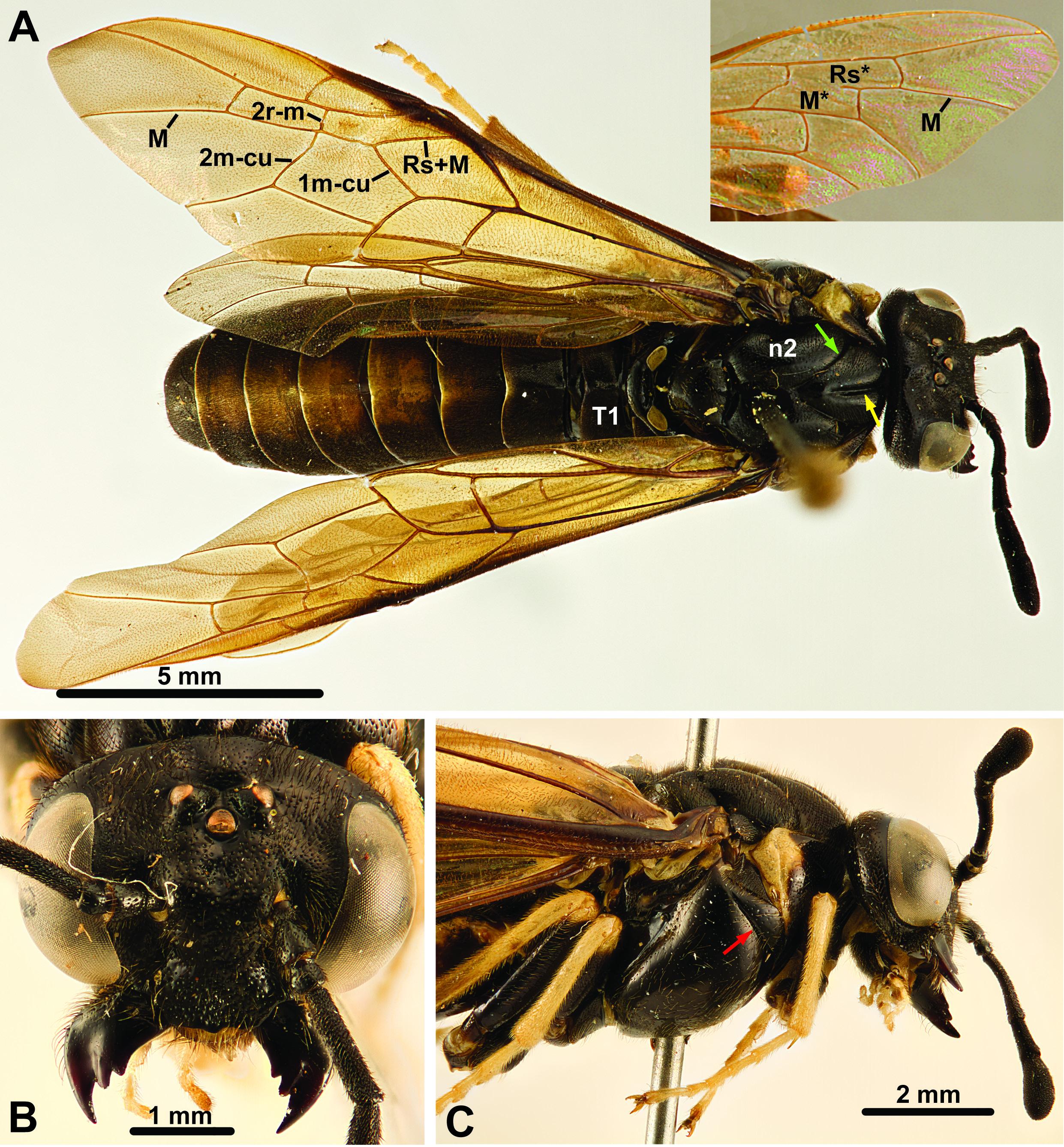
COLOR. Coloration uniformly black to dark brown with a few lighter brown to creamy-white areas on lateral parts of pronotum, posteriorly on dorsal part of abdominal terga and on lateroterga ( Fig. 13A View Fig ). Legs proximally dark brown to black, creamy white from distal part of femora. Wings weakly and uniformly infuscate.
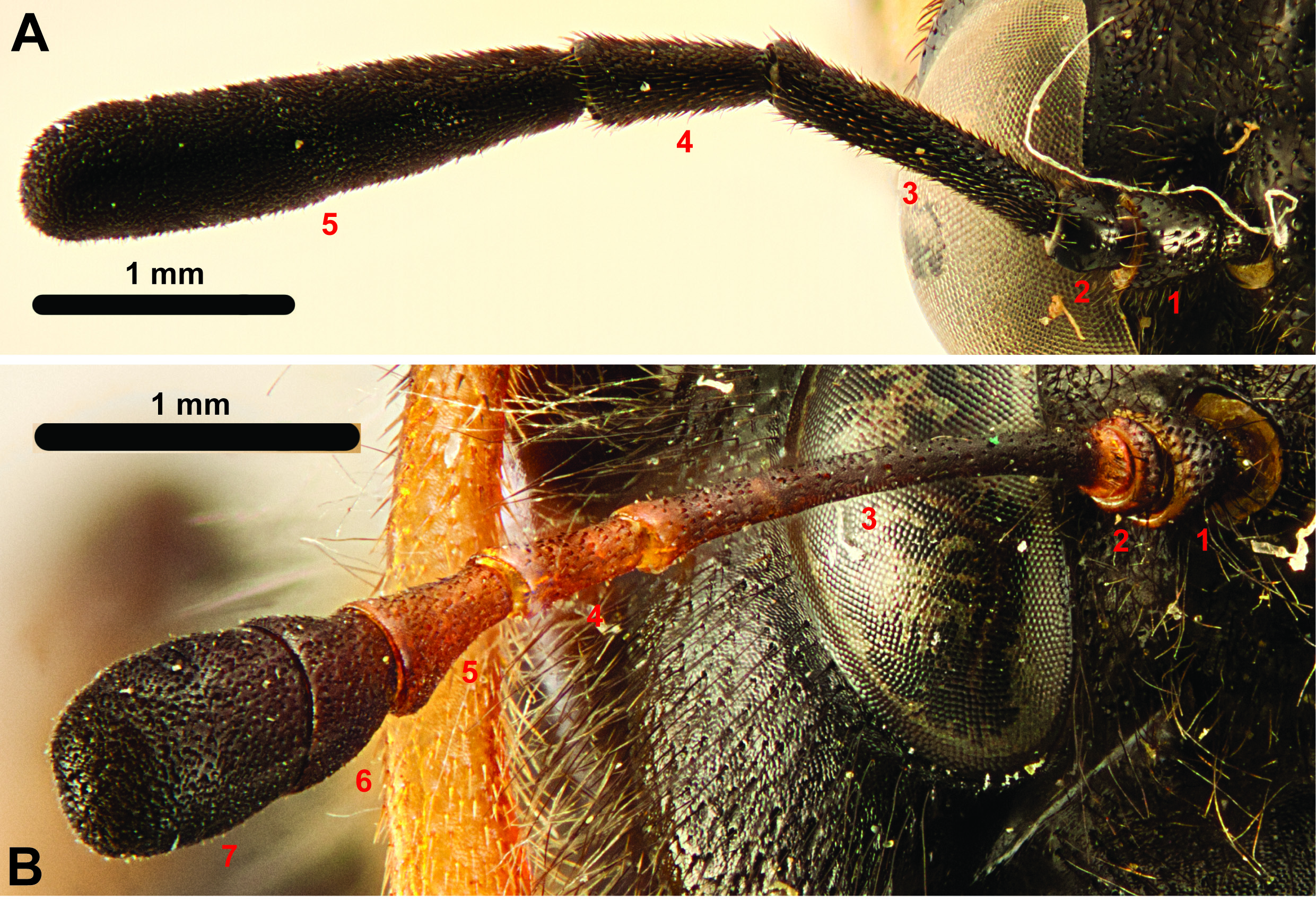
HEAD. Eyes slightly converging ventrally, inner margins slightly incurved. Posterior ocelli at level with dorsal margin of eyes. Toruli in middle of face, slightly closer to median ocellus than to ventral margin of clypeus ( Fig. 13B View Fig ). Epistomal sulcus absent. Clypeus ventral margin straight, with slight protrusion in the middle. Gena medially less wide than width of eye, wider dorsally than ventrally. Malar space very low. Occipital carina absent. Sclerotisation between occipital and oral foramina present. Antennae with five antennomeres ( Figs 1A View Fig , 13C View Fig ); antennomere 1 approx. 2× as long as wide, antennomere 2 approx. as wide as long, antennomere 3 1.8× as long as antennomere 4, antennomere 5 slightly expanded distally. Labrum broad, convex, hairy, flat, distal margin slightly curved. Mandibles less than ½ the height of head, with three teeth each, inner margin not serrated ( Fig. 13B View Fig ). Maxilla with stipes of approx. equal width throughout, palps with six palpomeres, longer than labial palps. Labial palps with four palpomeres, palps inserting at level with maxillary palps. Postmentum drop-shaped, approx. 2× as long as wide.
THORAX. Pronotum comparatively high medially, with transverse groove extending from submedially on pronotum to posterodorsal corner; lateral groove continuous with transverse, extending ventrally on pronotum until ventral to anterior thoracic spiracle where it terminates; pronotum fused with mesopleuron ventrolaterally of spiracle from halfway between spiracle and ventrolateral corner of pronotum. Dorsal cervical sclerite present. Propectus without lateral projection, propleural sulcus present, medioventral margins of propectus widely separated, posteriorly extended into narrow points. Prosternum laterally extended, continuous with katepisternum which articulate, but does not fuse with propleuron at lateral coxal articulation point. Anterior fore tibial spur straight, simple, not much longer than posterior spur, spurs pointed and sclerotized apically. Mesonotum with distinct median sulcus and deep notauli ( Fig. 13A View Fig ), laterophragmal apodeme not observed, postscutellum absent. Small anterodorsal part of mesopleuron separated from rest by vertical groove ( Fig. 13C View Fig ), prepectus absent as separate sclerite. Horizontal carina absent laterally on mesopleuron. Posterior thoracic spiracle visible in lateral view, situated in incurvation in dorsal margin of mesopleuron. Median midcoxal articulations adjacent, only separated by small wedge of cuticle. Mesofurca not observed. Insertion of mesonotometanotal muscle on small projection on anterior margin of metanotum; cenchri approx. 2× as broad as long. Anapleural cleft present, small. Metapleuron fused with abdominal tergum 1 along dorsal margin; posteroventral metapleural apodeme not observed; paracoxal sulcus curving posteriorly, terminating in the middle of the metapleural sulcus; metacoxal foramina open dorsally, without metapleural inflection laterally. Metafurca not observed. Hind coxa less than twice as long as wide, median carina or spine not observed. Hind femoral ventral spur absent. Hind tibial apical spurs shorter than apical width of tibia. Hind basitarsomere shorter than tarsomeres 2–4, tarsal claws bifid, teeth of subequal length.
WINGS. Fore wing with vein M joining Sc+R close to Rs+M; vein 2r-m posteriorly inserts on cell 2M, very close to anterior end of 2m-cu ( Fig. 13A View Fig ); vein 1m-cu oriented obliquely, inserting on Rs+M some distance from 2r-m (distance 1m-cu–2r-m at least ½ distance M–1m-cu on Rs+M); posterior anal vein present proximally and distally but discontinuous in the middle. Hind wing cell R1 closed; vein M discontinuous, making cells Rs and M partly confluent ( Fig. 13A View Fig insert), cell M not reaching vein Rs; cross vein 2a absent.
ABDOMEN. Tergum 1 not subdivided medially ( Fig. 13A View Fig ), short median carina present, lateral carina absent, posterior margin with slight incurvation medially; tergum 1 not brighter colored than other abdominal terga. Metaphragma not observed. Lateroterga around abdominal spiracles separated from median terga as demarcated by conspicuous fold. Cerci triangular, length approx. equals width at base. Ovipositor apparatus not observed; Smith (1988: fig. 46) illustrates 1 st valvula, with longitudinal line, vertically oriented sawteeth with serrulae, and tip slightly curving dorsally.
Comments
The holotype appears to be lost ( Smith 1988), according to Malaise (1939: 27) “All the types of Schrottky have been destroyed in a civil war”. The type locality given by Schrottky (1910) is Puerti [sic, probably Puerto] Bertoni, Paraguay. Malaise (1939) also listed Nova Teutonia, Brazil, as a locality. Jörgensen (1913) described Enslinia holmbergi from Bonpland, Misiones, Argentina; Conde (1932) synonymized this species with Pseudabia fusca , a decision that was followed by Smith (1988). In contrast, Malaise (1939) considered the two taxa should be kept separate, citing differences from the descriptions of Schrottky (1910) and Jörgensen (1913) in relative antennomere proportions head width, mesonotal sculpture and sawsheaths (3 rd valvulae). The present authors follow Smith (1988).
The specimens examined here fit well with the description of Schrottky (1910). The NHML specimen has a broad creamy-white band along each side of the abdomen, covering most of the lateroterga; in contrast, the NMNH specimens have only a narrow stripe along the bend between the median terga and the lateroterga. Jörgensen (1913) and Smith (1988) considered the abdomen/body to have a metallic sheen (“obscure metalico”,'mostly metallic green'). The specimens examined by us look more brownish black, with at most a faint metallic sheen on the abdomen; perhaps the color changes as the specimen ages. Schrottky (1910) and Jörgensen (1913) described Pseudabia fusca / Enslinia holmgreni as having eight or seven antennomeres, respectively, with the apical 3–4 forming the apical club; Smith (1988: fig. 33) illustrated P. fusca as having five antennomeres, with a subdivision in the middle of the club. Indeed several subdivisions can be discerned in the specimens we have examined, but these are never distinct enough to separate antennomeres within the club as clearly as between it and the four proximal antennomeres. Hence the antenna is regarded as comprising five antennomeres, as in all other South American Cimbicidae .
The most distinctive feature of Pseudabia fusca is the interruption of vein M in the hind wings, which makes cells Rs and M partly continuous; this is a unique feature within Cimbicidae . The general habitus is also slightly different from the other South American species in that P. fusca , even though it has a body length similar to Pachylosticta spp., appears to be more slender than the comparatively compact and/or shorter members of the other cimbicid species. Among the South American Cimbicidae , P. fusca is the only species to have a well-developed median carina on abdominal tergum 1.
No known copyright restrictions apply. See Agosti, D., Egloff, W., 2009. Taxonomic information exchange and copyright: the Plazi approach. BMC Research Notes 2009, 2:53 for further explanation.
|
Kingdom |
|
|
Phylum |
|
|
Class |
|
|
Order |
|
|
Genus |
Pseudabia fusca Schrottky, 1910
| Vilhelmsen, Lars, Smith, David R. & Malagón-Aldana, Leonardo A. 2018 |
Enslinia holmbergi Jörgensen, 1913: 254
| Jorgensen P. 1913: 254 |
Pseudabia fusca
| Schrottky C. 1910: 168 |