
Pachylosticta sp., Klug, 1824
|
publication ID |
https://doi.org/ 10.5852/ejt.2018.482 |
|
publication LSID |
lsid:zoobank.org:pub:6F3B12C7-2311-48EA-8727-5B90489E26E3 |
|
DOI |
https://doi.org/10.5281/zenodo.3846163 |
|
persistent identifier |
https://treatment.plazi.org/id/723287A9-572E-F00F-B3C8-B164974CF45D |
|
treatment provided by |
Valdenar |
|
scientific name |
Pachylosticta sp. |
| status |
|
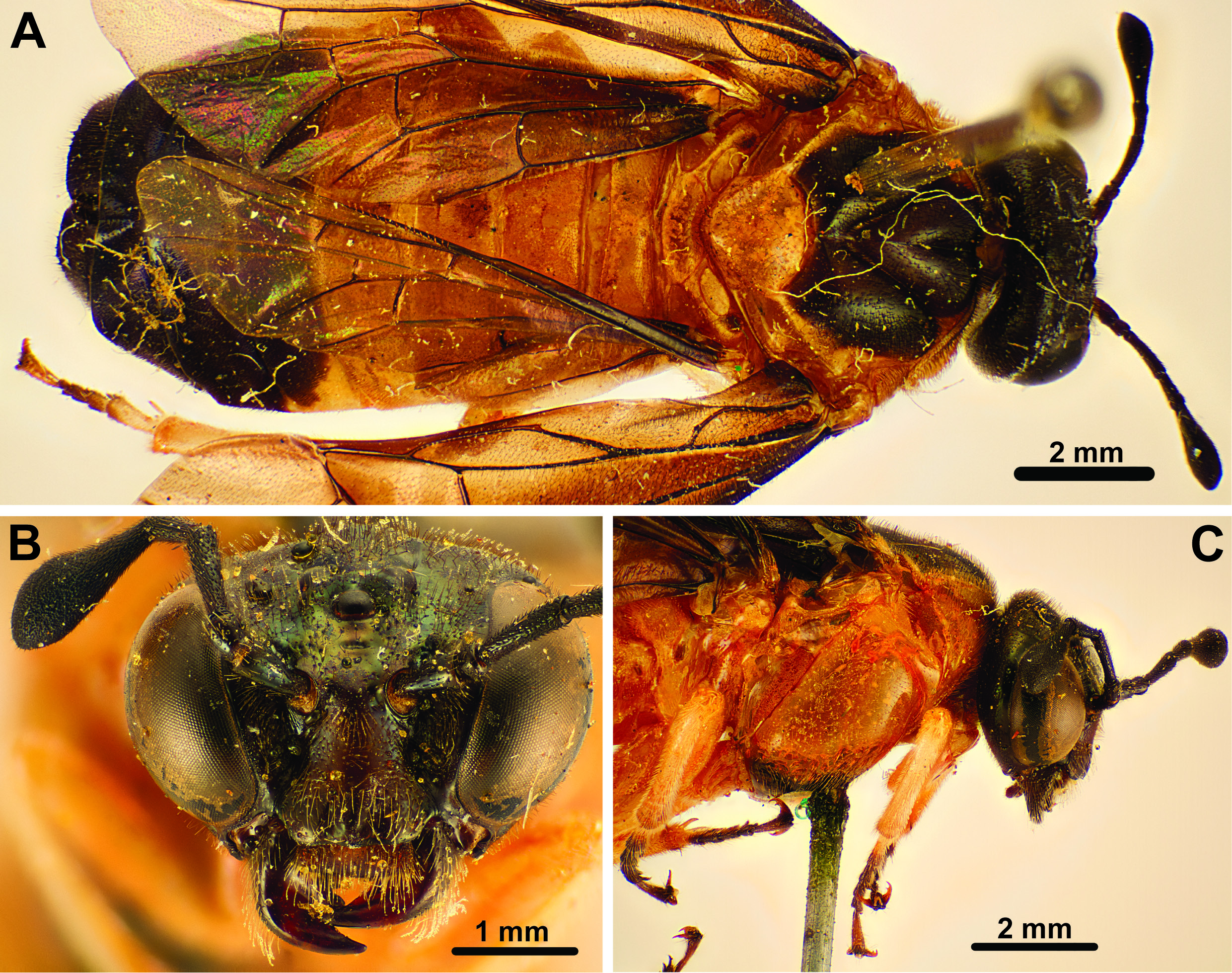
Fig 12 View Fig
Males with partly reddish coloration on thorax or abdomen.
Material examined
BRAZIL: – Espirito Santo: 2 ƋƋ, Córrego do Itá, Nov. 1956, W. Zikan leg. ( MNRJ). – São Paulo: 1 Ƌ, Valle du Rio Pardo, Dec. 1898, E. Gounelle leg. ( A. plaumanni ? det. D.R. Smith, 1986) ( MNHN).
Redescription (female, male structures only mentioned when differing from females).
MEASUREMENTS. Comparatively large and robust sawflies, body length 12–16 mm.
COLOR. Males ( Figs 9 View Fig , 12 View Fig ) with dark blue metallic coloration on most of body, rarely with parts of thorax ( Fig. 12B View Fig ) or abdominal tergum 4 to 6 reddish brown ( Fig. 12D View Fig ). Females ( Figs 8 View Fig , 10–11 View Fig View Fig ) with extensive reddish-brown coloration on large parts of thorax and also often anterior abdomen, remaining body parts dark blue metallic. Wings mostly darkly infuscated in both sexes, in female P. apicalis infuscation distally contrast with more hyaline wing coloration proximally ( Fig. 10A View Fig ).
HEAD. Eyes at most slightly converging ventrally, inner margins at most slightly incurved. Posterior ocelli at level with dorsal margin of eyes ( Figs 8B View Fig , 10B View Fig , 11B View Fig ). Toruli in middle of face, approx. halfway between median ocellus and ventral margin of clypeus. Epistomal sulcus absent. Clypeus with ventral margin straight. Gena medially less wide than width of eye, wider dorsally than ventrally. Malar space very low. Occipital carina absent. Sclerotisation between occipital and oral foramina present. Antennae with five antennomeres; antennomere 1 approx. 2× as long as wide, antennomere 2 approx. as wide as long, antennomere 3 1.2–1.5× as long as antennomere 4, antennomere 5 slightly expanded distally; females of P. albiventris and P. apicalis with elongate, lighter colored area on ventral side of antennomere 5. Labrum broad, convex, hairy, raised in the middle under ventral margin of clypeus, distal margin straight or slightly curved. Mandibles less than ½ the height of head in both sexes, with three well developed teeth on right, proximal tooth less developed on left mandible, inner margin not serrated. Maxilla with stipes of approx. equal width throughout, palps with four palpomeres, shorter than labial palps. Labial palps with three palpomeres, palps inserting at level with maxillary palps. Postmentum narrow, at least 3× as long as wide.
THORAX. Pronotum usually comparatively high medially, with transverse groove extending from submedially on ventral margin to posterodorsal corner of pronotum; lateral groove continuous with transverse, extending parallel to lateral margin of pronotum until ventral to anterior thoracic spiracle where it diverges medially to ventral margin of pronotum; pronotum fused with mesopleuron for short distance ventrolaterally. Dorsal cervical sclerite present. Propectus without lateral projection, propleural sulcus present, medioventral margins of propectus widely separated, posteriorly extended into narrow points. Prosternum laterally extended, katepisterna separate sclerites not reaching lateral coxal articulation point. Anterior fore tibial spur straight, simple, not much longer than posterior spur, spurs pointed and sclerotized apically. Mesonotum with distinct median sulcus and deep notauli ( Figs 8A View Fig , 11A View Fig ), laterophragmal apodeme short, postscutellum absent. Small anterodorsal part of mesopleuron separated from rest by vertical groove ( Figs 8C View Fig , 9A View Fig , 12A View Fig , C–D), prepectus absent as separate sclerite. Horizontal carina absent laterally on mesopleuron. Posterior thoracic spiracle visible in lateral view, situated in incurvation in dorsal margin of mesopleuron ( Figs 8C View Fig , 10C View Fig ). Median midcoxal articulations adjacent, only separated by small wedge of cuticle. Mesofurca with mesospina and elongate anterior and lateral arms. Insertion of mesonoto-metanotal muscle on metanotum not on conspicuous structure; cenchri approx. 2.2–3× as broad as long. Anapleural cleft present, small. Metapleuron fused with abdominal tergum 1 along dorsal margin; posteroventral metapleural apodeme absent; paracoxal sulcus curving posteriorly, terminating in the middle of the metapleural sulcus; metacoxal foramina open dorsally, with slight metapleural inflection laterally. Metafurca with anterior arms hardly developed, lateral arms elongate. Hind coxa less than twice as long as wide, median carina or spine absent. Hind femoral ventral spur absent. Hind tibial apical spurs shorter than apical width of tibia. Hind basitarsomere longer than tarsomeres 2–4 ( Figs 9A View Fig , 12A, C View Fig ), tarsal claws bifid, inner tooth variable.
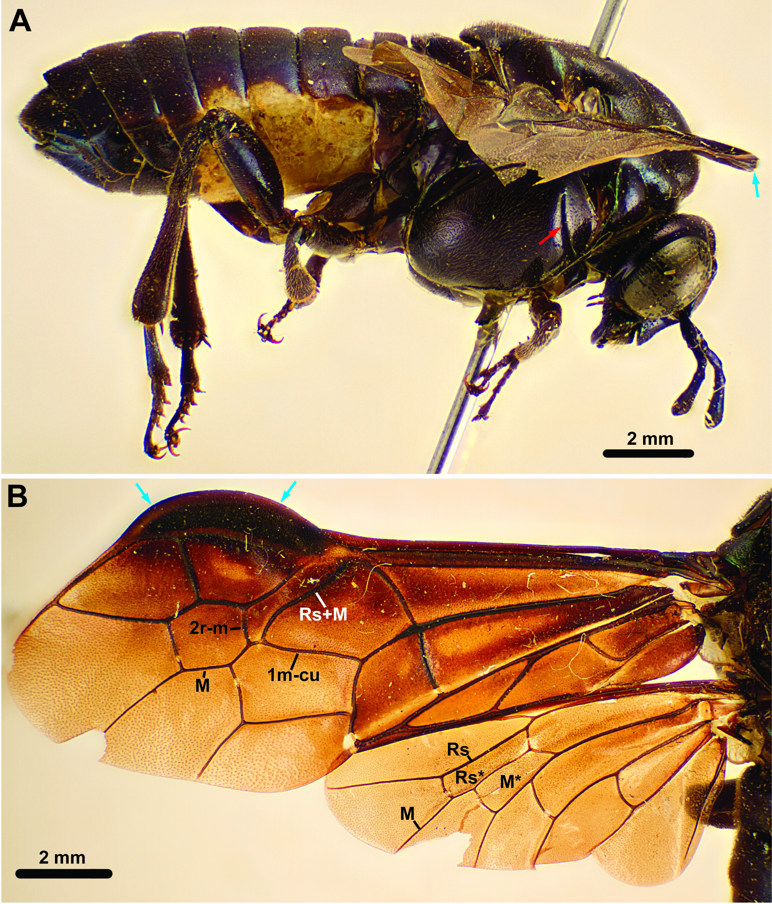
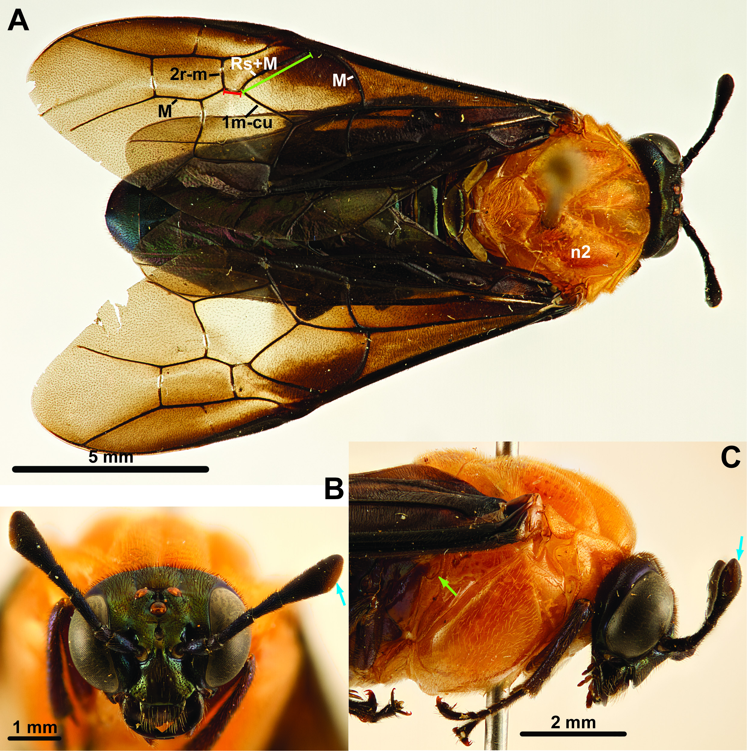
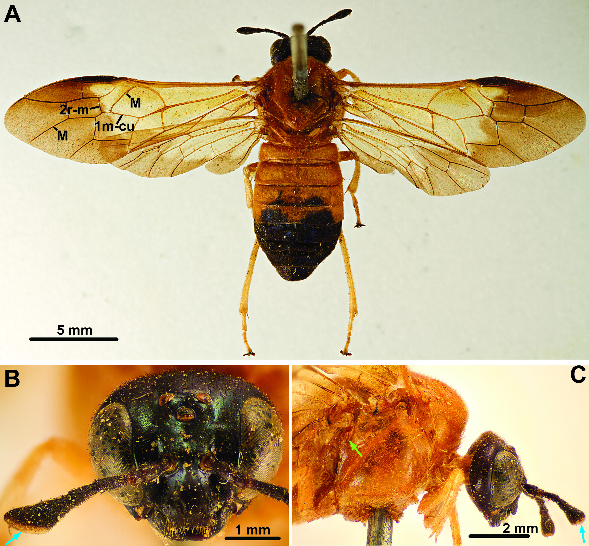
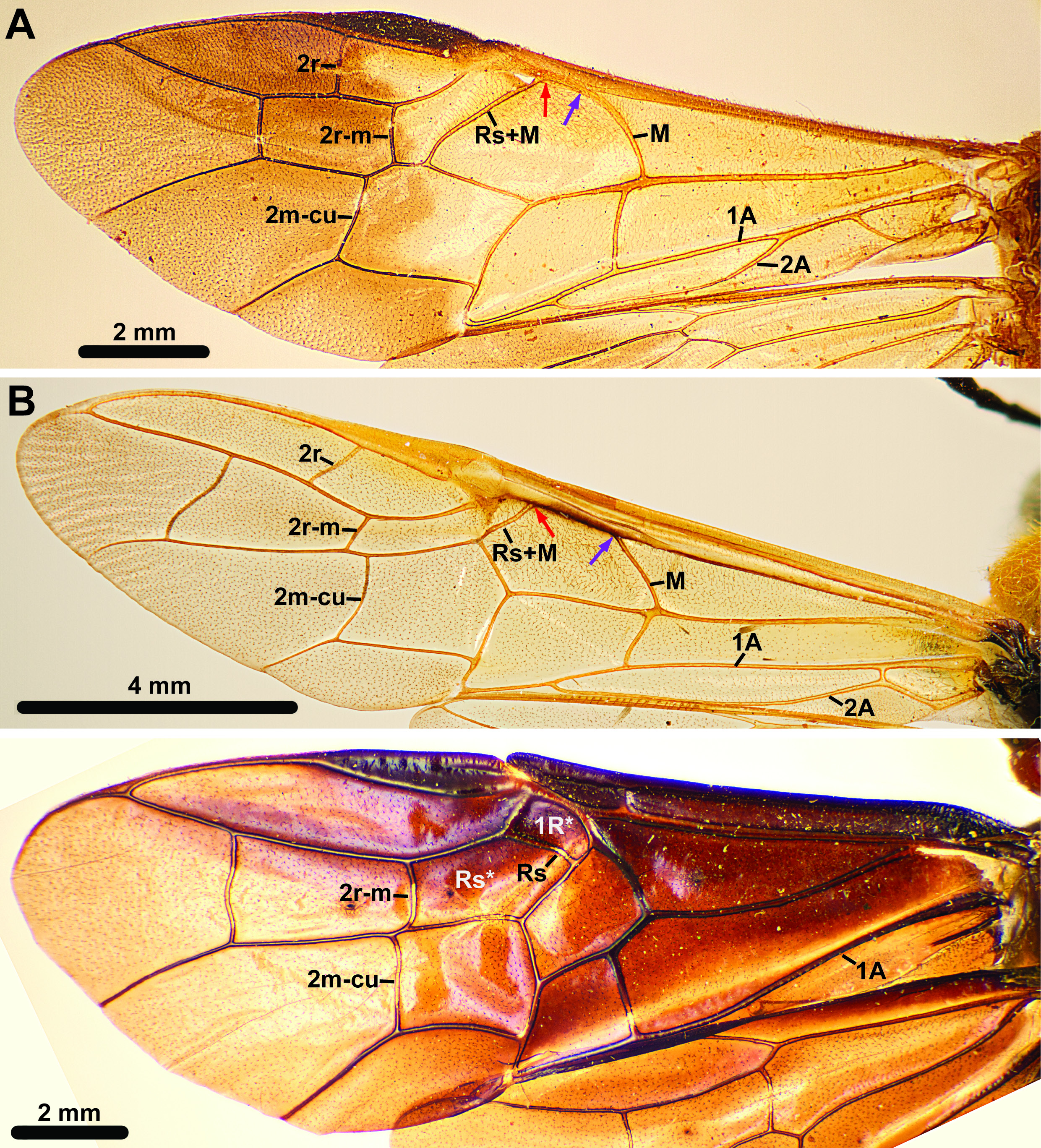
WINGS. Fore wing with pterostigma extended anteriorly and thickened in male ( Figs 9A View Fig , 12 View Fig ); vein M joins Sc+R close to Rs+M; vein 2r-m posteriorly inserts on cell 2M, some distance proximally to anterior end of 2m-cu; vein 1m-cu oriented obliquely to almost parallel with longitudinal axis of wing, inserting on Rs+M close to posterior insertion of 2r-m (distance 1m-cu – 2r-m at most ¼ distance M – 1m-cu on Rs+M; Figs 2A View Fig , 8A View Fig , 9A View Fig ); posterior anal vein present proximally and distally but discontinuous in the middle. Hind wing cell R1 open, except in P. plaumanni and some females of P. albiventris ; vein M continuous, separating cells Rs and M, cell M not reaching vein Rs; cross vein 2a absent.
ABDOMEN. Tergum 1 not subdivided medially ( Fig. 10A View Fig ), median carina absent, lateral carina absent, posterior margin slightly curved; tergum 1 not brighter colored than other abdominal terga. Metaphragma continuous medially, of approx. equal height throughout, median foramen absent. Lateroterga around abdominal spiracles separated from median terga as demarcated by conspicuous fold ( Fig. 3A View Fig ). Cerci variable, length less than 2× width mid length. Ovipositor apparatus with 1 st valvula having longitudinal line, sawteeth strongly asymmetric, broadened in ventral view, slanted proximally, serrulae absent ( Smith 1988: figs 42–44; Vilhelmsen 2018: fig. 19B); tip of 1 st and 2 nd valvulae only slightly curving dorsally; ventral margin with tufts of setae along its length, tufts not on extended lobes ( Vilhelmsen 2018: figs 18D, 19E).
Comments
Pachylosticta Klug, 1824 is the first genus of South American Cimbicidae described and hence the nominal genus of the Pachylostictinae . The most striking feature of the genus is the thickened and anteriorly extended pterostigma in the males. The absence of this feature in the females resulted in the genus Plagiocera Klug, 1834 being proposed for Plagiocera thoracica ; judging from the illustration in Klug (1834: plate II, fig. 5) the description was based on what appears to be a female of Pachylosticta albiventris . Only much later was the connection between the two genera made; the earliest reference to Plagiocera as a synonym of Pachylosticta we have come across is in Konow (1907). An autapomorphy for the genus displayed by both sexes is the orientation of fore wing vein 1m-cu, which inserts on vein Rs+M distally, so that the distance between the insertions of 2r-m and 1m-cu on Rs+M is at most ¼ of the distance between the insertions of M and 1m-cu on Rs+M.
Pachylosticta is most similar to Pseudopachylosticta among the South American Cimbicidae . Both have striking coloration patterns, blue-black metallic body sometimes with paler coloration on some parts of the males and extensive reddish-brown areas on the thorax and sometimes abdomen in the females. The other South American genera are comparatively drab in appearance. Furthermore, Pachylosticta and Pseudopachylosticta both have the labrum raised in the middle and reduced maxillary and labial palps, with four and three palpomeres, respectively. Apart from the fore wing characters, Pachylosticta differs from Pseudopachylosticta in having the hind wing cell RS larger and cell M not reaching vein Rs, and the hind basitarsomere is longer than tarsomeres 2–4 combined in Pachylosticta spp. Finally, Pachylosticta spp. are larger (body length may exceed 1.5 cm) and more compact than Pseudopachylosticta or any other South American Cimbicidae . Pseudabia fusca reaches a body length similar to Pachylosticta spp., but is more slender in appearance.
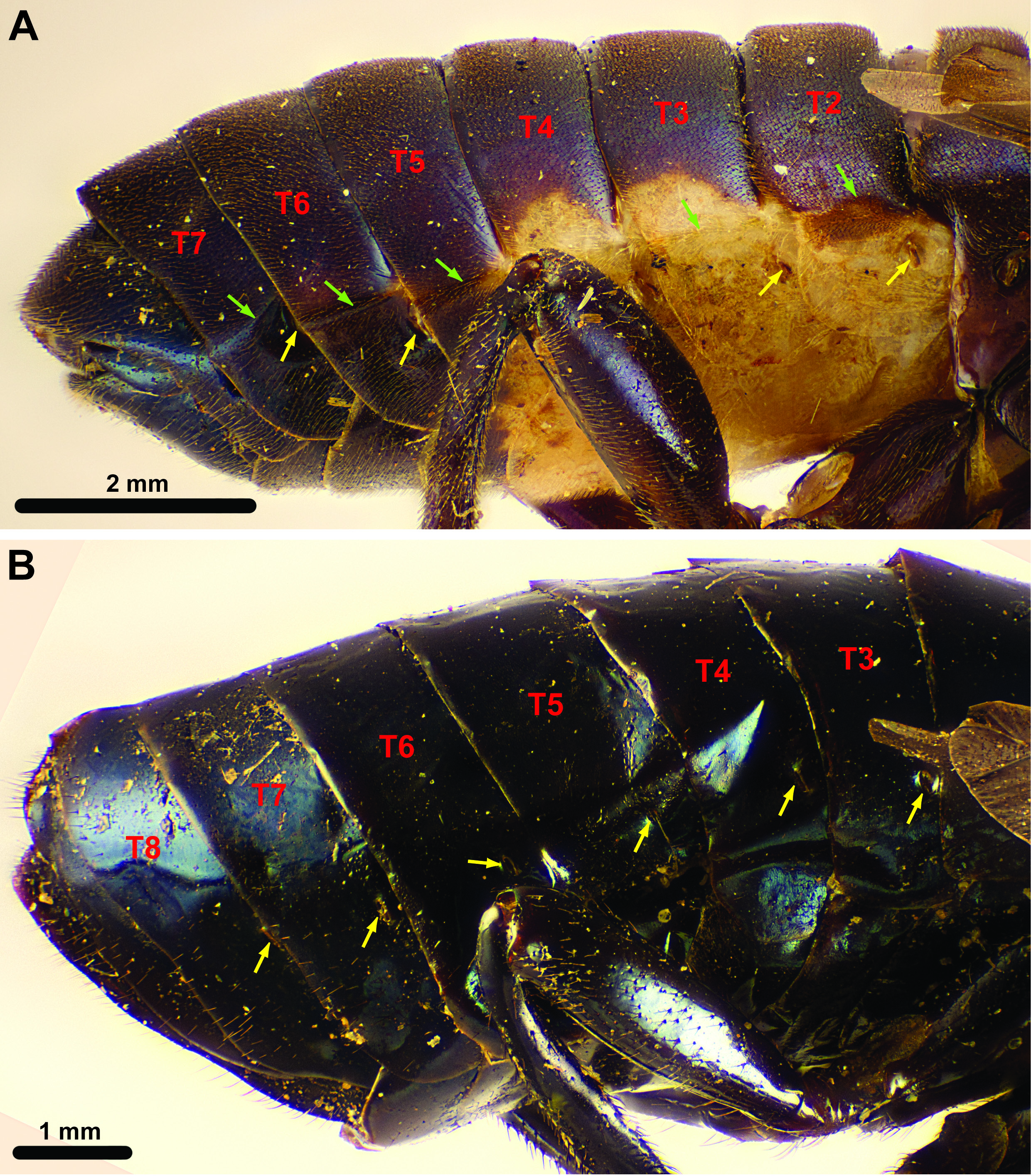
Conde (1937, 1940) and Malaise (1939) disagreed strongly on the separation of males in Pachylosticta . Conde (1937) regarded all three species ( P. albiventris , P. tibialis , P. violacea ) originally described by Klug (1824) as one species, perceiving no significant differences in the male genitalia and regarding the color variation between them as a variation within P. tibialis , the type species. In contrast, Malaise (1939) correlated the color differences with a variation in the male genitalia and recognized three species; this scheme was confirmed by Smith (1988) and is also followed here (see also the key). Pachylosticta violacea is characterized by being completely darkly colored with no pale markings ( Fig. 12C View Fig ); male P. albiventris has an extensive creamy-white to orange patch on the underside of the abdomen (sternum and laterotergum 2 to 4/5; Fig 9A View Fig ); P. tibialis has paler coloration on the upper part of the mesopleuron and the distal part of the legs (tibiae and most of the tarsi; Fig. 12A View Fig ).
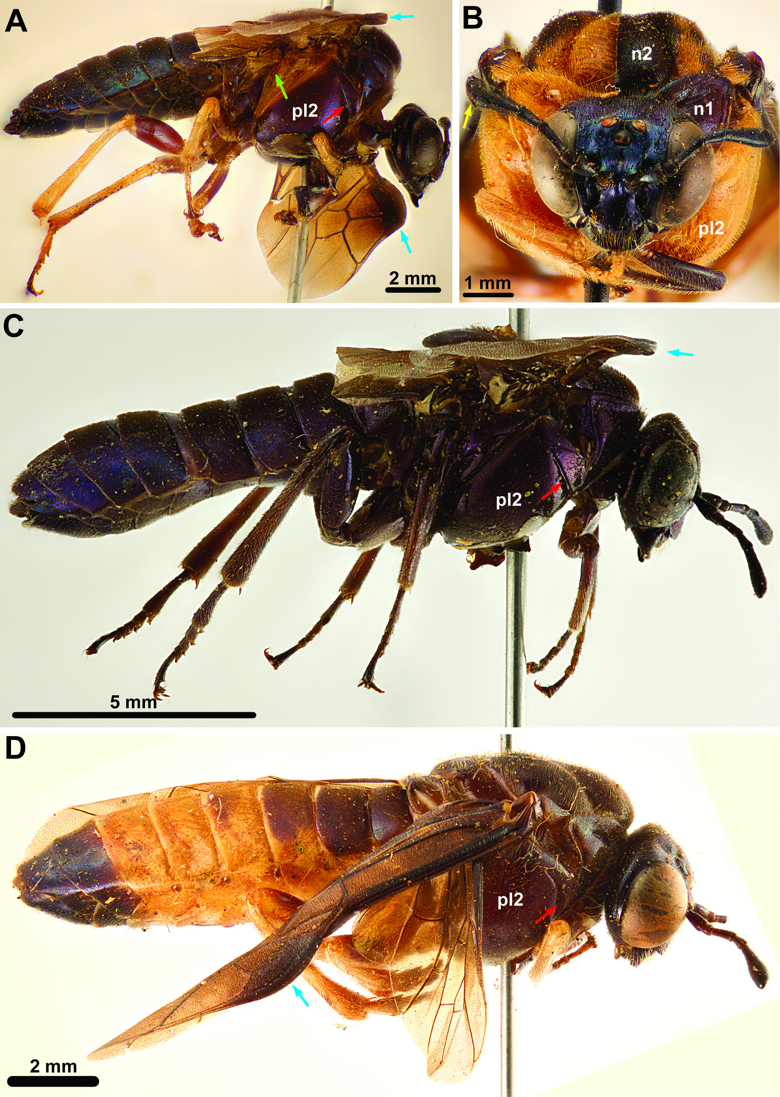
The two male Pachylosticta sp. from Espirito Santo, Córrego do Itá (in MNRJ) differ from all other males examined here in having a reddish-brown band formed by the different color of abdominal terga 4–6 across the abdomen ( Fig. 12D View Fig ); we have not placed them to species. The male from São Paulo, Valle du Rio Pardo (in MNHN) was identified by D.R. Smith as ‘ P. plaumanni ?’ (see also Smith 1988: p. 219). The thorax of this specimen is predominantly reddish brown with the following parts dark brown to black ( Fig. 12B View Fig ): left half of pronotum, left half of propectus, left hind leg, most of mesonotum (including mesoscutellum) except area between the median mesoscutal sulcus and right notaulus, posterior half of both tegulae, both postspiracular sclerites, posterodorsal margin of left mesepimeron (adjacent to the posterior thoracic spiracle), left ¾ of metanotum (including the metascutellum). The right half of the 1 st abdominal tergum is reddish brown, the left dark brown. In addition, on the right apical antennomere there is an elongate lightly colored area ventrally ( Fig. 12B View Fig , yellow arrow), similar to the one observed in female P. albiventris an P. apicalis (see below); the left apical antennomere is uniformly darkly colored as in all other male Pachylosticta spp. observed. The asymmetrical, harlequin-like coloration of this specimen is probably aberrant and we refrain from suggesting a species identification based on the coloration alone. Another aberrantly colored specimen is a P. albiventris male from Caetité, Bahia, Brazil (NHML); it has the distinctive pale anterior abdominal sternites but reddish brown markings posteriorly on the pronotum, in the middle of the mesonotum along the notauli and dorsally on the meso- and metapleuron as well as the proximal part of the hind tibia creamy white.
Female Pachylosticta spp. can also be separated by color differences on the body and wings. Pachylosticta albiventris and P. apicalis usually have the pro- and mesothorax reddish brown throughout ( Figs 8A View Fig , 10A View Fig ), whereas P. plaumanni has the median part of the pronotum and the mesoscutum darkened ( Fig. 11A View Fig ). However, the female Pachylosticta albiventris from Linhares has the mesonotum as well as the lower parts of the mesopleura darkened; Smith (1988) reported observing a similar color variation in the females of this species. Pachylosticta albiventris has the metathorax and abdomen uniformly blue-black metallic ( Fig. 8A View Fig ), whereas the other two species have only the apex of the abdomen (from segment 4 or 5) darkened ( Figs 10A View Fig , 11A View Fig ). In the fore wing, P. apicalis has the tip darkly infuscate contrasting with the hyaline base ( Fig. 10A View Fig ), whereas the other two species have the fore wings more or less uniformly infuscate ( Fig. 8A View Fig ). Finally, in P. albiventris and P. apicalis there is an elongate lightly colored area on the ventral side of the apical antennomere ( Figs 8 View Fig B–C, 10B–C); this is missing from P. plaumanni ( Fig. 11B View Fig ) and male Pachylosticta spp. (but see above); the function is unknown. Interestingly, this feature is illustrated in Klug (1834: plate II, fig. 5g) in the description of Plagiocera thoracica , but not mentioned in the text.
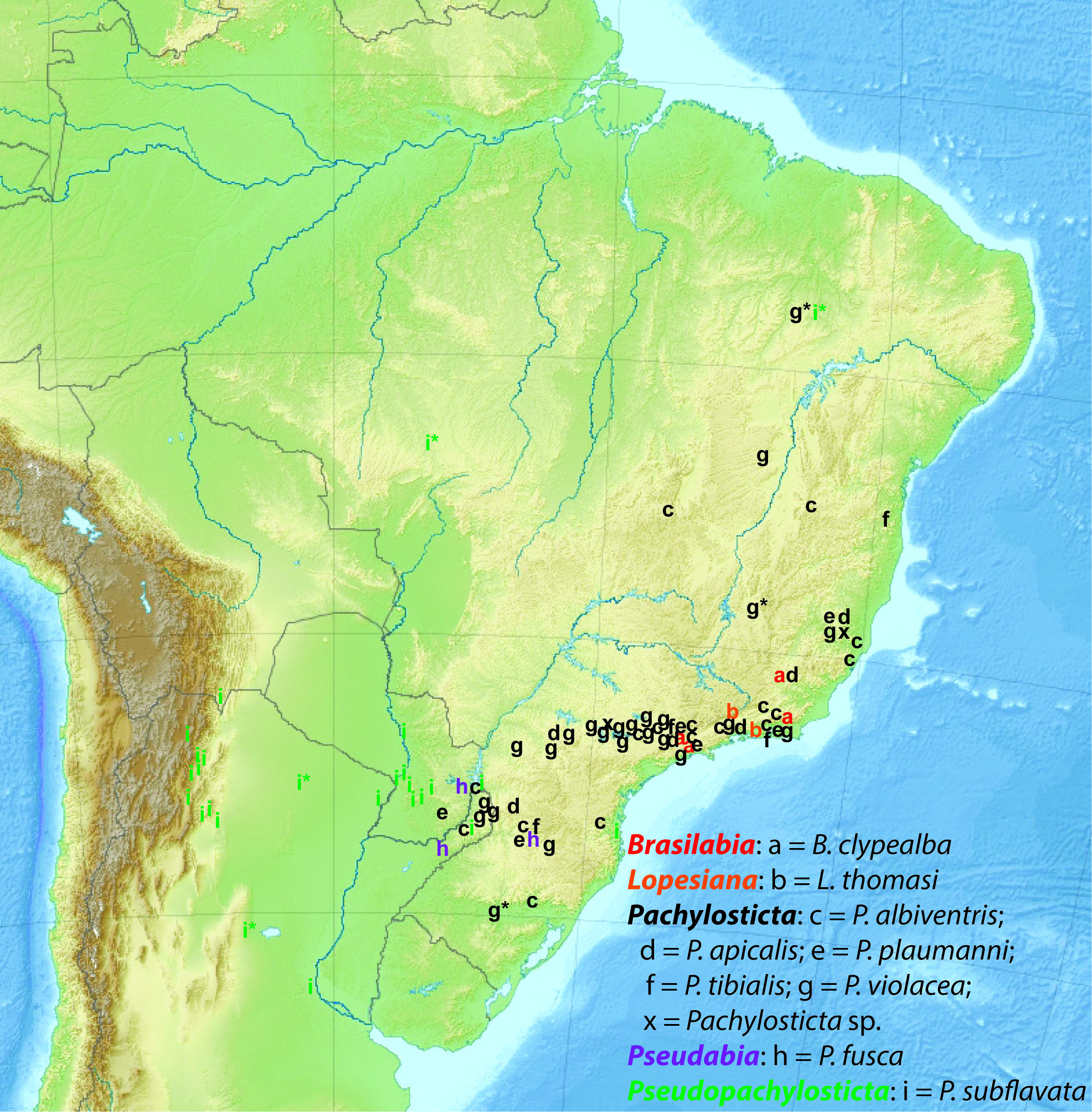
With five currently recognized species, Pachylosticta is the only non-monotypic genus of South American Cimbicidae , comprising over half the species. Nevertheless, there is a possibility that the diversity of the genus is overestimated, as males and females have only been associated for the most common species, P. albiventris . Of the remaining species, P. apicalis and P. plaumanni are known only from females, whereas P. tibialis and P. violacea are known only from males. Unfortunately, no morphological characters have been discovered that can be used to link any of these species to each other. In addition, the affinity of males with coloration more resembling that of the females has not been decided. Pachylosticta violacea males have been collected in the same localities as both P. apicalis (Parana, Rolândia) and P. plaumanni (Espirito Santo, Córrego do Itá), thus providing little evidence for associating P. violacea with any of the females of the two other species. The aberrantly colored Pachylosticta spp. males were also collected in Espirito Santo, Córrego do Itá. In general, the distributions of the described species of Pachylosticta are broadly overlapping ( Fig. 16 View Fig ), thus providing no help for associating the sexes. DNA barcoding could potentially help to associate males and females in Pachylosticta spp. and decide if some species have different color morphs in one sex or the other. Unfortunately, the material available for this study is somewhat dated and it might not be possible to make adequate extractions from it.
No known copyright restrictions apply. See Agosti, D., Egloff, W., 2009. Taxonomic information exchange and copyright: the Plazi approach. BMC Research Notes 2009, 2:53 for further explanation.