
Brasilabia clypealba ( Conde, 1932 )
|
publication ID |
https://doi.org/ 10.5852/ejt.2018.482 |
|
publication LSID |
lsid:zoobank.org:pub:6F3B12C7-2311-48EA-8727-5B90489E26E3 |
|
DOI |
https://doi.org/10.5281/zenodo.3846143 |
|
persistent identifier |
https://treatment.plazi.org/id/723287A9-5735-F007-B33B-B3359046F28E |
|
treatment provided by |
Valdenar |
|
scientific name |
Brasilabia clypealba ( Conde, 1932 ) |
| status |
|
Brasilabia clypealba ( Conde, 1932)
Fig. 5 View Fig
Material examined
Holotype
BRAZIL: ♀, São Paulo, Ypiranga , Jan. 1906, Luederwaldt leg. ( MZSP).
Other material
BRAZIL: – Minas Gerais: 1 ♀, Viçosa, Universidad Federal de Viçosa, Areo do Deparatmento de Apicultura, 651 m a.s.l., 20°45’ S, 42°57’ W, 4 Nov. 2007, A. I. A Pereira leg. ( UFVB); 1 ♀ same collecting data as preceding but 15 Nov. 2007 ( UFVB); 1 ♀ same collecting data as preceding but 20 Nov. 2007 ( UFVB). – Rio de Janeiro: 1 ♀, Nova Friburgo, Mury, 1– 31 Jan. 1965, Gred and Guimaraes leg. ( NMNH). – São Paulo: 1 ♀, São Paulo, Cidade de Universidade, 15 Oct. 1971, C.G. Froehlic leg. ( MZSP).
Redescription (female, male unknown)
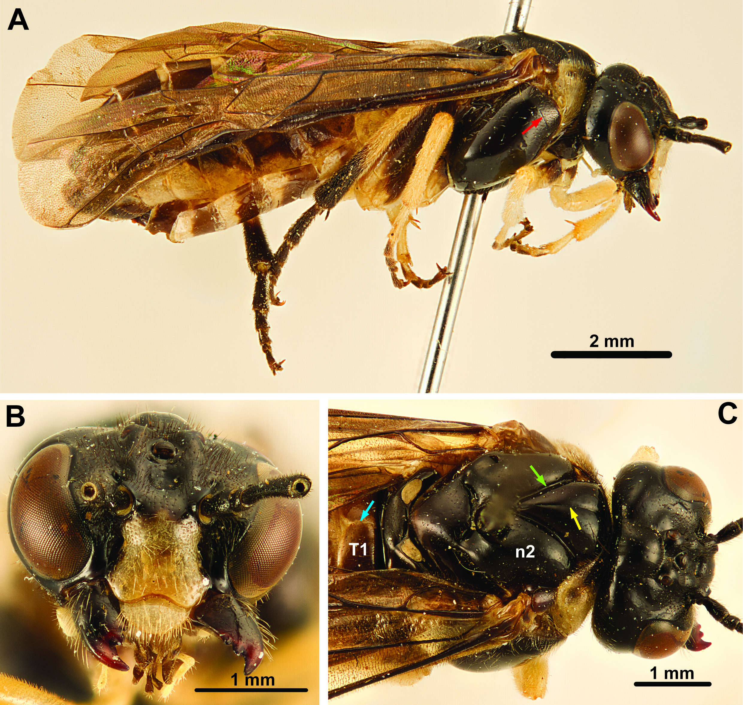
MEASUREMENTS. Medium-sized sawfly, body length 10.5 mm.
COLOR. Coloration predominantly black to dark brown ( Fig. 5 View Fig ), creamy white markings of various size on all body parts (e.g., clypeus on head ( Fig. 5 B View Fig ), lateral parts of pronotum). Legs creamy white proximally, black to dark brown distally. Wings predominantly hyaline.
HEAD. Eyes converging ventrally, inner margins slightly incurved ( Fig. 5 B View Fig ). Posterior ocelli at level with dorsal margin of eyes. Toruli in middle of face, slightly closer to median ocellus than to ventral margin of clypeus. Epistomal sulcus absent. Clypeus with shallow incurvation medially. Gena medially less wide than width of eye, wider dorsally than ventrally. Malar space very low ( Fig. 5 B View Fig ). Occipital carina absent. Sclerotisation between occipital and oral foramina present. Antennae with five antennomeres [from Smith 1988: fig. 34; specimen examined missing antennomeres 3–5 and 4–5, respectively]; antennomere 1 approx. 2× as long as wide, antennomere 2 approx. as wide as long, antennomere 3 approx. 2× as long as antennomere 4 [from Smith 1988], antennomere 5 slightly expanded distally [from Smith 1988]. Labrum broad, convex, flat, hairy, distal margin evenly curved. Mandibles less than ½ the height of head, with three teeth each ( Fig. 5 B View Fig ), inner margin not serrated. Maxilla with stipes of approx. equal width throughout, palps with six palpomeres, longer than labial palps. Labial palps with four palpomeres, palps inserting at level with maxillary palps. Postmentum narrow, at least 3× as long as wide.
THORAX. Pronotum comparatively high medially, with transverse grove ending in small depression laterally, no lateral groove; pronotum articulating with mesopleuron for short distance ventrolaterally. Dorsal cervical sclerite present. Propectus without lateral projection, propleural sulcus present, medioventral margins of propectus widely separated, posteriorly extended into narrow points. Prosternum laterally extended, continuous with katepisternum which articulate, but does not fuse with propleuron at lateral coxal articulation point. Anterior fore tibial spur straight, simple, not much longer than posterior spur, spurs pointed and sclerotized apically. Mesonotum with distinct median sulcus and deep notauli ( Fig. 5 C View Fig ), laterophragmal apodeme not observed, postscutellum absent. Small anterodorsal part of mesopleuron separated from rest by vertical groove ( Fig. 5 A View Fig ), prepectus absent as separate sclerite. Horizontal carina absent laterally on mesopleuron. Posterior thoracic spiracle visible in lateral view, situated in incurvation in dorsal margin of mesopleuron. Median midcoxal articulations adjacent, only separated by small wedge of cuticle. Mesofurca not observed. Insertion of mesonoto-metanotal muscle on metanotum not observed; cenchri approx. 2× as broad as long. Anapleural cleft present, small. Metapleuron fused with abdominal tergum 1 along dorsal margin; posteroventral metapleural apodeme not observed; paracoxal sulcus curving posteriorly, terminating in the middle of the metapleural sulcus; metacoxal foramina open dorsally, without metapleural inflection laterally. Metafurca not observed. Hind coxa less than 2× as long as wide, median carina or spine not observed. Hind femoral ventral spur absent. Hind tibial apical spurs not much longer than apical width of tibia. Hind basitarsomere slightly longer than tarsomeres 2–4, tarsal claws bifid, teeth of subequal length.
WINGS. Fore wing with vein M joining Sc+ R close to Rs+ M; vein 2r-m posteriorly inserts on cell 2 M, very close to anterior end of 2m-cu; vein 1m-cu oriented obliquely, inserting on M some distance from 2r-m (distance 1m-cu–2r-m at least ½ distance Rs+M–1m-cu on M); posterior anal vein present proximally and distally but discontinuous in the middle. Hind wing cell R 1 closed; vein M continuous, separating cells Rs and M, cell M not reaching vein Rs; cross vein 2a absent.
ABDOMEN. Tergum 1 subdivided medially by narrow longitudinal membranous line ( Fig. 5 C View Fig ), lateral carina absent, posterior margin straight; tergum 1 not brighter colored than other abdominal terga. Metaphragma not observed. Separation between median terga and lateroterga weakly demarcated, fold most conspicuous on tergum 2. Cerci triangular, length approx. 1.5× width mid length. Ovipositor apparatus not observed; Smith (1988: fig. 47) illustrates 1 st valvula, with longitudinal line, vertically oriented sawteeth with serrulae, and tip curving dorsally.
Comments
According to Smith (1988), the holotype was collected in Ypiranga (possibly Ipiranga, São Paulo, Brazil). The other material examined here fits well with the original description of Conde (1932). Conde originally described the species as Pseudabia clypealba , but later transferred it to a separate genus, Brasilabia ( Conde 1937) ; the justification for removing the species from Pseudabia was the medially subdivided abdominal tergum 1 and the small size. The tergum 1 configuration is indeed unique among all Cimbicidae ; it might represent a retained plesiomorphy ( Blasticotomidae , Diprionidae , most Tenthredinidae and some Argidae and Pergidae display this feature; see Vilhelmsen 2015: character 80) or a reversal compared to other cimbicids. Also diagnostic of Brasilabia clypealba is the eponymous creamy-white clypeus, contrasting with the blackish-brown color of the remainder of the head capsule; all other South American cimbicids have the head capsule uniformly darkly colored in anterior view.
| MZSP |
Sao Paulo, Museu de Zoologia da Universidade de Sao Paulo |
| S |
Department of Botany, Swedish Museum of Natural History |
| W |
Naturhistorisches Museum Wien |
| A |
Harvard University - Arnold Arboretum |
| I |
"Alexandru Ioan Cuza" University |
| UFVB |
Vicosa, Universidade Federal de Vicosa, Museum of Entomology |
| NMNH |
Smithsonian Institution, National Museum of Natural History |
| B |
Botanischer Garten und Botanisches Museum Berlin-Dahlem, Zentraleinrichtung der Freien Universitaet |
| C |
University of Copenhagen |
| M |
Botanische Staatssammlung M�nchen |
| R |
Departamento de Geologia, Universidad de Chile |
No known copyright restrictions apply. See Agosti, D., Egloff, W., 2009. Taxonomic information exchange and copyright: the Plazi approach. BMC Research Notes 2009, 2:53 for further explanation.