
Dictyosporium duliujiangense L.L. Liu & Z.Y. Liu, 2023
|
publication ID |
https://doi.org/10.11646/phytotaxa.606.4.2 |
|
DOI |
https://doi.org/10.5281/zenodo.8202738 |
|
persistent identifier |
https://treatment.plazi.org/id/7269B674-294F-FFE0-21E5-FF76FB213ECB |
|
treatment provided by |
Plazi |
|
scientific name |
Dictyosporium duliujiangense L.L. Liu & Z.Y. Liu |
| status |
sp. nov. |
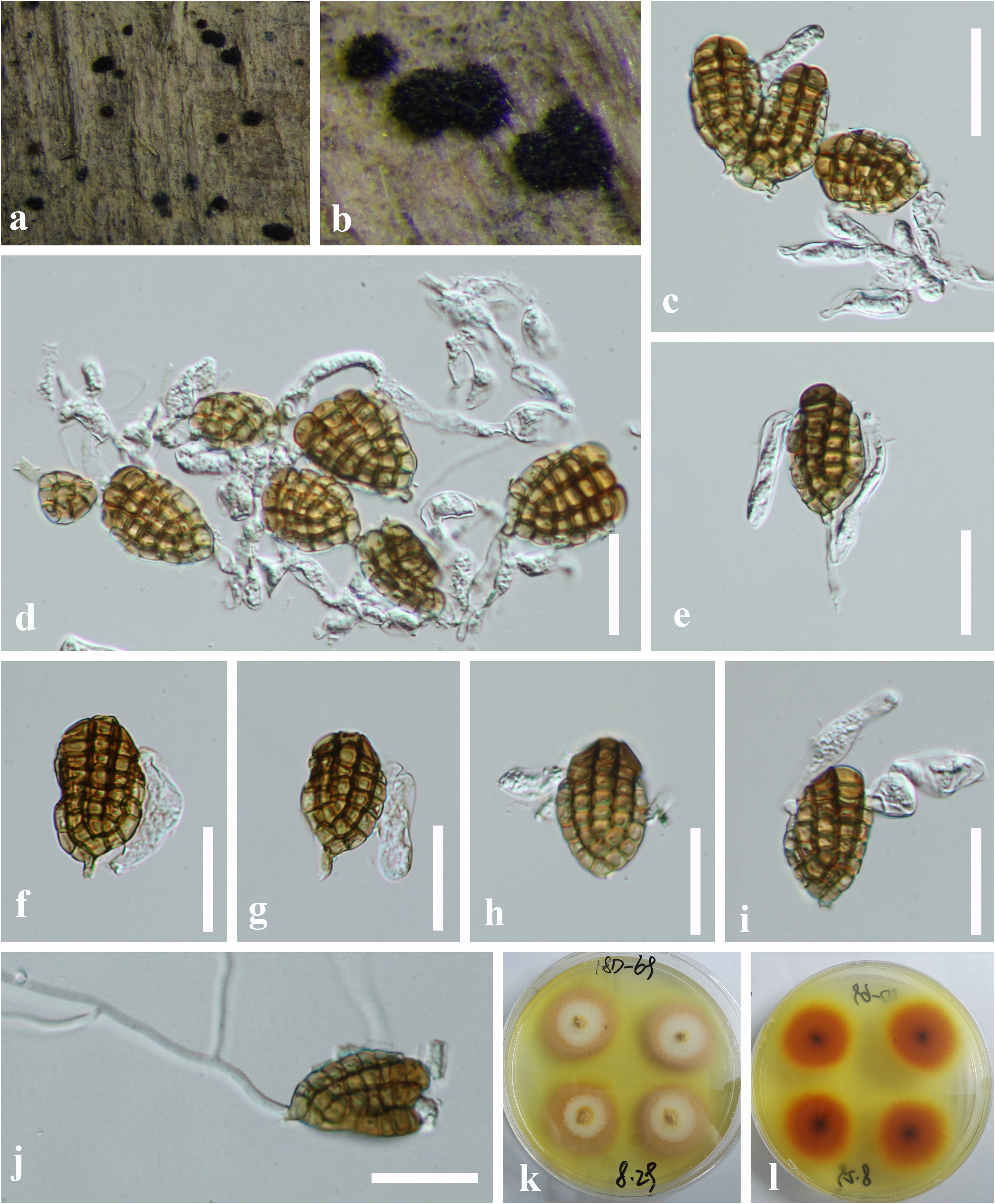
Dictyosporium duliujiangense L.L. Liu & Z.Y. Liu , sp. nov. Figure 3 View FIGURE 3 .
Index Fungorum number: IF 900403, Facesoffungi number: FoF 14119
Etymology: referring to the collecting site of Duliu River.
Saprobic on decaying plant substrates. Asexual morph: Colonies punctiform, sporodochial, scattered, dark brown to black, glistening. Mycelium mostly immersed, composed of smooth, septate, branched, hyaline to pale brown hyphae. Conidiophores macronematous, mononematous, septate, cylindrical, hyaline to pale yellow, smooth-walled, 4–17 × 2–5 μm, sometimes reduced to conidiogenous cells. Conidiogenous cells monoblastic, integrated, terminal, determinate, pale yellow to yellow. Conidia acrogenous, solitary, cheiroid, smooth-walled, complanate, yellowishbrown to pale brown, (17–) 31–36 ×17–22 μm ( x = 33 × 20 μm, n = 30), mostly consisting of five arms closely compact with side arms lower than middle arms, rarely with four arms, 4–8-euseptate in each arm, constricted at septa, one or two or more, hyaline, tubular, elongated appendages which are 16–38 × 3.5–10 μm ( x = 27 × 6 μm, n = 20) and mostly attached at the apical part of two or more outer arms. Sexual morph: Undetermined.
Cultural characteristics: Conidia germinating on PDA within 24 h and germ tubes produced from the basal cell. Colonies on PDA reaching 17–23 mm diam. in 2 weeks at 25 °C, in natural light, circular, divide into two layers on the surface, with fluffy, dense, white mycelium on the center layer and with sparse, yellow, entire margin, in reverse pale brown in the middle and white at the margin.
Material examined: China, Guizhou Province, Dushan County, Duliu river Deep ditch scenic spot, 25°55′ N, 107°37′ E, at an altitude of 1205 m, on decaying branch submerged in a freshwater River, 5 July 2018, L.L. Liu, 18D-69 ( HKAS 129172 View Materials , holotype, GZAAS 20–0321 , isotype), ex-type culture GZCC 19–0426 GoogleMaps .
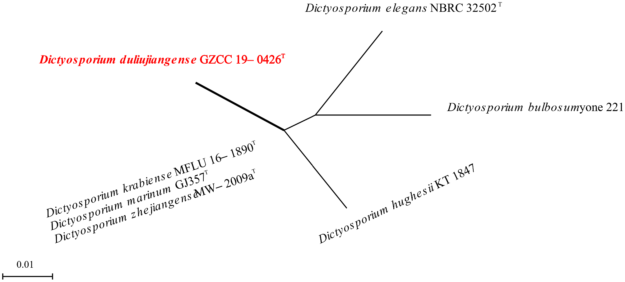
Notes: Dictyosporium duliujiangense ( GZCC 19–0426) has sporodochial colonies, macronematous conidiophores, and cheiroid, digitate conidia with numerous parallel rows of cells, which are typical of Dictyosporium ( Goh et al. 1999) . Dictyosporium duliujiangense shows a close phylogenetic affinity to D. marinum ( MFLU 19–1229) which was isolated from buried wood in sand from UK, Carmarthenshire ( Dayarathne et al. 2020) ( FIGURE 1 View FIGURE 1 ). A comparison of LSU sequence bases between Dictyosporium duliujiangense and D. marinum is 2.48% (785/805) difference. Furthermore, the two species are morphologically distinct, especially in their conidial morphology. Conidia of Dictyosporium duliujiangense are smaller ((17–) 31–36 × 17–22 μm) and comprise outer rows with one or two or more hyaline appendage, whereas D. marinum has relatively larger conidia (34–50 × 22–28 µm) that lacks appendages ( Dayarathne et al. 2020). Dictyosporium duliujiangense shares similar size of the conidia with D. tubulatum ( MFLU 15–1166) and D. zhejiangense ( MW –2009a). In addition, Dictyosporium duliujiangense can be distinguished from D. tubulatum in the cells of former are usually arranged in 5 rows while the cells of D. tubulatum are mostly in 4 rows, well distinguishable from D. zhejiangense by smaller appendages (16–38 × 3.5–10 μm vs. 29–54 × 5.5–8 µm), and inconspicuous septum, while D. zhejiangense constricts at the septa ( Wongsawas et al. 2009, Yang et al. 2018).The PHI analysis further confirms that Dictyosporium duliujiangense has no significant genetic recombination with closely related species (Fw> 0.05, FIGURE 4 View FIGURE 4 ). We thus introduced D. duliujiangense as a new species.
No known copyright restrictions apply. See Agosti, D., Egloff, W., 2009. Taxonomic information exchange and copyright: the Plazi approach. BMC Research Notes 2009, 2:53 for further explanation.
|
Kingdom |
|
|
Phylum |
|
|
Class |
|
|
Order |
|
|
Family |
|
|
Genus |