
Paradoxivena, Wei, Cong, Zhang, Yalin & Webb, M. D., 2006
|
publication ID |
https://doi.org/ 10.5281/zenodo.174939 |
|
DOI |
https://doi.org/10.5281/zenodo.6257026 |
|
persistent identifier |
https://treatment.plazi.org/id/745EBC71-F527-4628-CB5F-052FD2ADFDA2 |
|
treatment provided by |
Plazi |
|
scientific name |
Paradoxivena |
| status |
gen. nov. |
Paradoxivena View in CoL gen. n.
Type species. Paradoxivena zhamuensis sp. n.
Etymology. The generic name is after the bicoloured veins of the forewing. Gender is feminine.
Diagnosis. This new genus forms a group with other Oriental stegelytrine genera (see Intruduction) but can be distinguished from them by: 1) the forewing with veins bicoloured, brown intervened by yellowish white regularly; 2) male pygofer elongate and tapering apically with apex pointed, and pygofer phragma with a strongly developed lateral shelf-like sclerotised area (dorsal connective) from pygofer side to apex of dorsal apodeme of aedeagus; 3) valve broad, almost as long as wide, semicircle-shaped; 4) style with apical process strong, tapered to moderately acute apex, with a few ventral setae subapically; 5) aedeagal shaft with pair of short acute processes subapically on ventral surface and a triangular-like process on each side slightly distad of midlength.
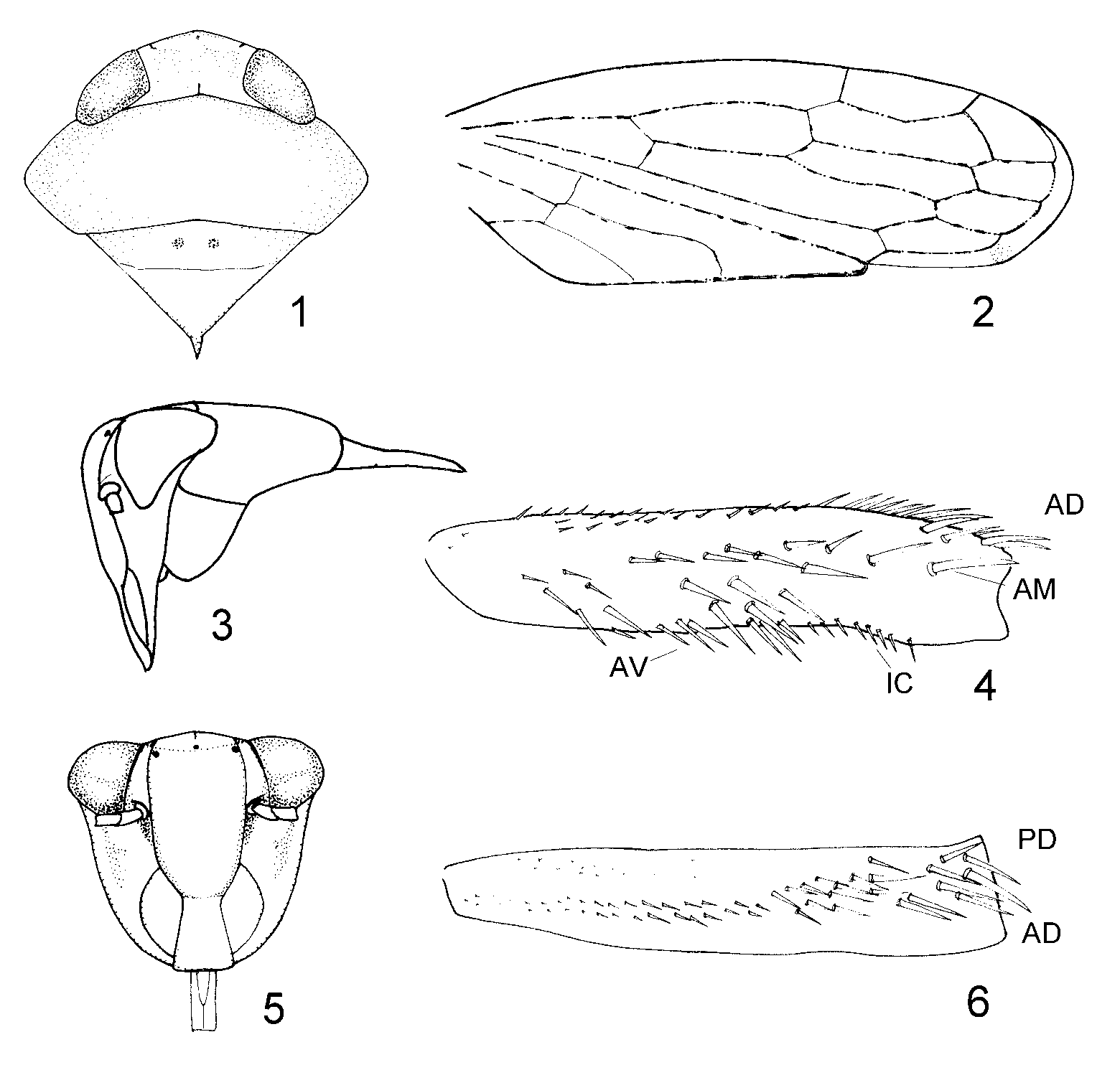
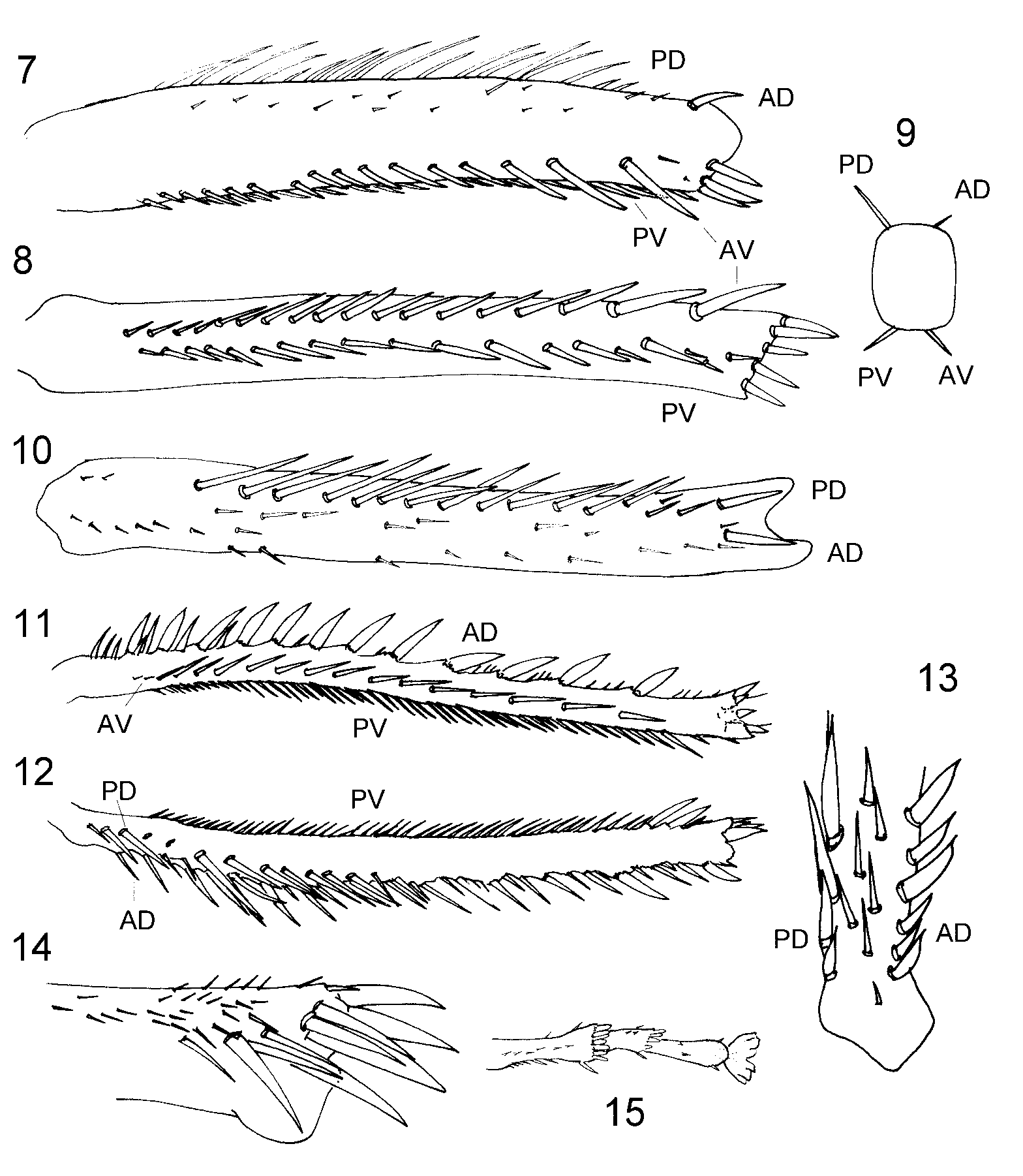
Description. Veins brown intervened by yellow-white ( Fig. 2 View FIGURES 1 – 6 ). Head small, distinctly narrower than pronotum, equal in width to scutellum ( Fig. 1 View FIGURES 1 – 6 ); fore margin slightly angularly rounded; eyes overlapping pronotum laterally; vertex nearly as long as width between eyes, sloping to front; coronal suture short ( Fig. 1 View FIGURES 1 – 6 ); face similar in length to width ( Fig. 5 View FIGURES 1 – 6 ); ocelli on anterior margin of vertex, situated approximately 1.5x their own diameter from corresponding eye; lateral frontal suture extending onto vertex, incurved apically, touching lateral margin of corresponding ocellus; transclypeal suture somewhat indistinct; clypellus very narrow basally, broadening apically with apical margin very slightly concave; gena nearly flat; lora broad; rostrum narrow, extending to apex of trochanter, labrum about half length of labium; antennae long, extending to near apex of forewing, arising adjacent to lower corner of eye; antennal ledge present; antennal pit shallow. Pronotum approximately 3x broader than median length; posterior margin slightly concave; lateral margin long; lateral carina present, curved dorsally toward adjacent eye ( Figs 1, 3, 5 View FIGURES 1 – 6 ). Proepisternum apparent ( Fig. 3 View FIGURES 1 – 6 ). Scutellum approximately equal in length to pronotum, with transversal depression distinct; basal width longer than medial length; posterior half weakly elevated and inclined from transverse suture; posterolateral carina absent; postnotum short, median carina absent ( Figs 1, 3 View FIGURES 1 – 6 ). Forewing transparent, with 5 apical cells; 5th apical cell normal; foremargin convex throughout length; middle and outer subapical cells closed, inner subapical cell open; outer subapical cell slightly shorter than medial subapical cell; crossvein present basally between inner claval vein and claval suture, situated at one third distance from base to apex of clavus; appendix moderately broad, slightly truncate apically, extended to fourth apical cell; claval margin strongly elevated and crimped at apex ( Fig. 2 View FIGURES 1 – 6 ). Legs densely setose. Fore femur with AM setae arranged irregularly, with numerous macrosetae basad of AM1; IC setae long, comblike; dorsal surface with two groups of very short setae over basal three fifths (AD and PD rows) and more irregularly arranged setae distally (AD row) ( Figs 4, 6 View FIGURES 1 – 6 ). Fore tibia rounded dorsally; AD setal row with apical seta thick and others small, short, and irregularly arranged; PD setae thick and in distinctly different length near apex; AV and PV setal rows extending from base to apex ( Figs 7–10 View FIGURES 7 – 15 ). Hind femur broadened distally and slightly bowed; several irregularly arranged distal setae elevated on strong bases; numerous relatively shorter setae subapically ( Fig. 14 View FIGURES 7 – 15 ). Hind tibia moderately flattened and bowed; PD setae long; AD setae very stout and with some short setae between stout ones; several supranumeral setae present between AD and PD rows ( Figs 11, 12 View FIGURES 7 – 15 ).
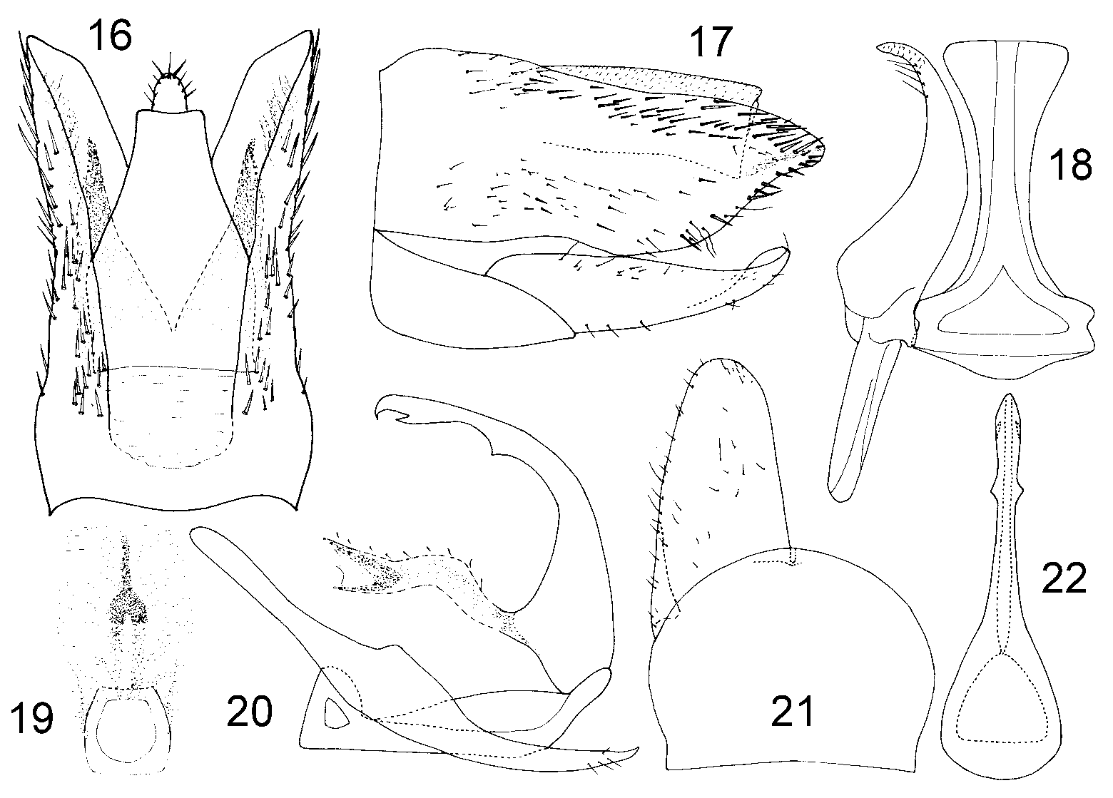
Male pygofer side longer than broad, with short dorsal and apical macrosetae; dorsal margin nearly horizontal adjacent to segment X; vertical hyaline band absent; phragma with a strongly developed shelf-like sclerotised area from pygofer side to apex of dorsal apodeme of aedeagus ( Figs 16, 17 View FIGURES 16 – 22 ), and a nearly Y-shaped medial sclerotised area (in dorsal/ventral view) between segment X and basal apodeme of aedeagus ( Fig. 19 View FIGURES 16 – 22 ). Valve broad and almost as long as wide, semicircular, similar to length of subgenital plate in ventral view ( Fig. 21 View FIGURES 16 – 22 ). Subgenital plates long, extended to near posterior margin of pygofer; inner margins slightly diverging, apex rounded, outer margin slightly convex, with a dorsal elongate lobe laterobasally holding style preapical lobe; a few irregularly arranged short to moderately long slender setae laterally and apically ( Figs 17, 21 View FIGURES 16 – 22 ). Connective somewhat T-shaped; stem moderately long; arms short; stem not extended beyond apex of medial sclerotised area ( Figs 18–20 View FIGURES 16 – 22 ). Style with anterior medial lobe very short, anterior lateral apodeme straight and elongate; lateral preapical lobe slight, gradually merging into apical process, the latter strong, elongate, tapered to moderately acute apex, a few fine ventral setae subapically ( Figs 18, 20 View FIGURES 16 – 22 ). Aedeagus simple, shaft elongate, cylindrical, strongly curved dorsally and anteriorly, tapering apically with a short acute processes subapically on dorsal surface and a triangular processes on each side slightly distad of midlength; basal apodeme very short; gonopore indistinct, probably apical ( Figs 20–22 View FIGURES 16 – 22 ). Segment X very long ( Figs 16, 17 View FIGURES 16 – 22 ).
Female unknown.
Distribution. China (Tibet).
No known copyright restrictions apply. See Agosti, D., Egloff, W., 2009. Taxonomic information exchange and copyright: the Plazi approach. BMC Research Notes 2009, 2:53 for further explanation.