
Tachycines (Gymnaeta) parvus, Qin & Liu & Li, 2019
|
publication ID |
https://doi.org/ 10.11646/zootaxa.4560.2.3 |
|
publication LSID |
lsid:zoobank.org:pub:3867A9CC-9CA9-4512-A84C-ADC45CA47D80 |
|
DOI |
https://doi.org/10.5281/zenodo.5941643 |
|
persistent identifier |
https://treatment.plazi.org/id/74632A58-FFBD-FFA3-2C95-0674FA0B5029 |
|
treatment provided by |
Plazi |
|
scientific name |
Tachycines (Gymnaeta) parvus |
| status |
|
Key to the species of subgenus (Gymnaeta) genus Tachycines View in CoL
1(26) Hind metatarsus with bristles beneath.
2(7) Supra internal spur of hind tibiae almost exceeding ventral apex of hind metatarsus.
3(4) Hind tibiae above on each side with less than 50 spines.............................. T. (G.) tonkinensis Chopard, 1929
4(3) Hind tibiae above on each side with more than 50 spines.
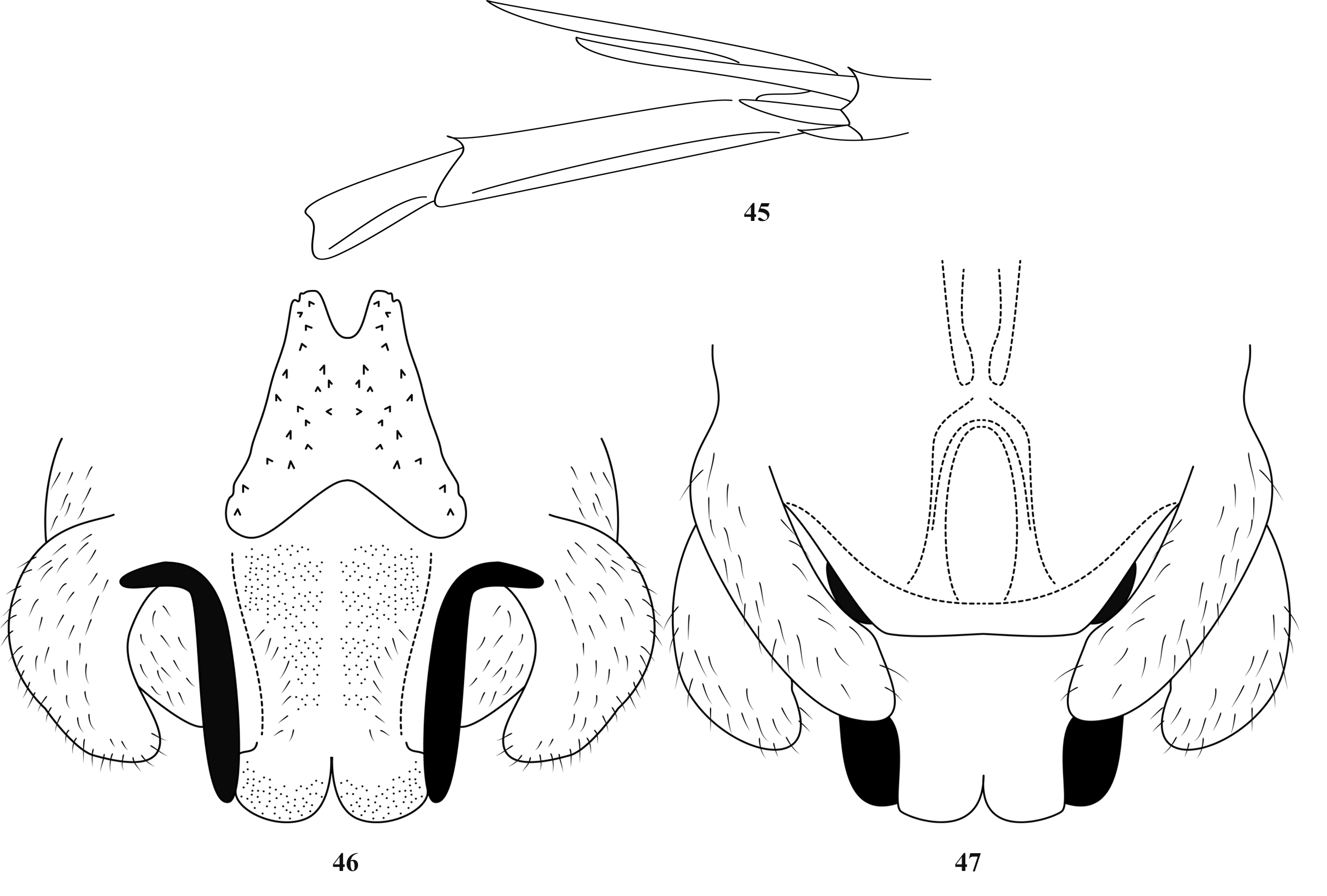
5(6) Dorsal surface of body without longitudinal band; male genitalia as Figs. 3–4 View FIGURES 2–5 ; female subgenital plate as Fig. 5............................................................................. T View FIGURES 2–5 . (G.) gonggashanicus ( Zhang & Liu, 2009)
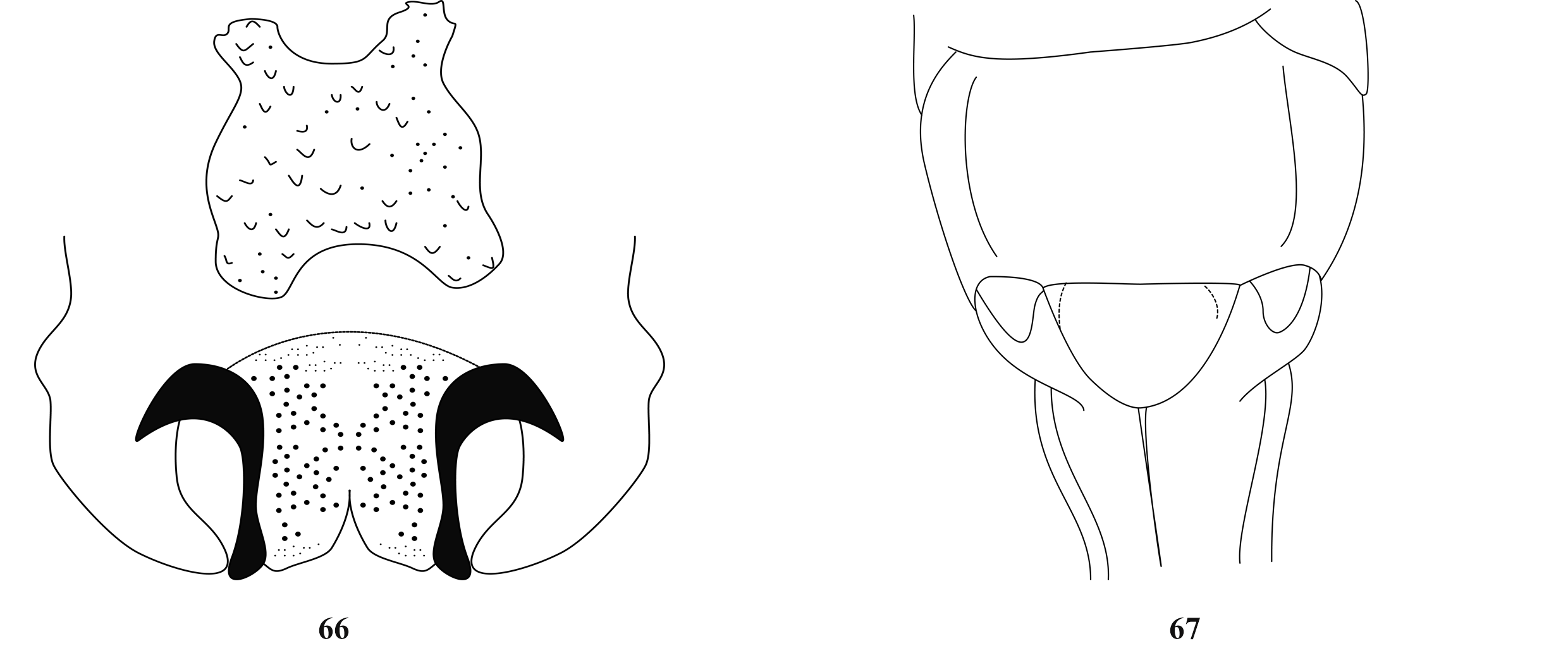
6(5) Dorsal surface of body with light longitudinal band; male genitalia as Fig. 7 View FIGURES 6–8 ; female subgenital plate as Fig. 8........................................................................................ T View FIGURES 6–8 . (G.) brevicaudus Karny, 1934
7(2) Supra internal spur of hind tibiae not exceeding ventral apex of hind metatarsus.
8(13) Internal genicular lobe of fore femur without spine; hind tibiae above on each side with more than 50 spines.
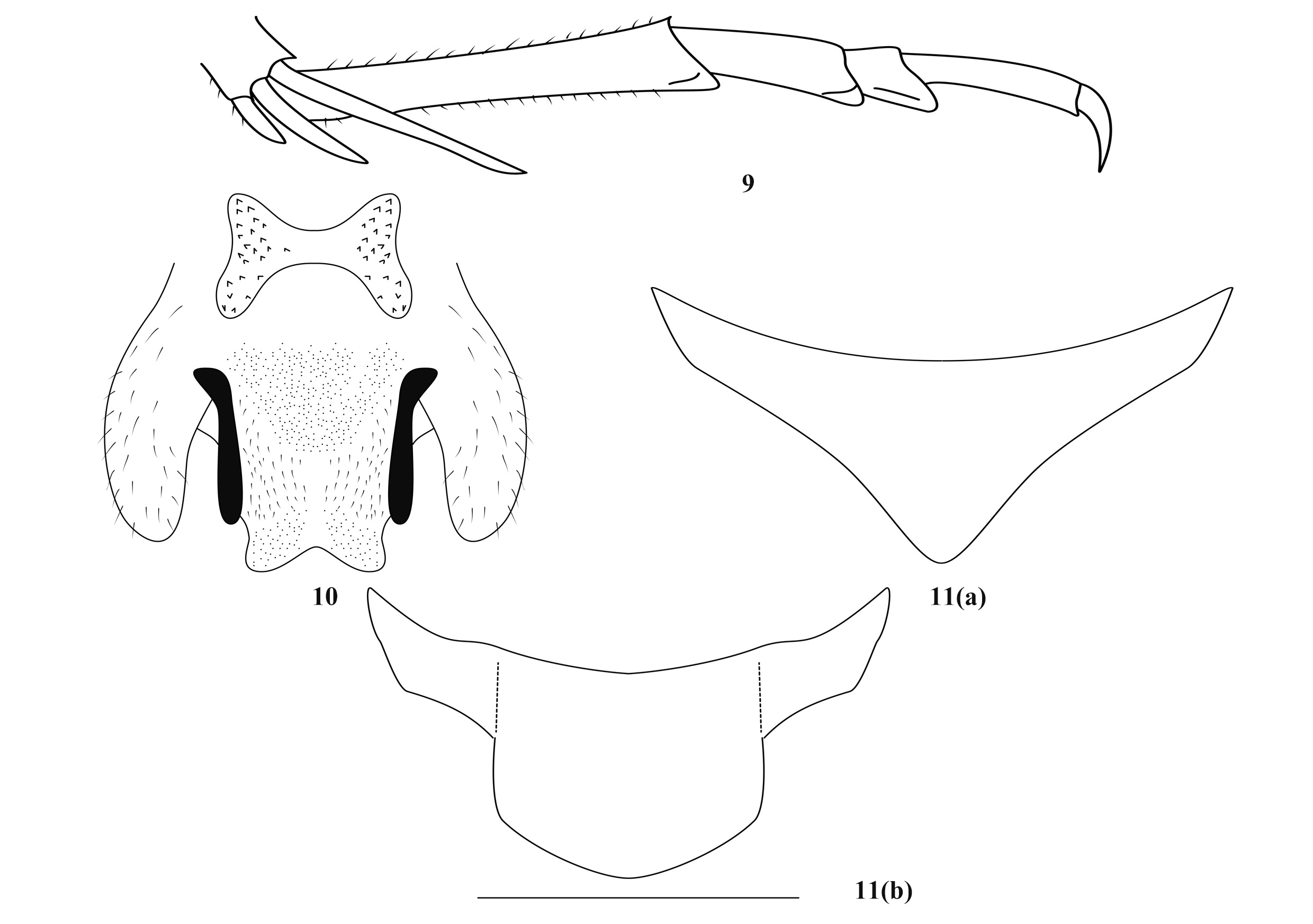
9(10) Subgenital plate of female transverse ( Fig. 11 View FIGURES 9–11 ); male genitalia as Fig. 10......... T View FIGURES 9–11 . (G.) wuyishanicus ( Zhang & Liu, 2009)
10(9) Subgenital plate of female not transverse.
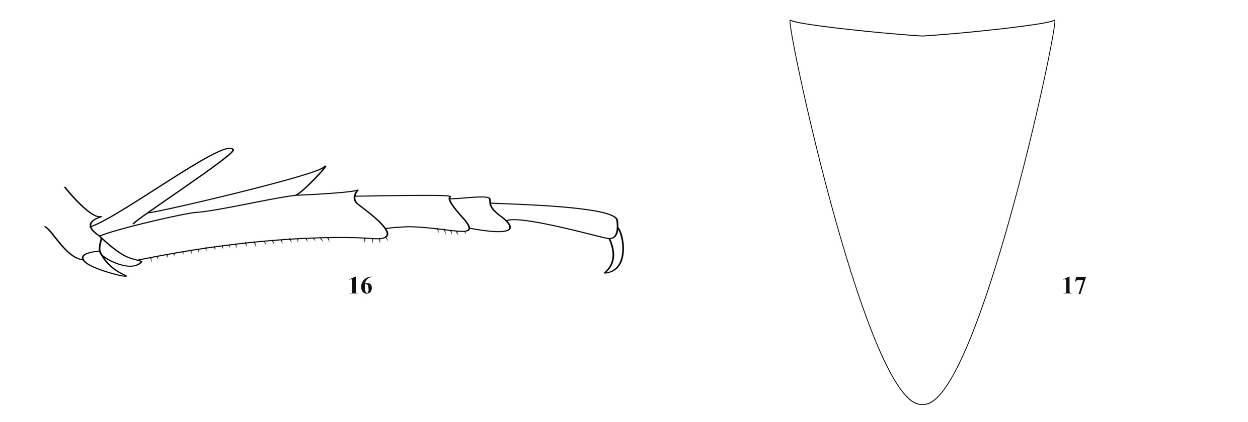
11(12) Subgenital plate of female as long as wide ( Fig. 15 View FIGURES 12–15 ); male genitalia as Figs. 13–14................. T View FIGURES 12–15 . (G.) parvus sp. nov.
12(11) Subgenital plate of female distinctly longer than wide ( Fig. 17 View FIGURES 16–17 )................. T. (G.) longilaminus ( Zhang & Liu, 2009)
13(8) Internal genicular lobe of fore femur with a short spine; hind tibiae above on each side with less than 50 spines.
14(19) Fore and mid tibiae beneath with spurs.
15(18) Mid tibiae beneath with 1–2 external spurs and 1 internal spur; 3–8th abdominal sternites with cone-like projections.
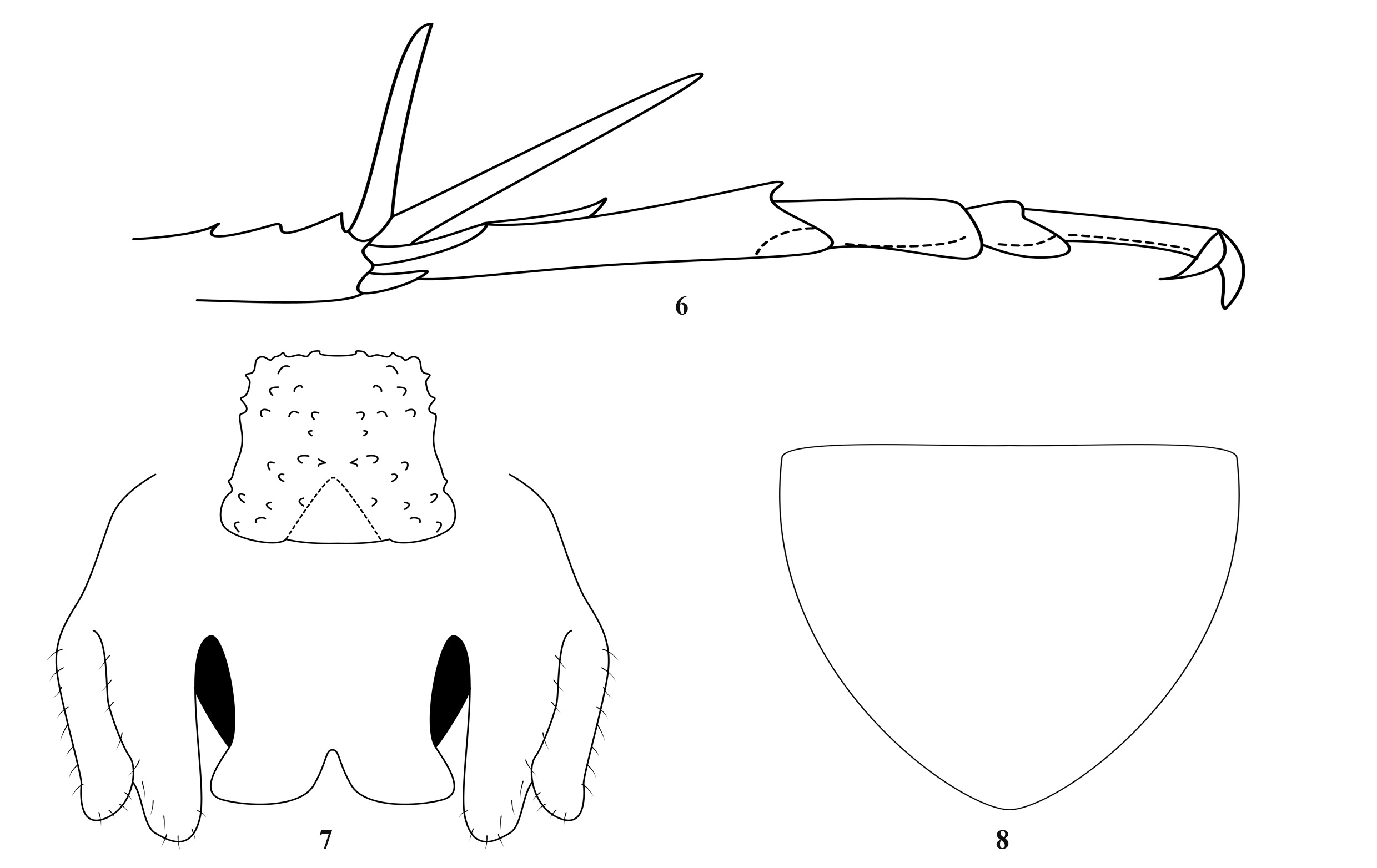
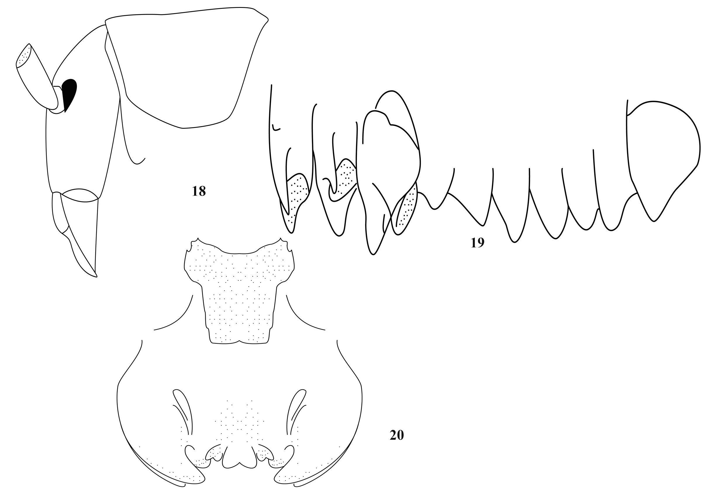
16(17) Eyes somewhat reduced ( Fig.18 View FIGURES 18–20 ); epiphallus of male as Fig. 20............... T View FIGURES 18–20 . (G.) semicrenatus ( Gorochov et al., 2006)
17(16) Eyes well developed; epiphallus of male as Fig. 21.............................. T View FIGURES 21–22 . (G.) chenhui ( Rampini et al., 2008)
18(15) Mid tibiae beneath with 1 internal spur only; 3–8th abdominal sternites without cone-like projections................................................................................................. T. (G.) cuenoti Chopard, 1929
19(14) Fore and mid tibiae beneath without spurs.
20(21) Eyes less reduced; female subgenital plate as Fig. 27........................... T View FIGURES 27–28 . (G.) caudatus ( Gorochov et al., 2006)
21(20) Eyes strongly reduced.
22(25) Eyes presented by a line.
23(24) Hind tibiae above on each side with 26–39 spines............................. T. (G.) proximus ( Gorochov et al., 2006)
24(23) Hind tibiae above on each side with 9–11 spines............................. T. (G.) ferecaecus ( Gorochov et al., 2006)
25(22) Eyes absent....................................................... T. (G.) omninocaecus ( Gorochov et al., 2006)
26(1) Hind metatarsus keeled beneath.
27(68) Supra internal spur of hind tibiae not exceeding ventral apex of hind metatarsus.
28(51) Hind tibiae above on each side with 50–90 spines.
29(38) Body smaller (hind femur shorter than 20mm).
30(37) Body unicolor; frons without dark longitudinal bands.
31(34) Epiphallus of male A-shaped.
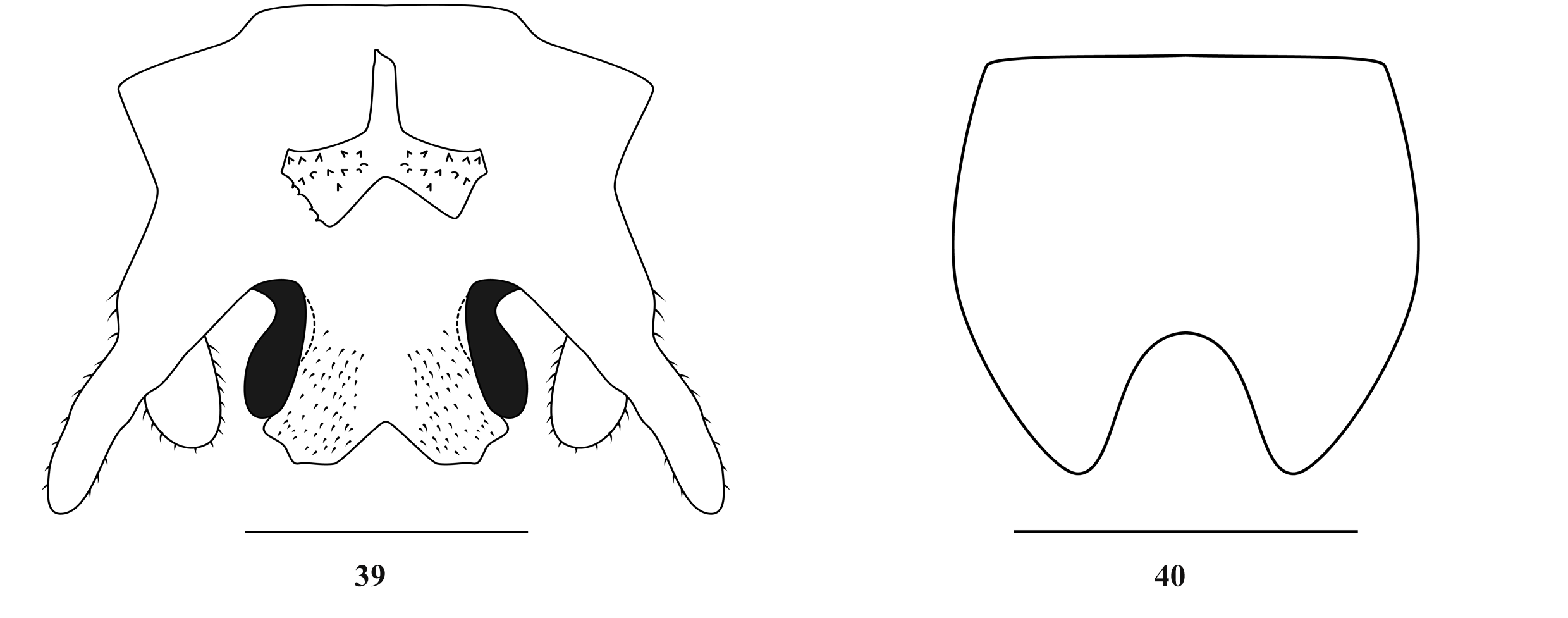
32(33) Male genitalia as Figs. 36–37 View FIGURES 36–38 ; female subgenital plate as Fig. 38.................... T View FIGURES 36–38 . (G.) beresowskii ( Adelung, 1902)
33(32) Male genitalia as Fig. 39 View FIGURES 39–40 ; female subgenital plate as Fig. 40......................... T View FIGURES 39–40 . (G.) belousovi (Gorochov, 2010)
34(31) Epiphallus of male not A-shaped.
35(36) Male genitalia as Figs. 42–43.................................................. T View FIGURES 41–44 . (G.) fallax ( Zhang & Liu, 2009)
36(35) Male genitalia as Figs. 46–47.............................................. T View FIGURES 45–47 . (G.) femoratus ( Zhang & Liu, 2009)
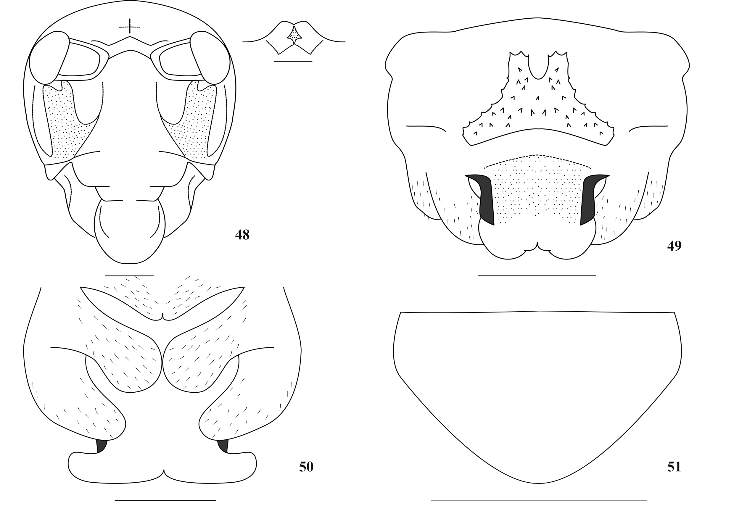
37(30) Body light brown, mottled with dark blotches; frons with 2 dark longitudinal bands ( Fig. 48 View FIGURES 48–51 ); male genitalia as Figs. 49–50 T View FIGURES 48–51 . (G.) lushuicus sp. nov.
38(29) Body larger (hind femur longer than 20mm).
39(40) Epiphallus of male distinctly transverse ( Fig. 52 View FIGURE 52 ).................................. T. (G.) bifurcatus (Gorochov, 2010)
40(39) Epiphallus of male not transverse.
41(46) Subgenital plate of female with triangular median lobe.
42(43) Epiphallus of male nearly rectangular............................................. T. (G.) racovitzai Chopard, 1915
43(42) Epiphallus of male H-shaped.
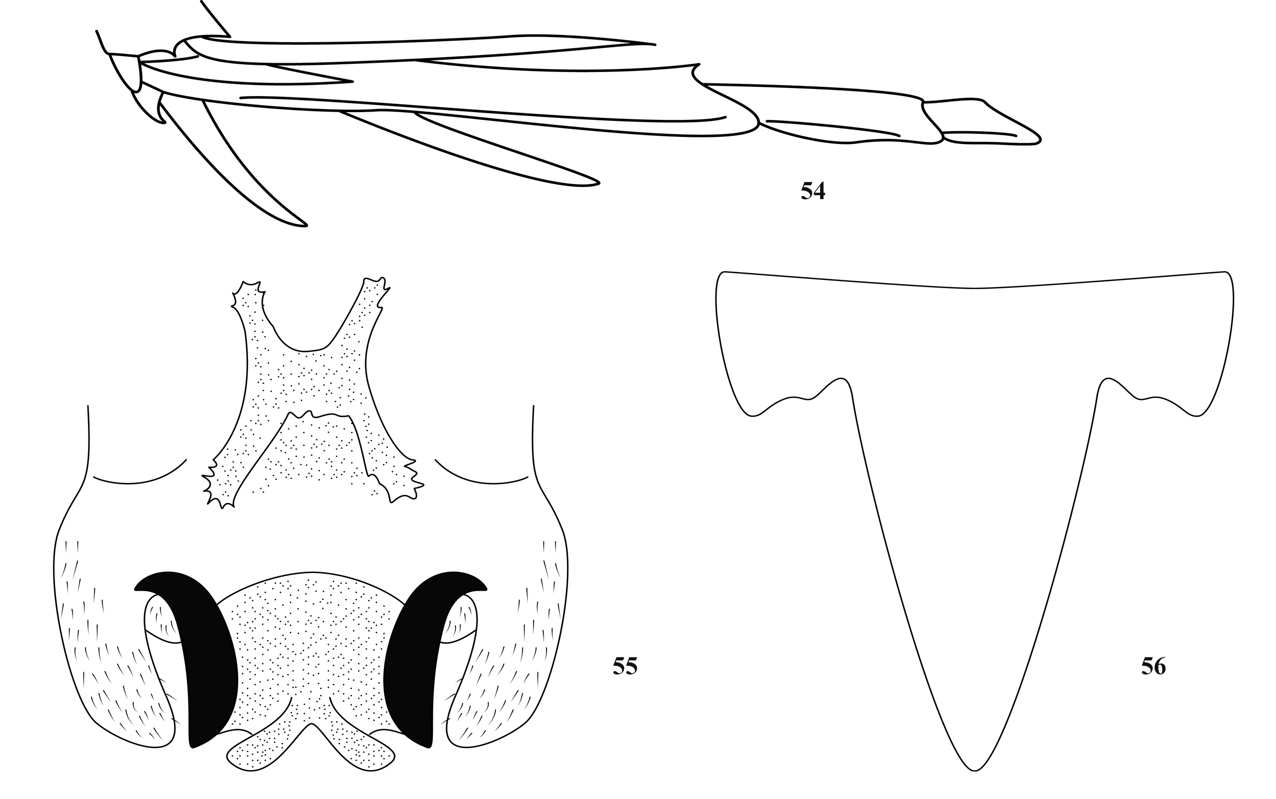
44(45) Male genitalia as Fig. 55 View FIGURES 54–56 ; female subgenital plate as Fig. 56...................... T View FIGURES 54–56 . (G.) solidus ( Gorochov et al., 2006)
45(44) Male genitalia as Fig. 57 View FIGURES 57–58 ; female subgenital plate as Fig. 58....................... T View FIGURES 57–58 . (G.) latellai ( Rampini et al., 2008) 46(41) Subgenital plate of female with rounded median lobe.
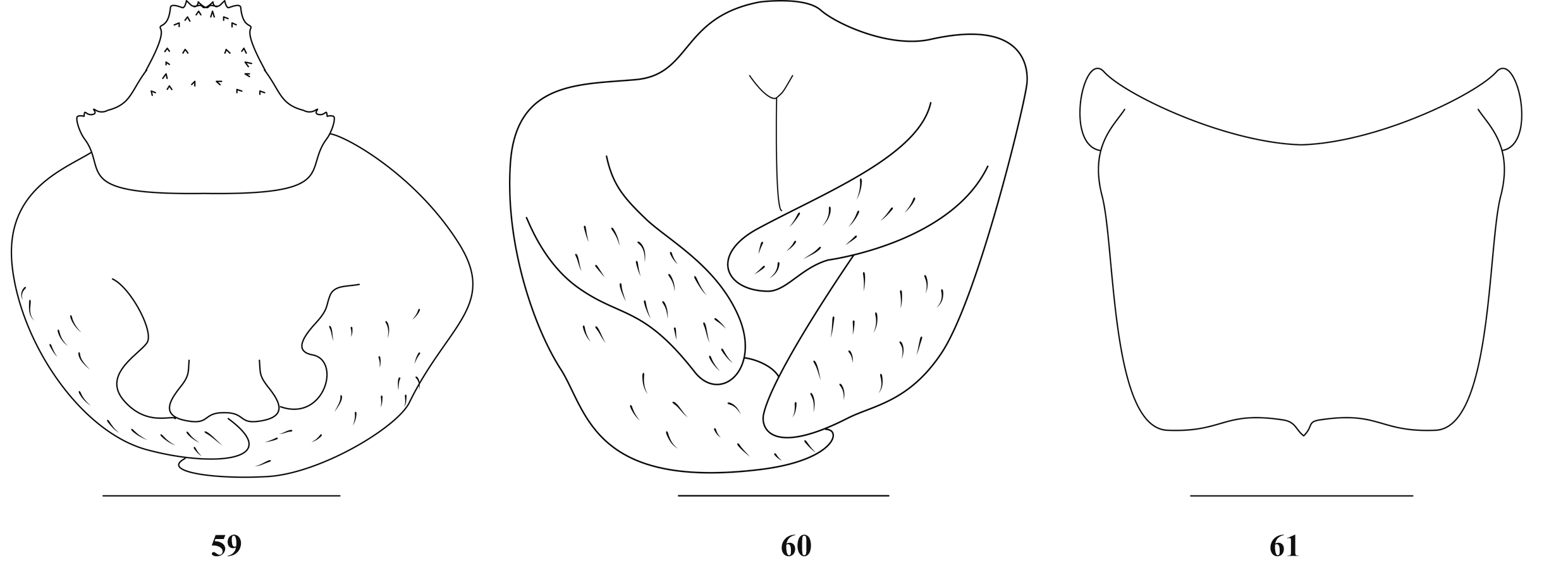
47(48) Body unicolor; male genitalia as Figs. 59–60 View FIGURES 59–61 ; female subgenital plate as Fig. 61.............. T View FIGURES 59–61 . (G.) yueyangensis sp. nov.
48(47) Body light brown, mottled with dark blotches.
49(50) Apex of female subgenital plate bluntly projected ( Fig. 63 View FIGURES 62–63 )........................... T. (G.) latus ( Zhang & Liu, 2009)
50(49) Apex of female subgenital plate bluntly truncate ( Fig. 65 View FIGURES 64–65 )........................ T. (G.) roundatus ( Zhang & Liu, 2009)
51(28) Hind tibiae above on each side with 30–50 spines.
52(57) Fore femur about 2.4–2.5 times longer than the pronotum.
53(54) Abdominal sternites with short ventral projections; male genitalia as Fig. 66........... T View FIGURES 66–67 . (G.) zorzini ( Rampini et al., 2008)
54(53) Abdominal sternites without ventral projections.
55(56) Male genitalia as Figs. 69–70 View FIGURES 68–71 ; subgenital plate of female as Fig. 71................... T View FIGURES 68–71 . (G.) borutzkyi ( Gorochov, 1994)
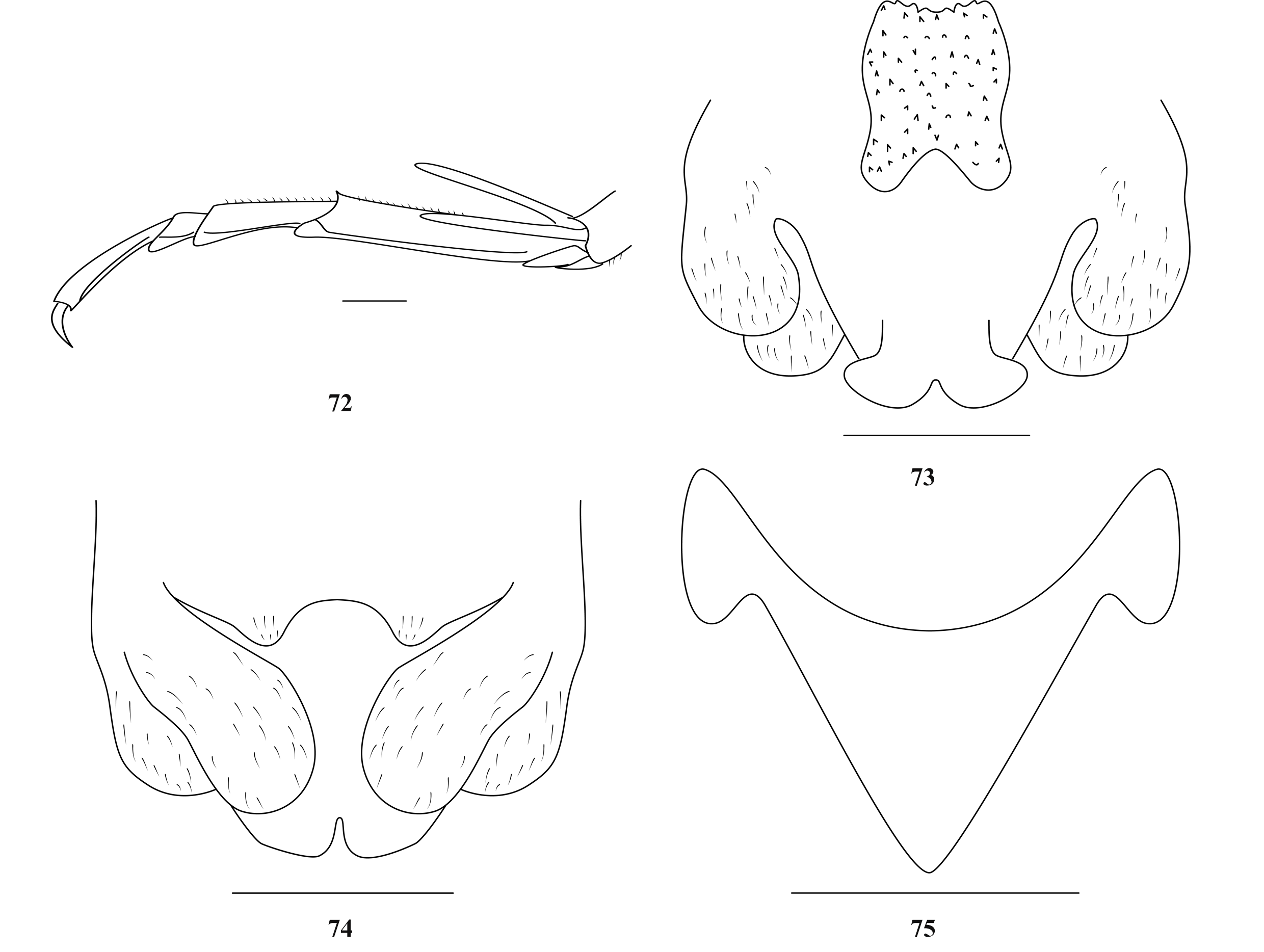
56(55) Male genitalia as Figs. 73–74 View FIGURES 72–75 ; subgenital plate of female as Fig. 75................................. T View FIGURES 72–75 . (G.) lii sp. nov.
57(52) Fore femur about 2.0–2.2 times longer than the pronotum.
58(59) Body larger (hind femur longer than 20mm); epiphallus of male as Fig. 76................ T View FIGURES 76–77 . (G.) coomani Chopard, 1929
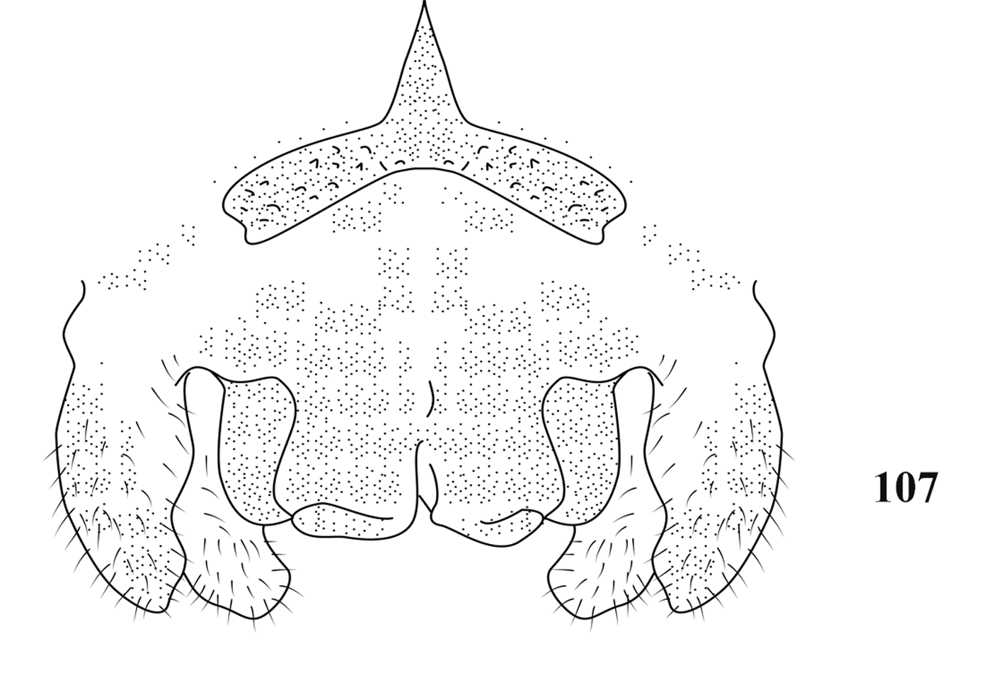
59(58) Body smaller (hind femur shorter than 20mm).
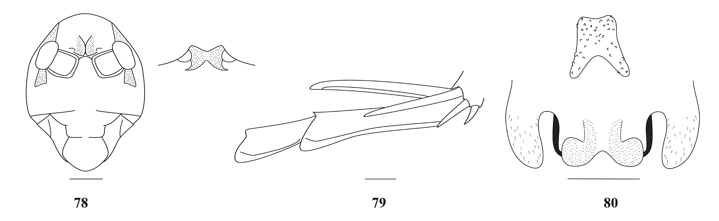
60(61) Fore tibiae beneath with 2 external spurs and 2 internal spurs; male genitalia as Fig. 80.......... T View FIGURES 78–80 . (G.) dianxicus sp. nov.
61(60) Fore tibiae beneath with 1–2 external spurs and 1 internal spur.
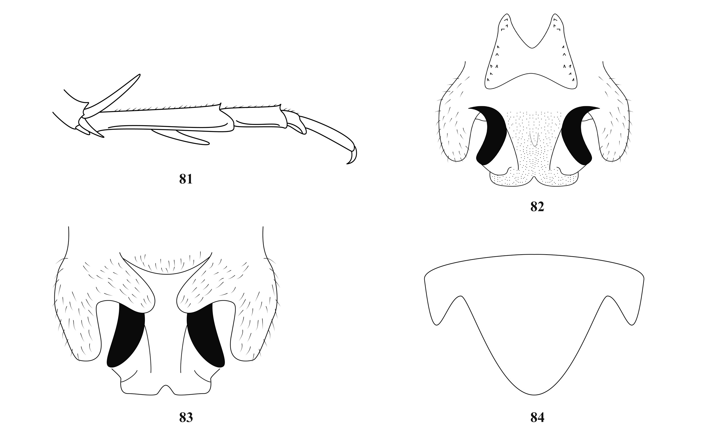
62(63) Epiphallus of male nearly H-shaped ( Fig. 82 View FIGURES 81–84 ); subgenital plate of female as Fig. 84....... T View FIGURES 81–84 . (G.) cavernus ( Jiao et al., 2008)
63(62) Epiphallus of male genitalia nearly subquadrate.
64(65) Apex of subgenital plate of female notched; male genitalia as Figs. 85–86.................... T View FIGURES 85–87 . (G.) bruneri Karny, 1934
65(64) Apex of subgenital plate of female blunt.
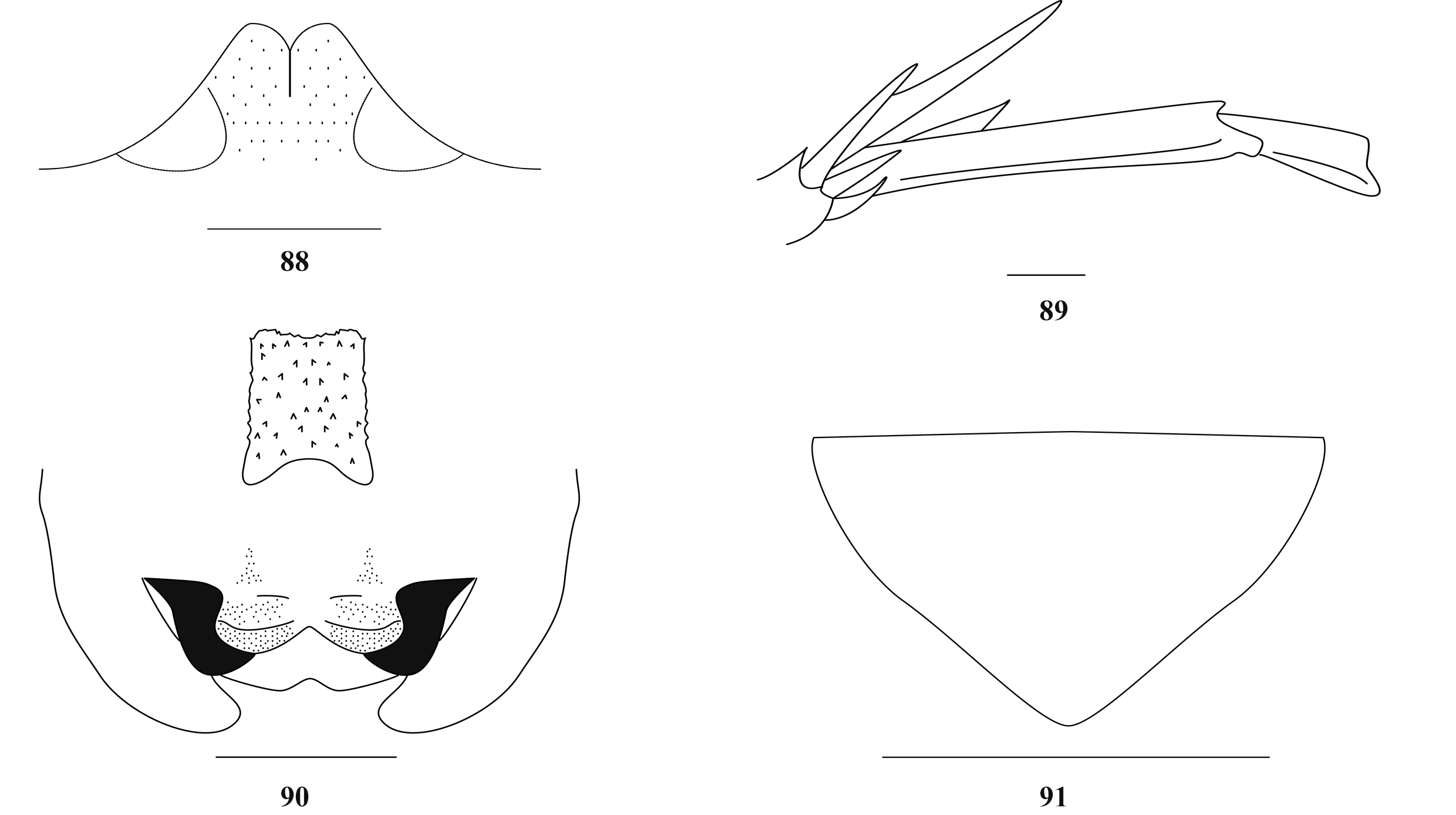
66(67) Male genitalia as Fig. 90 View FIGURES 88–91 ; subgenital plate of female as Fig. 91................................ T View FIGURES 88–91 . (G.) pallidus sp. nov.
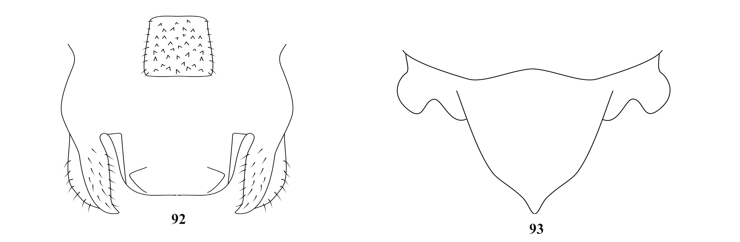
67(66) Male genitalia as Fig. 92 View FIGURES 92–93 ; subgenital plate of female as Fig. 93...................... T View FIGURES 92–93 . (G.) sonlaensis (Gorochov, 1990)
68(27) Supra internal spur of hind tibiae almost exceeding ventral apex of hind metatarsus.
69(76) Body larger (hind femur longer than 20mm).
70(73) Hind tibiae above on each side with 70–90 spines.
71(72) Male genitalia as Fig. 94........................................................ T View FIGURES 94–96 . (G.) adelungi Chopard, 1921
72(71) Male genitalia as Fig. 98..................................................... T View FIGURES 97–99 . (G.) nocturnus ( Gorochov, 1992)
73(70) Hind tibiae above on each side with 50–70 spines.
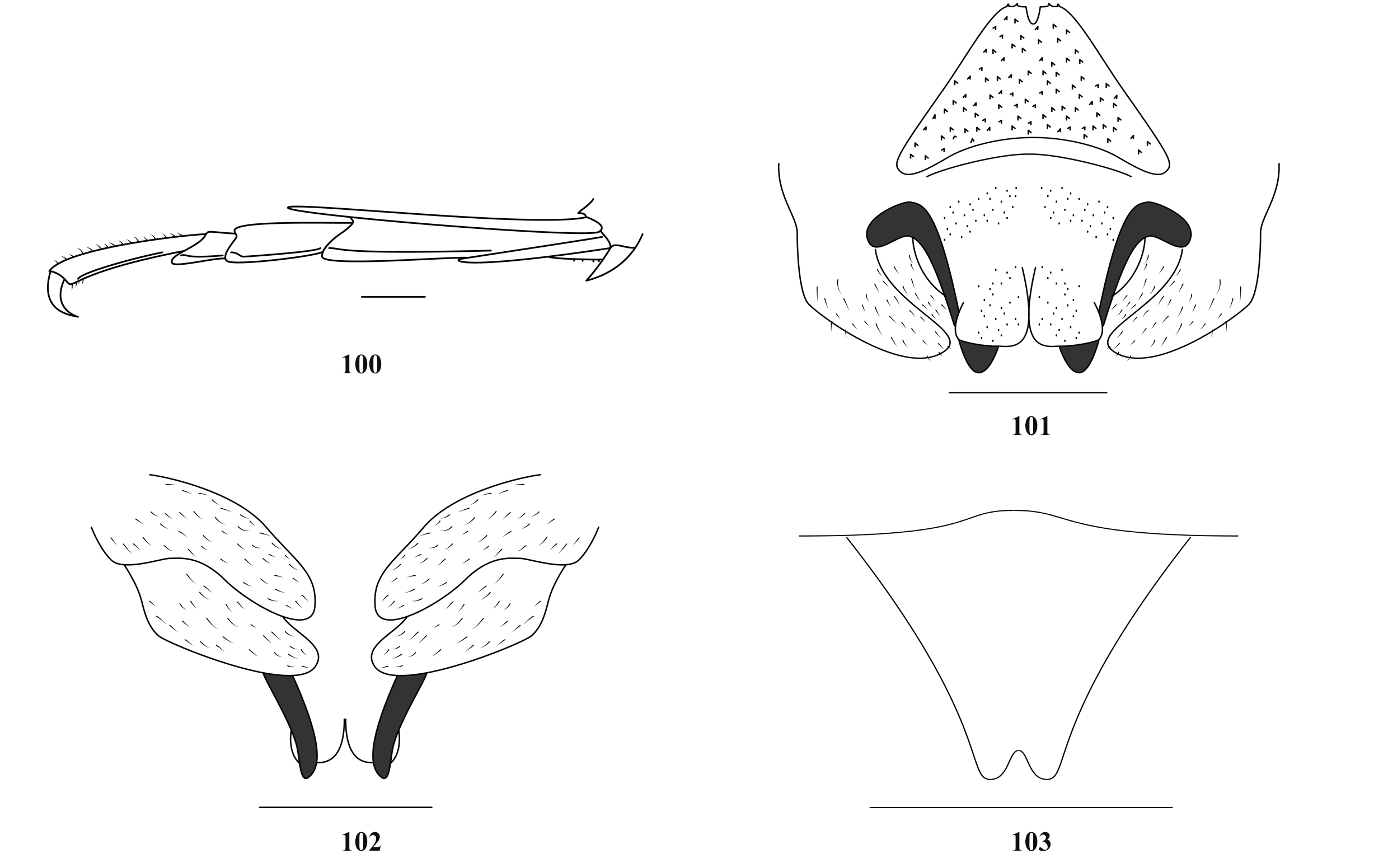
74(75) Hind tibiae above on each side with 61–67 spines; male genitalia as Figs. 101–102................. T View FIGURES 100–103 . (G.) dispar sp. nov.
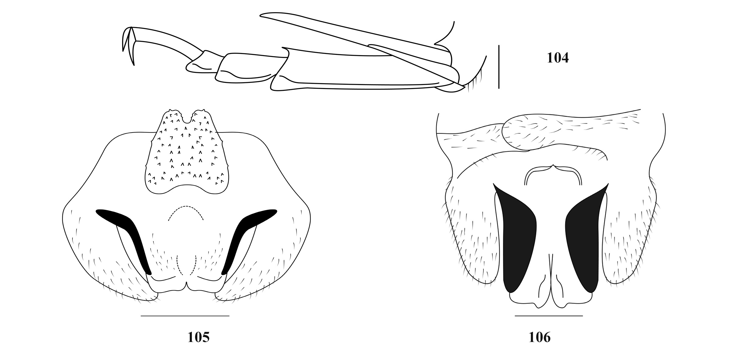
75(74) Hind tibiae above on each side with 50–51 spines; male genitalia as Figs. 105–106.................. T View FIGURES 104–106 . (G.) verus sp. nov.
76(69) Body smaller (hind femur shorter than 20mm).
77(82) Epiphallus of male A-shaped.
78(81) Fore tibiae beneath with 2 external spurs and 2 internal spurs.
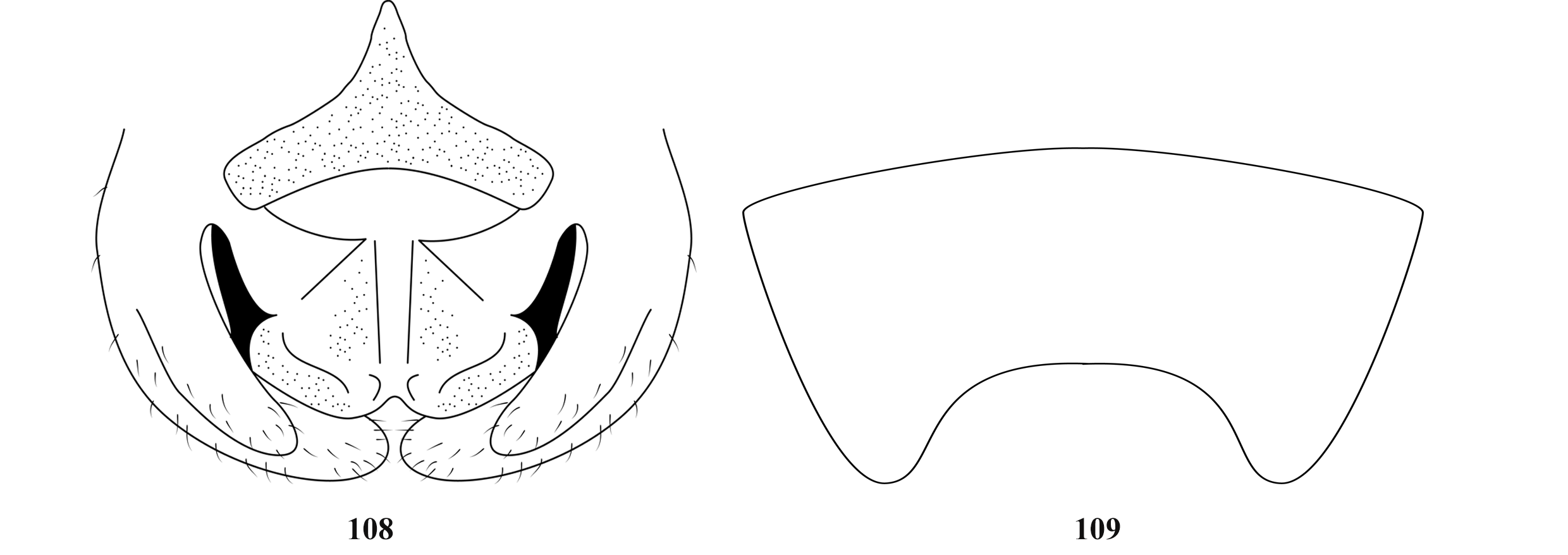
79(80) Male genitalia as Fig. 107...................................................... T View FIGURE 107 . (G.) gansu (Gorochov, 2010)
80(79) Male genitalia as Fig. 108 View FIGURES 108–109 ; subgenital plate of female as Fig. 109....................... T View FIGURES 108–109 . (G.) longicaudus Karny, 1934
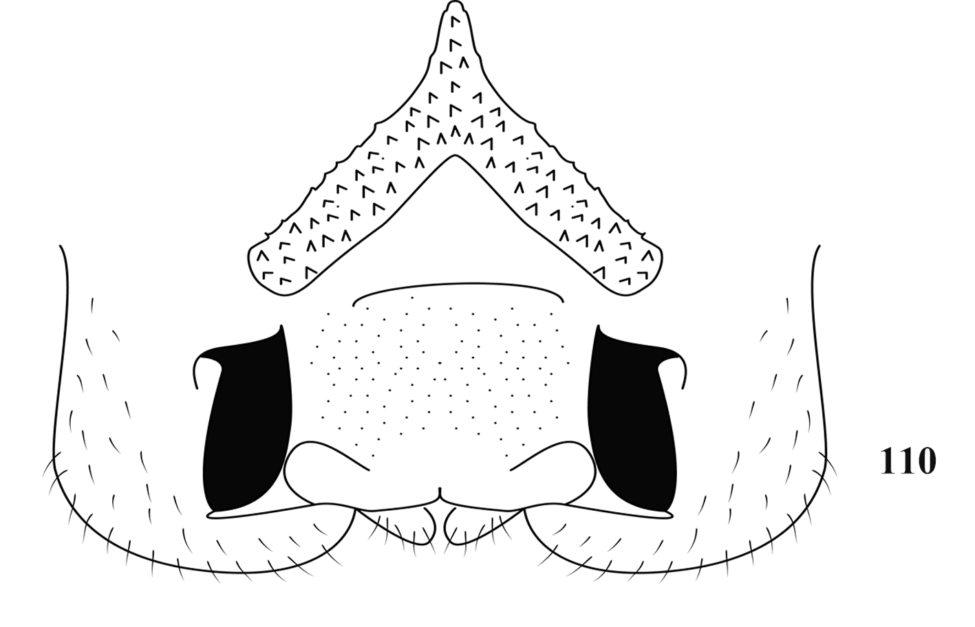
81(78) Fore tibiae beneath with 2 external spurs and 1 internal spur; male genitalia as Fig. 110..... T View FIGURE 110 . (G.) kabaki (Gorochov, 2010)
82(77) Epiphallus of male not A-shaped.
83(86) Epiphallus of male nearly triangular.
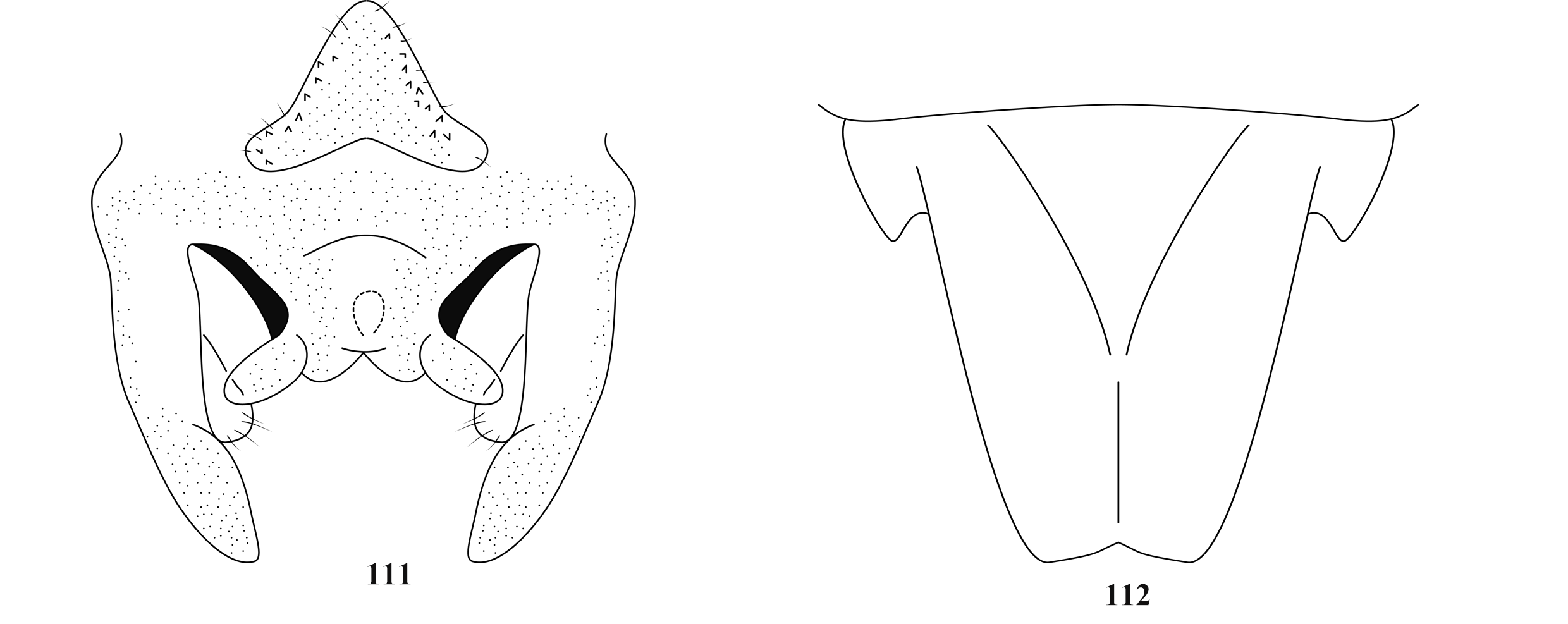
84(85) Male genitalia as Fig. 111.................................................... T View FIGURES 111–112 . (G.) sichuanus (Gorochov, 2010)
85(84) Male genitalia as Fig. 113................................................. T View FIGURE 113 . (G.) altimontanus (Gorochov, 2010)
86(83) Epiphallus of male not triangular-shaped.
87(88) Male genitalia as Fig. 115.............................................................. T View FIGURES 114–116 . (G.) vicinus sp. nov.
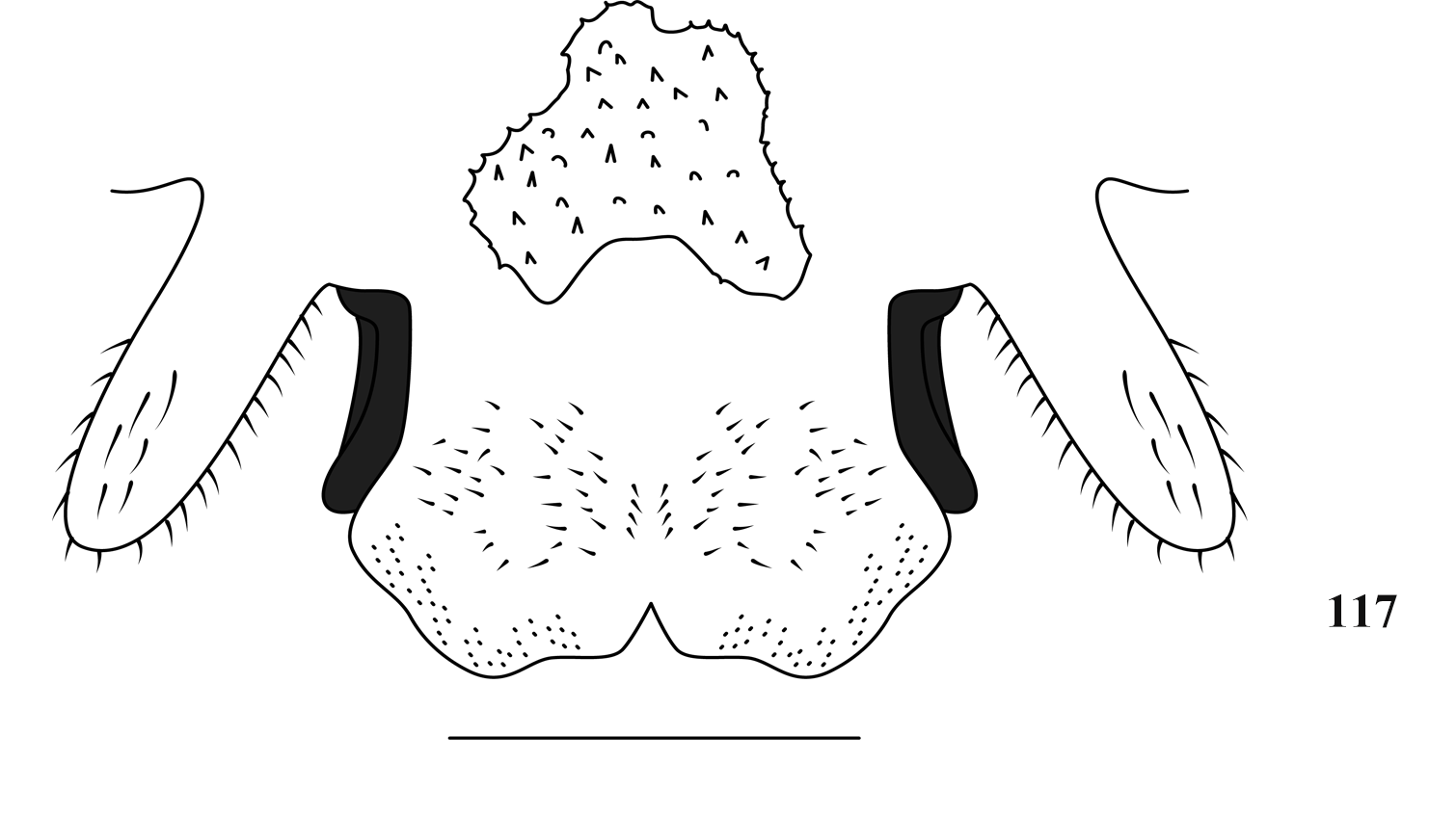
88(87) Male genitalia as Fig. 117...................................................... T View FIGURE 117 . (G.) maoershanensis sp. nov.


No known copyright restrictions apply. See Agosti, D., Egloff, W., 2009. Taxonomic information exchange and copyright: the Plazi approach. BMC Research Notes 2009, 2:53 for further explanation.
|
Kingdom |
|
|
Phylum |
|
|
Class |
|
|
Order |
|
|
Family |
|
|
SubFamily |
Aemodogryllinae |
|
Tribe |
Aemodogryllini |
|
Genus |
|
|
SubGenus |
Tachycines |
|
Kingdom |
|
|
Phylum |
|
|
Class |
|
|
Order |
|
|
Family |
|
|
SubFamily |
Aemodogryllinae |
|
Tribe |
Aemodogryllini |
|
Genus |
|
|
SubGenus |
Tachycines |
|
Kingdom |
|
|
Phylum |
|
|
Class |
|
|
Order |
|
|
Family |
|
|
SubFamily |
Aemodogryllinae |
|
Tribe |
Aemodogryllini |
|
Genus |
|
|
SubGenus |
Tachycines |
|
Kingdom |
|
|
Phylum |
|
|
Class |
|
|
Order |
|
|
Family |
|
|
SubFamily |
Aemodogryllinae |
|
Tribe |
Aemodogryllini |
|
Genus |
|
|
SubGenus |
Tachycines |
|
Kingdom |
|
|
Phylum |
|
|
Class |
|
|
Order |
|
|
Family |
|
|
SubFamily |
Aemodogryllinae |
|
Tribe |
Aemodogryllini |
|
Genus |
|
|
SubGenus |
Tachycines |
|
Kingdom |
|
|
Phylum |
|
|
Class |
|
|
Order |
|
|
Family |
|
|
SubFamily |
Aemodogryllinae |
|
Tribe |
Aemodogryllini |
|
Genus |
|
|
SubGenus |
Tachycines |
|
Kingdom |
|
|
Phylum |
|
|
Class |
|
|
Order |
|
|
Family |
|
|
SubFamily |
Aemodogryllinae |
|
Tribe |
Aemodogryllini |
|
Genus |
|
|
SubGenus |
Tachycines |
|
Kingdom |
|
|
Phylum |
|
|
Class |
|
|
Order |
|
|
Family |
|
|
SubFamily |
Aemodogryllinae |
|
Tribe |
Aemodogryllini |
|
Genus |
|
|
SubGenus |
Tachycines |
|
Kingdom |
|
|
Phylum |
|
|
Class |
|
|
Order |
|
|
Family |
|
|
SubFamily |
Aemodogryllinae |
|
Tribe |
Aemodogryllini |
|
Genus |
|
|
SubGenus |
Tachycines |
|
Kingdom |
|
|
Phylum |
|
|
Class |
|
|
Order |
|
|
Family |
|
|
SubFamily |
Aemodogryllinae |
|
Tribe |
Aemodogryllini |
|
Genus |
|
|
SubGenus |
Tachycines |