
Doryphoribius dawkinsi, Michalczyk, Łukasz & Kaczmarek, Łukasz, 2010
|
publication ID |
https://doi.org/ 10.5281/zenodo.193905 |
|
DOI |
https://doi.org/10.5281/zenodo.5668448 |
|
persistent identifier |
https://treatment.plazi.org/id/751687E9-B953-FFEC-FF00-FF40E964FB59 |
|
treatment provided by |
Plazi |
|
scientific name |
Doryphoribius dawkinsi |
| status |
sp. nov. |
Doryphoribius dawkinsi sp. nov.
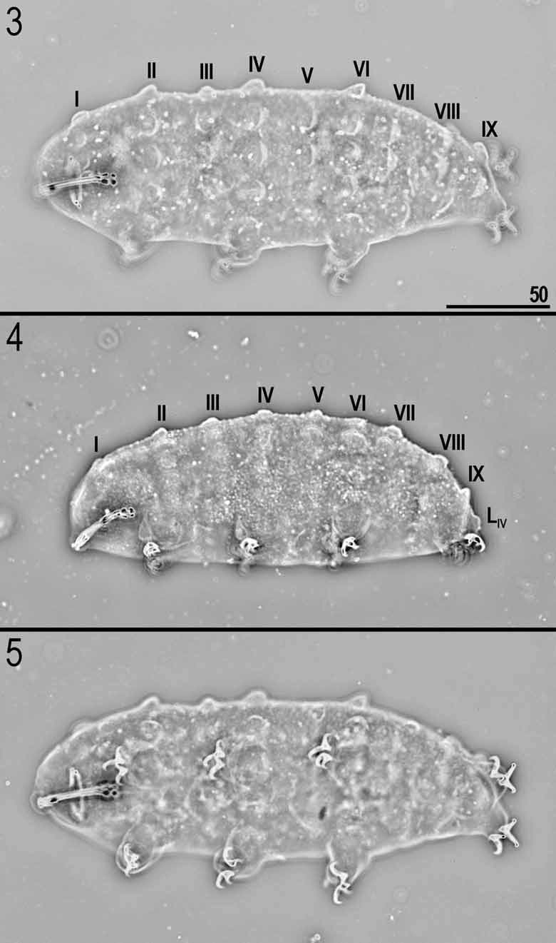
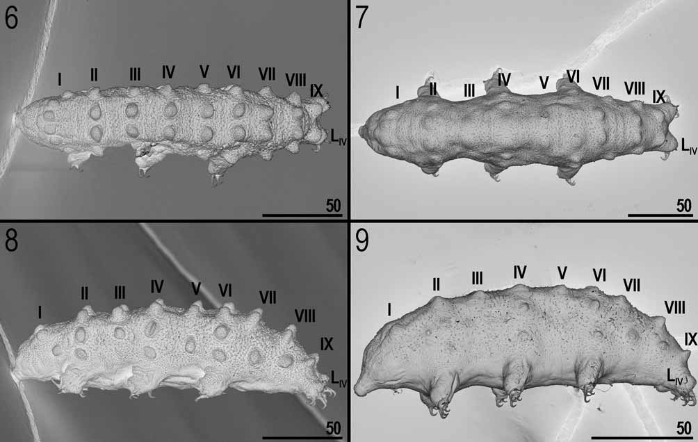
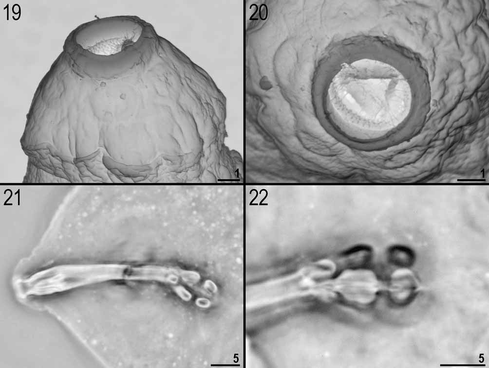
FIGURES 3–26 View FIGURES 3 – 5 View FIGURES 6 – 9 View FIGURES 19 – 22 View FIGURES 23 – 26. 23 – 25 , TABLE 2 View TABLE 2
Material examined: Holotype and 16 paratypes: Central America, Costa Rica: Cartago Province, Heredia, 84.11289°W and 10.00285°N, 1161 m asl, urban area, moss from tree, 1 specimen; Guanacaste Province, in the vicinity of Cenzosa Vista al Mar, the “Bosque Nacional Diria” forest, 85.63004°W and 10.12553°N, 960 m asl, premontane wet forest, liverworts from tree, 13 specimens; Puntarenas Province, San Vito, in the vicinity of the cemetery, 82.97075°W and 8.81742°N, 1020 m asl, urban area, liverworts from tree, 2 specimens; San Jose Province, Chirripo National Park, 83.60058°W and 9.46502°N, 1288 m asl, premontane wet forest, moss from stone, 1 specimen. All collected by the second author in December 2002.
Description (see Table 2 View TABLE 2 for measurements, pt values and basic statistics): Body yellow ( Figs. 3–9 View FIGURES 3 – 5 View FIGURES 6 – 9 ). Eyes absent in all examined specimens. Dorso-lateral cuticle covered with small (0.5–1.5 in diameter), irregular, often elongated tubercles (Figs. 10, 13–14, 16–18). Apart from evenly scattered tubercles, nine rows of gibbosities cover dorso-lateral parts of the body. Additionally, there is also a single gibbosity on each of the hind legs (Fig. 18). The gibbosity configuration is as follows: IX:6-6-4-6-4-6-4-4-2+2[L IV] ( Figs. 3–4 View FIGURES 3 – 5 , 6–9 View FIGURES 6 – 9 ). Each of the most lateral gibbosities in rows I, VII and VIII is placed slightly posteriorly relative to the dorsal ones (i.e., gibbosities I c, VII b and VIII b) ( Figs. 8–9 View FIGURES 6 – 9 , 12). Gibbosity rows II, IV and VI are in line with legs I, II and III, respectively. Gibbosities in row I are slightly smaller than all other gibbosities. Both the cuticular sculpture and gibbosities exhibit significant variation in their appearance. Namely, in some individuals cuticular tubercles are scattered and weakly visible (Fig. 15), whereas in others they are distinct and densely arranged (Fig. 13). Locally, elongated tubercles may merge and form a quasi-reticulum (Figs. 16–17). Also, in some animals gibbosities are clearly defined and easy to count ( Figs. 3–6, 8 View FIGURES 3 – 5 View FIGURES 6 – 9 ), whereas in others they are only weakly outlined and may be difficult to identify, especially under LM ( Figs. 7, 9 View FIGURES 6 – 9 ). Legs I–III without gibbosities. Ventral cuticle smooth (i.e., without tubercles and gibbosities).
On the dorsal side of the head there is a small round or triangular tubercle (ca. 2.0 in diameter) (Figs. 11– 12, see also Remarks). This structure is visible only in SEM.
Mouth antero-ventral. Bucco-pharyngeal apparatus of the Doryphoribius type ( Figs. 19–22 View FIGURES 19 – 22 ). Peribuccal lamellae and papulae absent. Oral cavity armature visible only in SEM ( Figs. 19–20 View FIGURES 19 – 22 ), consisting of a single band of very small (ca. 0.1 in diameter), cone shaped teeth arranged in ca. 5 rows placed on the ring fold in the centre of the oral cavity. Buccal tube with a single anterior bend (visible in lateral view only) and with a ventral lamina ( Fig. 21 View FIGURES 19 – 22 ). At the end of the buccal tube rounded pharyngeal apophyses present. Pharyngeal bulb spherical with two macroplacoids decreasing in length (i.e., configuration 2-1) ( Fig. 22 View FIGURES 19 – 22 ). The first macroplacoid in the shape of a short rod, with a slight central constriction. The second macroplacoid granular, without constrictions. Microplacoid and septulum absent.
Claws of the Isohypsibius type, similar in shape on all legs, with short basal claws ( Figs. 23–25 View FIGURES 23 – 26. 23 – 25 ). Lunules absent, but claw bases may be occasionally slightly widened. Internal claws I–III and anterior claws IV smaller than external claws I–III and posterior claws IV, respectively. However, the difference between the claws is smaller on hind legs than on legs I–III. Claws I–III similar in size and all smaller than claws IV. Primary branches of claws with thin, but well visible accessory points ending at the highest point of the primary branch. Bars and other cuticular thickenings on legs absent.
Eggs unknown (see also Remarks).
Remarks: Given that the head tubercle observed in SEM (Figs. 11–12) is in a location that corresponds with the base of the median cirrus in marine heterotardigrades (e.g., Stygarctus), it could be a homologous sensory organ. Interestingly, conspicuous cuticular structures in the same location were described also in other eutardigrades (e.g., cuticular depression in Macrobiotus derkai reported by Degma et al. 2008).
Although eggs are unknown, we should probably expect them to be smooth and deposited in exuvium as in all other known Doryphoribius species.
Locus typicus: Guanacaste Province, in the vicinity of Cenzosa Vista al Mar, the “Bosque Nacional Diria” forest, 85.63004°W and 10.12553°N, 960 m asl, Premontane wet forest, liverworts from tree.
Etymology: We take great pleasure in dedicating the new species to a distinguished scientist, Professor Richard Dawkins (Oxford University, UK), and by this we thank him for his input in evolutionary biology as well as for his tireless popularisation of science, rationalism, scepticism and critical thinking and all actions against pseudoscience, religion and other sorts of superstition.
CHARACTER N RANGE MEAN SD Holotype Type depositories: Holotype and five paratypes on one slide (no. CR935/1) and other four paratypes (slides no. CR28n/2, CR253/2, and CR168/4) mounted in Hoyer’s medium are preserved at the Department of Animal Taxonomy and Ecology, A. Mickiewicz University, Umultowska 89, 61-614 Poznań, Poland.

No known copyright restrictions apply. See Agosti, D., Egloff, W., 2009. Taxonomic information exchange and copyright: the Plazi approach. BMC Research Notes 2009, 2:53 for further explanation.