
Pheronemoides fungosus, , Latin, 2017
|
publication ID |
https://doi.org/ 10.11646/zootaxa.4337.1.7 |
|
publication LSID |
lsid:zoobank.org:pub:279ABC49-BEB5-4C5A-8FAD-3CCE205DAD43 |
|
DOI |
https://doi.org/10.5281/zenodo.6042444 |
|
persistent identifier |
https://treatment.plazi.org/id/75578790-9C3D-DD0A-EAB3-FDD8FE7771C0 |
|
treatment provided by |
Plazi |
|
scientific name |
Pheronemoides fungosus |
| status |
gen. et sp. nov. |
Pheronemoides fungosus View in CoL gen. et sp. nov.
( Figures 1–3 View FIGURE 1 View FIGURE 2 View FIGURE 3 )
Material examined. Holotype: YM30037, seamount near Yap Trench (8°51.7615549'N, 137°44.4825605'E), 15 December 2014, 906 m depth, foraminiferal ooze.
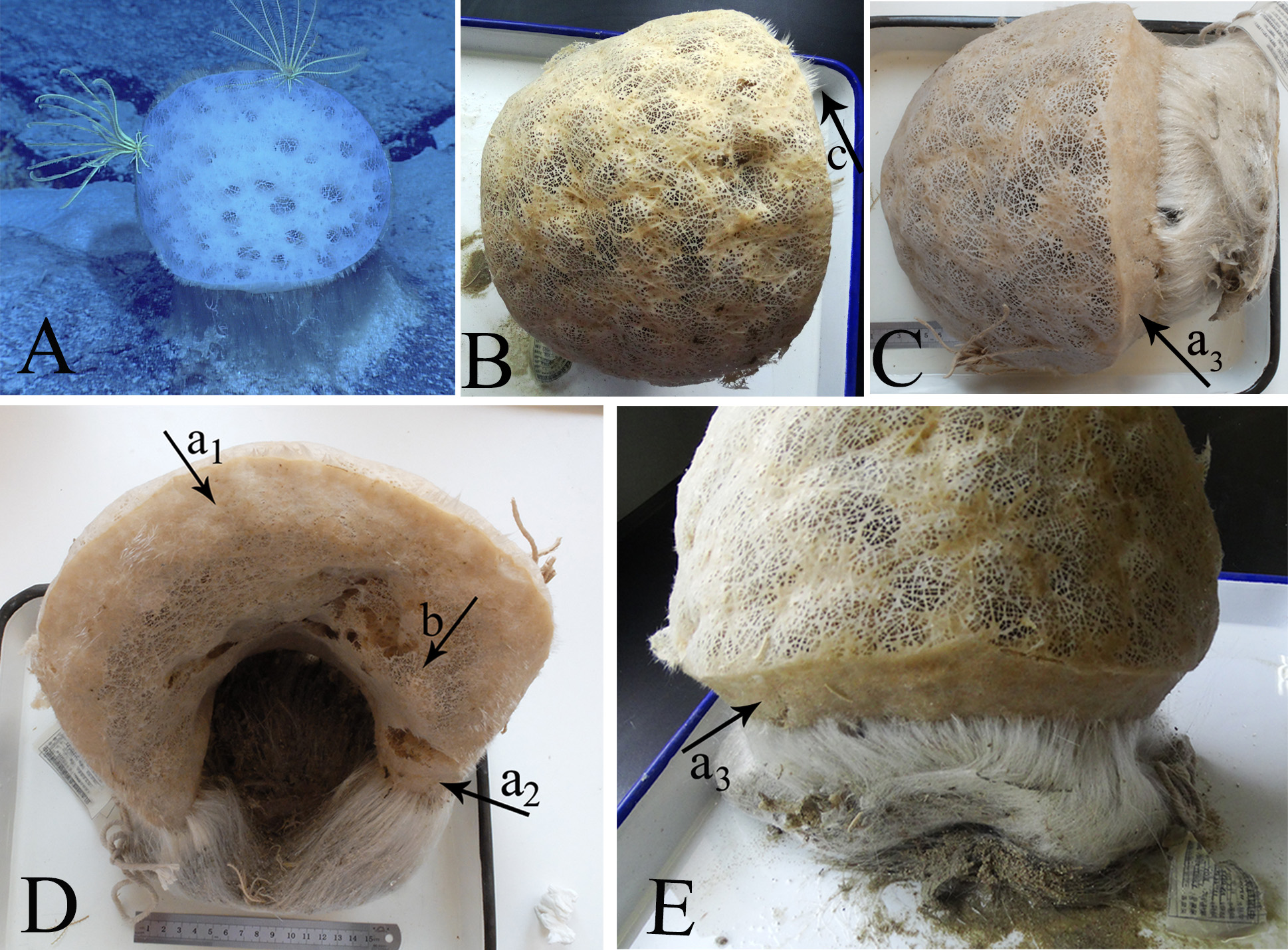
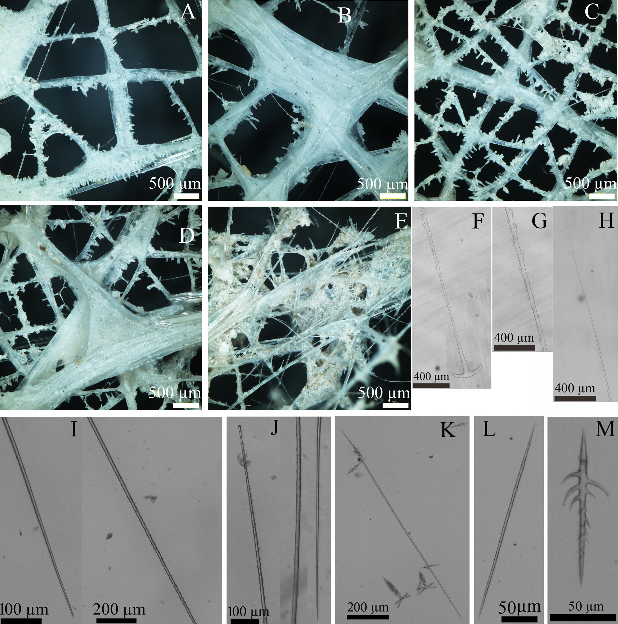
Description. Hemispherical, at least 260 mm in diameter, as observed in a living sponge viewed from above ( Fig. 1A View FIGURE 1 ). Live color white, but tan after collection. Atrial areas ( Fig. 1B View FIGURE 1 ) cover the upper surface of the sponge and dermal areas are on the lower surface ( Fig. 1D View FIGURE 1 ). The dermal areas consist of two parts, as follows. Part 1 possesses indistinctive meshes due to being covered by tissue ( Fig. 2E View FIGURE 2 ) and can be divided into three subparts ( Fig. 1D–E View FIGURE 1 , arrow a1–a3). Subpart a1 (<50 mm thick) and subpart a3 (<40 mm thick) are on the two sides around the atrial areas; subpart a2 (<22 mm thick) and subpart a3 are located on the two sides around the basalia. Part 2 ( Fig. 1D View FIGURE 1 , arrow b) has larger meshes compared with Part 1, and is located between subpart a1 and subpart a2. Observed laterally on one side (containing Part 2), the sponge is arched and hollow inside ( Fig. 1D View FIGURE 1 ); observed on the other side (containing subpart a3), it is spherical and appears mushroom-like ( Fig. 1C View FIGURE 1 ). The openings in the atrial-surface meshes and Part-2 dermal-surface meshes are easily discernible ( Fig. 2A–D View FIGURE 2 ), while those of Part-1 dermal surfaces are not as obvious as on Part-2 surfaces ( Fig. 2E View FIGURE 2 ). The atrial-surface meshes (of 0.8–2 mm diameter) ( Fig. 2A–B View FIGURE 2 ) are wider than the Part-2 dermal-surface meshes (of 0.4–0.8 mm diameter) ( Fig. 2C–D View FIGURE 2 ). Large exhalant canals are present underneath the atrial lattices. The basalia on the bottom of the sponge are located amesially, rather than exactly at the center on the dermal surface. Observed from the underside, the basalia attach to the dermal surface as a semicircle, thus leaving a large hollow between the basalia and Part-2 dermal areas ( Fig. 1D View FIGURE 1 ). Tufts of basalia,> 120 mm in length, consist of many small spicule tufts (2–8 mm in width). Some marginalia are present on the boundary between the atrial and dermal surfaces ( Fig. 1B View FIGURE 1 , arrow c).
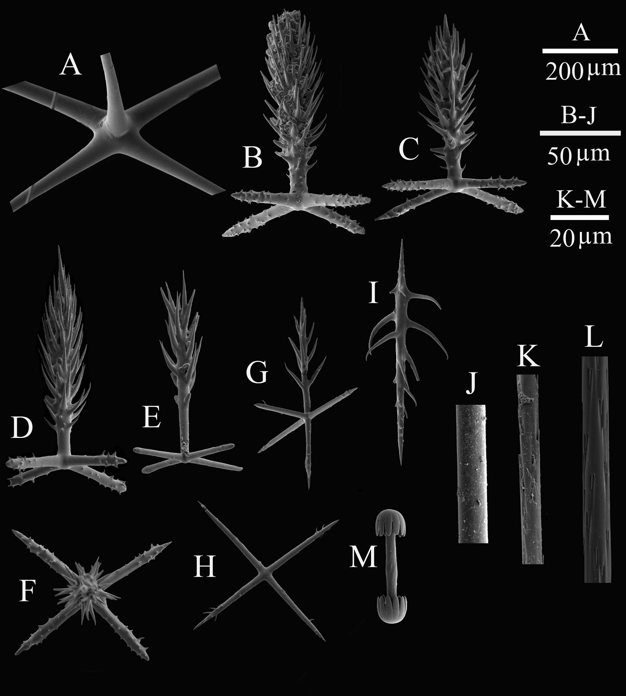
Spicules. The choanosomal skeleton consists of pentactins and uncinates. The choanosomal pentactins ( Fig. 3A View FIGURE 3 ) have smooth tangential rays (length: 216.6–4217.9 µm) and smooth proximal rays (length: 265.8–4000.1 µm). There are three types of uncinates: macrouncinates, mesouncinates and microuncinates. The macrouncinates ( Fig. 3L View FIGURE 3 ) are easily broken and reach several millimeters in length and 8.9–16.0 µm in width. The mesouncinates, covered by many tiny brackets and barbs on the shaft ( Fig. 2K View FIGURE 2 & Fig. 3K View FIGURE 3 ), are 879.7–1094.2 µm in length and 7.4– 7.6µm in width. Microuncinates ( Fig. 2L View FIGURE 2 ), are 140.5–312.7 µm in length and 2.4–3.2 µm in width. Dermalia are pinular pentactins ( Fig. 3D View FIGURE 3 ) which have tangential rays that are barely spined (length: 34.4–74.8 µm) and pinular rays (length: 91.8–214.9 µm) that are spindle-like, bushy with lateral spines and a terminal spine. Atrialia are pinular pentactins ( Fig. 3B–C View FIGURE 3 ), similar to but smaller than the dermalia; their pinular rays are 88.9–137.8 µm in length and 38.5–64.9 µm in total width. Choanosomal pinular pentactins invariably have fewer lateral spines on the pinular ray and are slender than the atrialia and dermalia. The shape of the tangential rays resembles an ‘X’ or ‘+’ ( Fig. 3E–F View FIGURE 3 ), and their length is variable: proximal rays are 69.7–268.5 µm, tangential rays are 27.3–61.6 µm. A small number of pinular hexactins can also be found ( Fig. 3G View FIGURE 3 ). Basalia have a two-toothed anchor ( Fig. 2F View FIGURE 2 ), spiny shaft ( Fig. 2G View FIGURE 2 ), and tapering terminus ( Fig. 2H View FIGURE 2 ). Marginalia are mainly diactins, with a smooth shaft ( Fig. 3J View FIGURE 3 ) and smooth or tiny spines terminals ( Fig. 2I View FIGURE 2 ), 0.02–0.32 mm in diameter. Sceptres ( Fig. 2J View FIGURE 2 ) also can be found as marginalia, with 2.0– 2.6 mm in length. Stauractins are similar to the pinular pentactins and possess tangential rays shaped as a ‘+’ ( Fig. 3H View FIGURE 3 ) under the LM; however, the stauractins are thinner and have fewer spines covering the rays, which are 39.8–54.8 µm long. Microscleres consist of micramphidiscs and microdiactins. Micramphidiscs ( Fig. 3M View FIGURE 3 ) have a smooth shaft of total length 23.2–37.8 µm, umbel length 4.8–7.1 µm, and umbel diameter 4.8–9.0 µm. Microdiactins ( Fig. 2M View FIGURE 2 & Fig. 3I View FIGURE 3 ) have irregular, long teeth, with a total length of 94.1–153.3 µm.
Etymology. The species is named fungosus, Latin for mushroom-shaped, in reference to the body shape of the new specimen.
Remarks. Pheronemoides fungosus gen. et sp. nov. has a hemispherical or spherical body shape and possesses spinous microuncinates. Based on external morphology, the new species is most similar to 6 of the 20 known species of Pheronema ( P. megaglobosum Tabachnick, 1988 ; P. nasckaniense Tabachnick, 1990 ; P. globosum Schulze, 1886 ; P. semiglobosum Lévi & Lévi, 1982 ; P. raphanus Schulze, 1895 ; and P. hemisphaericum Gray, 1873 ). However, the new species displays two distinct differences in its external shape: (1) it is bilaterally symmetrical with basalia emanating from one side of the body; and (2) marginalia are located on the boundary between the atrial and dermal surfaces. Several other differences between the new species and the six species of Pheronema were observed: (1) no concavity is found on the upper surface in the new specimen, unlike in P. hemisphaericum and P. globosum ; (2) the specimen could be distinguished from P. raphanus and P. semiglobosum in having only one category of amphidisc; (3) the new specimen differs from P. megaglobosum by having stauractins and pinular hexactins; and, (4) the specimen is distinct from P. nasckaniense in lacking microhexactins and possessing only one type of basalia.
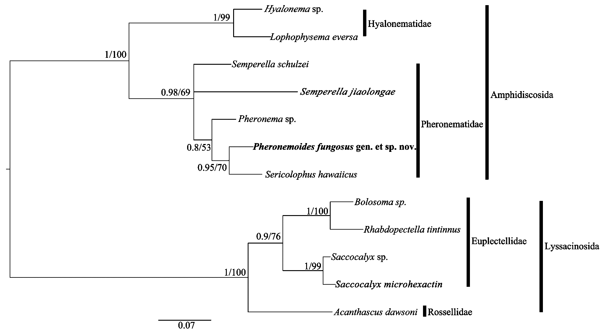
Molecular data. The phylogenies based on 16S rDNA ( Figure 4 View FIGURE 4 ) of ML and BI analyses were highly congruent. Pheronemoides fungosus sp. nov. was first grouped with Sericolophus hawaiicus ; then, gathered with Pheronema sp., it showed a closer relationship with Sericolophus than with Pheronema or Semperella . Thus, the ensuing phylogenic tree ensured the validity of our placement of Pheronemoides fungosus gen. et sp. nov. as a member of the Pheronematidae .
No known copyright restrictions apply. See Agosti, D., Egloff, W., 2009. Taxonomic information exchange and copyright: the Plazi approach. BMC Research Notes 2009, 2:53 for further explanation.