
Drusilla bifida, Assing, 2016
|
publication ID |
https://doi.org/ 10.21248/contrib.entomol.66.1.13-111 |
|
DOI |
https://doi.org/10.5281/zenodo.5903486 |
|
persistent identifier |
https://treatment.plazi.org/id/766F7C36-FFF0-FFAA-FF36-75D4DC89FC02 |
|
treatment provided by |
Felipe |
|
scientific name |
Drusilla bifida |
| status |
sp. nov. |
Drusilla bifida View in CoL spec. nov.
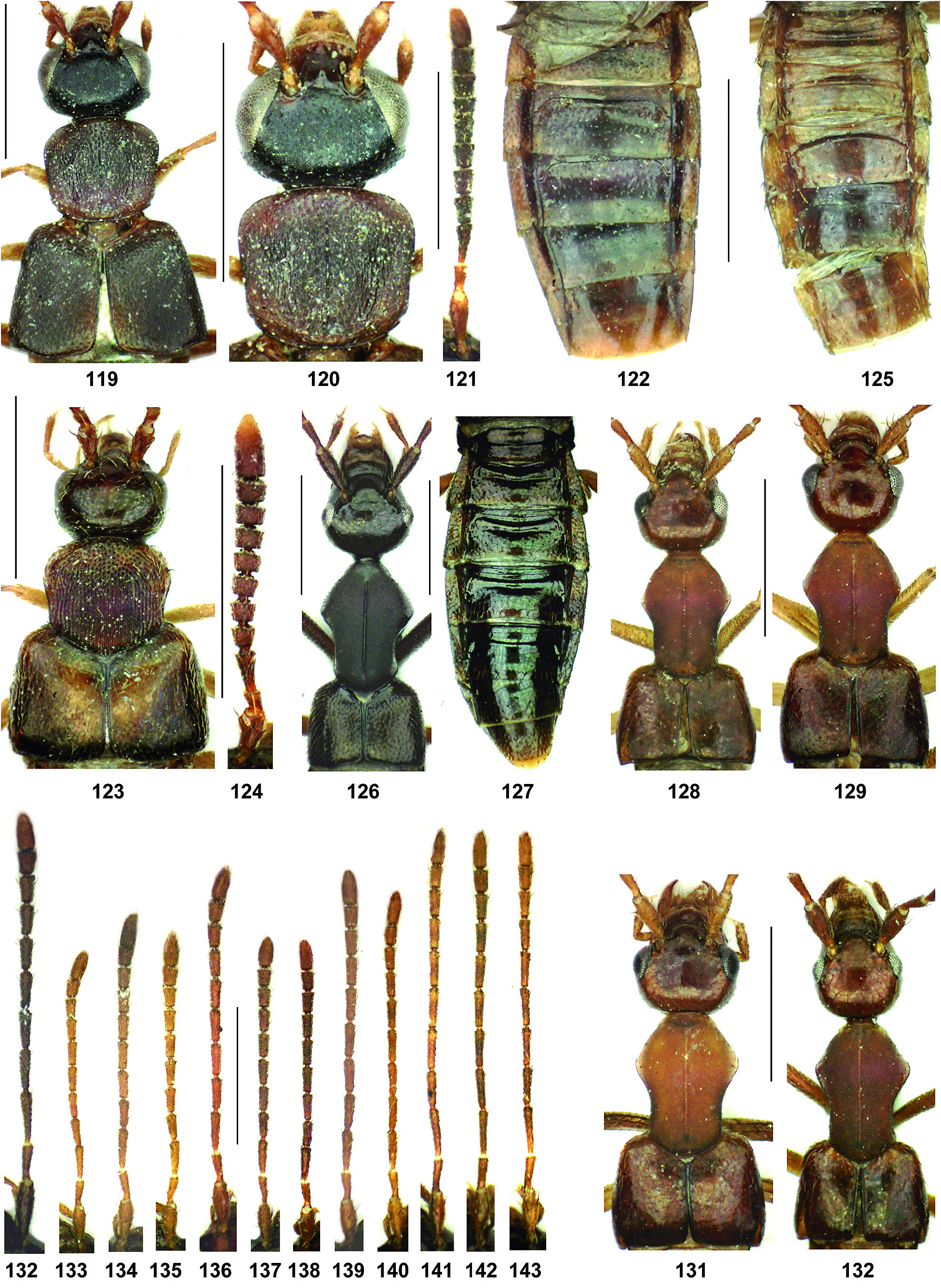
( Figs 119–122 View Figs 119–143 , 373–379 View Figs 359–378 View Figs 379–395 )
Type material: Holotype ♂: “ Thailand [47] – Doi Pha Hom Pok , Bhoo Muan waterf., 20°02'N, 99°14'E, 800 m, litter, 25.I.2014, leg. Ob / Holotypus ♂ Drusilla bifida sp. n., det. V. Assing 2015” (cAss). GoogleMaps
Etymology: The specific epithet (Latin, adjective) alludes to the bifid ventral process of the aedeagus.
Description: Body length 4.7 mm; length of forebody 2.2 mm. Coloration: head blackish; pronotum and elytra dark-brown; scutellum paler brown; abdomen darkbrown, with segments III and VIII–X reddish-brown; legs dark-yellowish; antenna dark-brown with antennomeres I–II and the base of III reddish; maxillary palpi brown with the apical palpomere yellowish.
Head ( Figs 119–120 View Figs 119–143 ) transverse, 1.21 times as broad as long; posterior angles weakly marked, nearly obsolete; frons and sclerotized portion of clypeus each with a distinct smooth elevation; punctation fine, practically invisible in the pronounced microreticulation ( Fig. 373 View Figs 359–378 ); dorsal surface nearly matt. Eyes large and strongly bulging, nearly twice as long as distance from posterior margin of eye to posterior constriction in dorsal view. Antenna ( Fig. 121 View Figs 119–143 ) 1.9 mm long and rather massive; antennomere III somewhat flattened, distinctly dilated apically, approximately twice as long as broad, and much longer than antennomere II; antennomeres IV distinctly and V–VI weakly oblong, VII aproximately as long as broad, VIII–X indistinctly transverse, and XI small, narrower than the preceding antennomeres and shorter than the combined length of IX and X.
Pronotum ( Figs 119–120 View Figs 119–143 ) 1.08 times as broad as long and as broad as head, very weakly convex in cross-section, broadest at anterior angles; disc coarsely and longitudinally rugosely sculptured, matt, only in antero-median portion less matt and with defined punctation; midline with very narrow sulcus reaching neither anterior nor posterior margins; disc with an indistinct elevation and a lateral oblong impression on either side.
Elytra ( Fig. 119 View Figs 119–143 ) 1.07 times as long as pronotum; punctation dense and moderately fine; interstices with pronounced microreticulation ( Fig. 379 View Figs 379–395 ). Hind wings present. Metatarsomere I slightly longer than the combined length of II and III.
Abdomen ( Fig. 121 View Figs 119–143 ) slightly narrower than elytra; sternites III–VI anteriorly with dense and posteriorly with sparser fine punctation; tergite VII with very fine and very sparse punctation; posterior margin of tergite VII with palisade fringe.
♂: tergite VIII ( Fig. 374 View Figs 359–378 ) transverse, posterior margin with distinct and broad concavity in the middle; sternite VIII ( Fig. 375 View Figs 359–378 ) transverse and with convex posterior margin; median lobe of aedeagus ( Figs 376–377 View Figs 359–378 ) 0.53 mm long; ventral process apically bifid ( Fig. 378 View Figs 359–378 ); internal structures of distinctive shapes; paramere 0.47 mm long. ♀: unknown.
Comparative notes and comment: Drusilla bifida is distinguished from other species recorded from Thailand and neighbouring regions by the bifid ventral process of the aedeagus, the characteristic sculpture of the pronotum, and the smooth elevations on the frons and the clypeus, from most species also by the coloration, the massive antennae, and the pronounced microsculpture on the head and elytra. The median lobe of the aedeagus slightly resembles that of the much larger D. umranicola PACE, 2005 from India (Meghalaya) and that of D. thaifuscicollis PACE, 2012 ( Thailand) , from which D. bifida additionally differs by the nearly obsolete posterior angles of the head, much larger eyes, and darker and distinctly more massive antennae with a relatively shorter antennomere XI.
Distribution and natural history: The type locality is situated in the extreme north of Thailand, close to the border with Myanmar. The holotype was sifted from leaf litter near a waterfall at an altitude of 800 m.


No known copyright restrictions apply. See Agosti, D., Egloff, W., 2009. Taxonomic information exchange and copyright: the Plazi approach. BMC Research Notes 2009, 2:53 for further explanation.
|
Kingdom |
|
|
Phylum |
|
|
Class |
|
|
Order |
|
|
Family |
|
|
Genus |