
Lophophysema eversa, Gong, Lin, Li, Xinzheng & Qiu, Jian-Wen, 2014
|
publication ID |
https://doi.org/ 10.11646/zootaxa.3884.6.3 |
|
publication LSID |
lsid:zoobank.org:pub:9302CD89-CF83-4762-A534-86A50B596832 |
|
DOI |
https://doi.org/10.5281/zenodo.6137733 |
|
persistent identifier |
https://treatment.plazi.org/id/780687D3-AB0F-4718-2EA2-DC298F9DB6A3 |
|
treatment provided by |
Plazi |
|
scientific name |
Lophophysema eversa |
| status |
sp. nov. |
Lophophysema eversa View in CoL sp. nov.
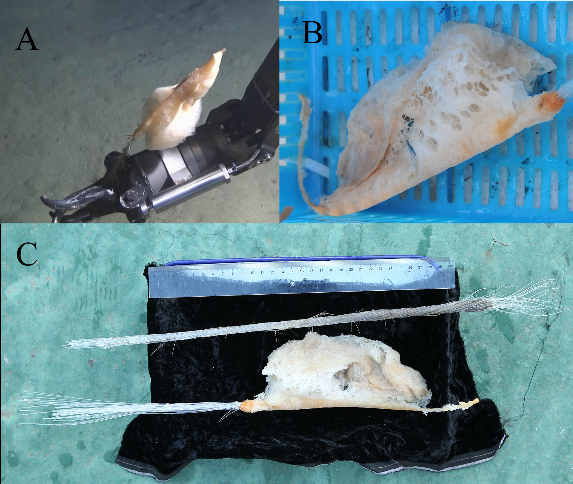
( Figures 1–3 View FIGURE 1 View FIGURE 2 View FIGURE 3 )
Lophophysema eversa Zhang, Sun, Li & Qiu, 2014 View in CoL : Mitochondrial DNA (in press) DOI: 10.3109/19401736.2014.945552 [nomen nudum]
Material examined. Holotype: MBM 179978, northeast of the Zhongsha Islands, South China Sea, 117°45.5'E, 17°36.7’N, 7 July 2013, 3683 m depth, muddy substrate, collected by Xinzheng Li and Jian-Wen Qiu using the “Jiaolong” submersible.
Description. The holotype is half-gourd shaped, composed of two opposite cones, with a smaller upper part and a larger lower part ( Figure 1 View FIGURE 1 ). It is different from the typical shape of genus Lophophysema which always have a larger upper cone. The body length (without stalk) is 182 mm, with a maximum diameter of 76 mm. An everted atrial surface extends from the whole apical cone to the flank of the lower cone diminishedly. The everted atrial surface composed of the upper cone and divide the flank of the body into two parts which is comparatively very closed. The dermal surface between the basalia and the everted atrial surface located at the lower cone. It has large holes and organized into sieve ( Figure 1 View FIGURE 1 B) with mesh size from 1.5 mm to 4.5 mm. The color of the atrial surface and dermal surface is darken yellow and beige, respectively. Furthermore, compared to the dermal surface, the color of the atrial surface looks darker. Large inhalant cavities can be found under the dermal surface. The basal exist on the side of the body. For other Lophophysema spcies, the basal may exist on the middle of body due to it has no special describe on it in Tabachnick & Lévi (1999) and the figure 4 of the Lophophysema body form in Tabachnick & Menshenina. Basalia with smooth surface tightly twisted in a tuft, and stretch from the bottom to protrude over the apex. These were broken as a result of their collection ( Figure 1 View FIGURE 1 C). The smallest diameter of the tuft is 7 mm; individual basalia vary from 0.1 mm to 1 mm in width. Sponge tissues are fleshy, light and compressible, with an orange rusty patch on the lower part wrapping the basalia ( Figure 1 View FIGURE 1 B). The basal stalk is long,> 650 mm, and the part protruding over the apex is 124 mm long. Marginalia were difficult to observe due to the fragmentation of the specimen after its preservation.
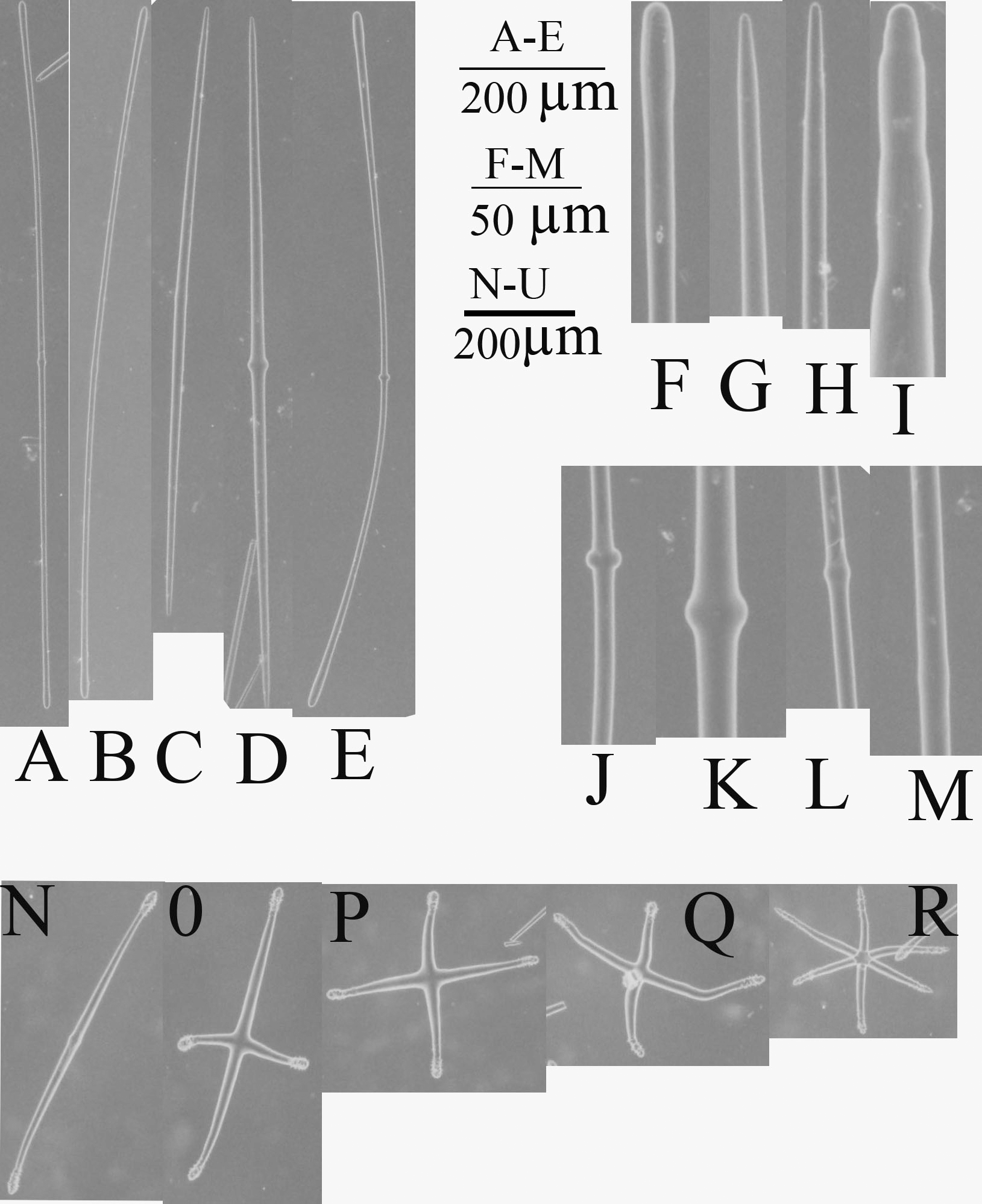
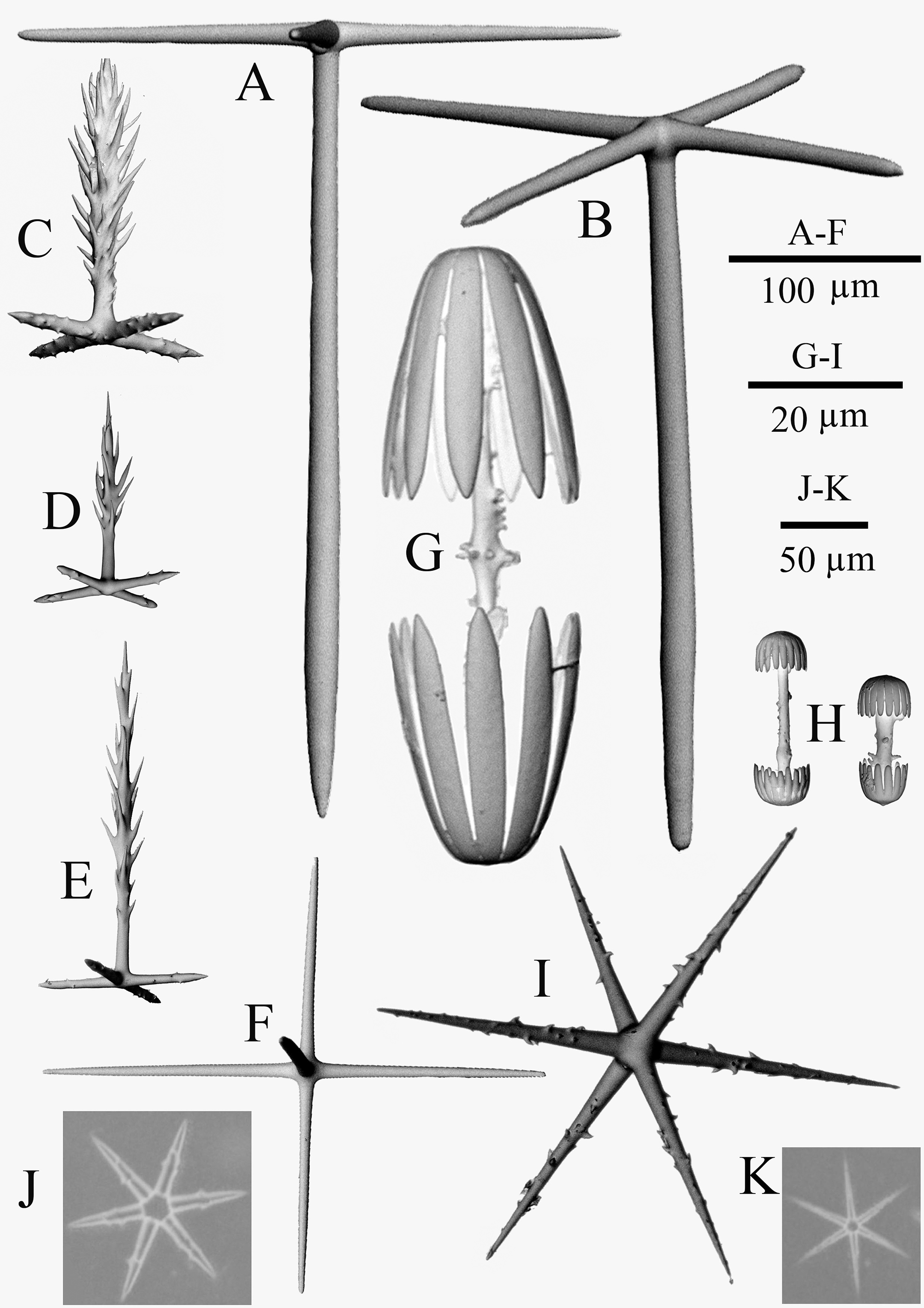
Spicules. The choanosomal skeleton has smooth and slightly curved diactins ( Figure 2 View FIGURE 2 ). Diactins are medially widened ( Figure 2 View FIGURE 2 J–L), terminally conical or slightly swollen ( Figure 2 View FIGURE 2 F–I). Acanthophores are present as diactins ( Figure 2 View FIGURE 2 N), stauractins ( Figure 2 View FIGURE 2 O –P), pentactins ( Figure 2 View FIGURE 2 Q), hexactins ( Figure 2 View FIGURE 2 R), with ends covered with small spines. Choanosomal hexactins ( Figure 3 View FIGURE 3 J) are covered with short spines and have relatively longer and thicker rays than microhexactins ( Figure 3 View FIGURE 3 K).
Dermalia, atrialia, and canalaria are pinular pentactins. The tangential rays of pinular pentactins are spiny. Atrialia ( Figure 3 View FIGURE 3 E) are similar to canalaria ( Figure 3 View FIGURE 3 D) but larger. The atrialia pinular rays are 204.9–402.2 µm long, while canalaria pinular rays are 28.8–129.7 µm long (Table 1). The dermalia pinular rays ( Figure 3 View FIGURE 3 C) are 104.3–174.2 µm long and usually spindle-like, with the teeth on the ray being longer and denser than the other two types. The hypodermal pentactins ( Figure 3 View FIGURE 3 B) have smooth rays of varying lengths: 187.6–674.9 µm long (proximal rays), and 75.6–220.7 µm long (tangential rays), pentactins with two reduced rays ( Figure 3 View FIGURE 3 A) may occasionally be present. The hypoatrial pentactins ( Figure 3 View FIGURE 3 F) have long tangential rays (137.3–406.1 µm long) and short proximal rays which are difficult to measure under light microscopy.
Microscleres consist of amphidiscs and microhexactins. Amphidiscs consist of two forms. Macramphidiscs are absent. Mesamphidiscs ( Figure 3 View FIGURE 3 G) have shafts covered by numerous spines, total length 55.2–89.2 µm, umbel length 20.1–34.5 µm, and umbel diameter 16.9–26.9 µm. Micramphidiscs ( Fig. 3 View FIGURE 3 H) have small spiny shafts similar to those of mesamphidiscs, with total length ranging from 12.4–21.2 µm. Micramphidiscs significantly outnumber the mesamphidiscs. Microhexactins ( Figure 3 View FIGURE 3 I, K) have numerous small spines on their rays. These rays ( Figure 3 View FIGURE 3 I) are 14.7–40.6 µm long, mostly straight, although some have slightly curved ends.
Etymology. The specific name eversa is derived from the Latin word "eversus", meaning everted, referring to the notably everted atrial surface.
Remarks. The genus Lophophysema Schulze, 1900 contains three described species: L. gilchristi Tabachnick & Lévi, 1999 , L. australicum Tabachnick & Lévi, 1999 and L. inflatum Schulze, 1900 . In a review of Lophophysema, Tabachnick & Lévi (1999) pointed out the distinct characters of the three species: L. inflatum contains microhexactins; L. australicum has rough monactin and the macramphidisc umbel is longer than half of the total length; and L. gilchristi has choanosomal hexactins and the umbel diameter in the mesamphidiscs is proportionally larger. Lophophysema eversa sp. nov. differs from all three described species by its unusual body shape with basalia on the side of the body and by lacking macramphidiscs. Specifically, the new species (1) can be differentiated from L. australicum by lacking rough monactin; (2) can be distinguished from L. gilchristi in that the pinular ray of its atrialia is longer than that of dermalia and canalaria; and (3) is distinct from L. inflatum by having spined choanosomal hexactins..
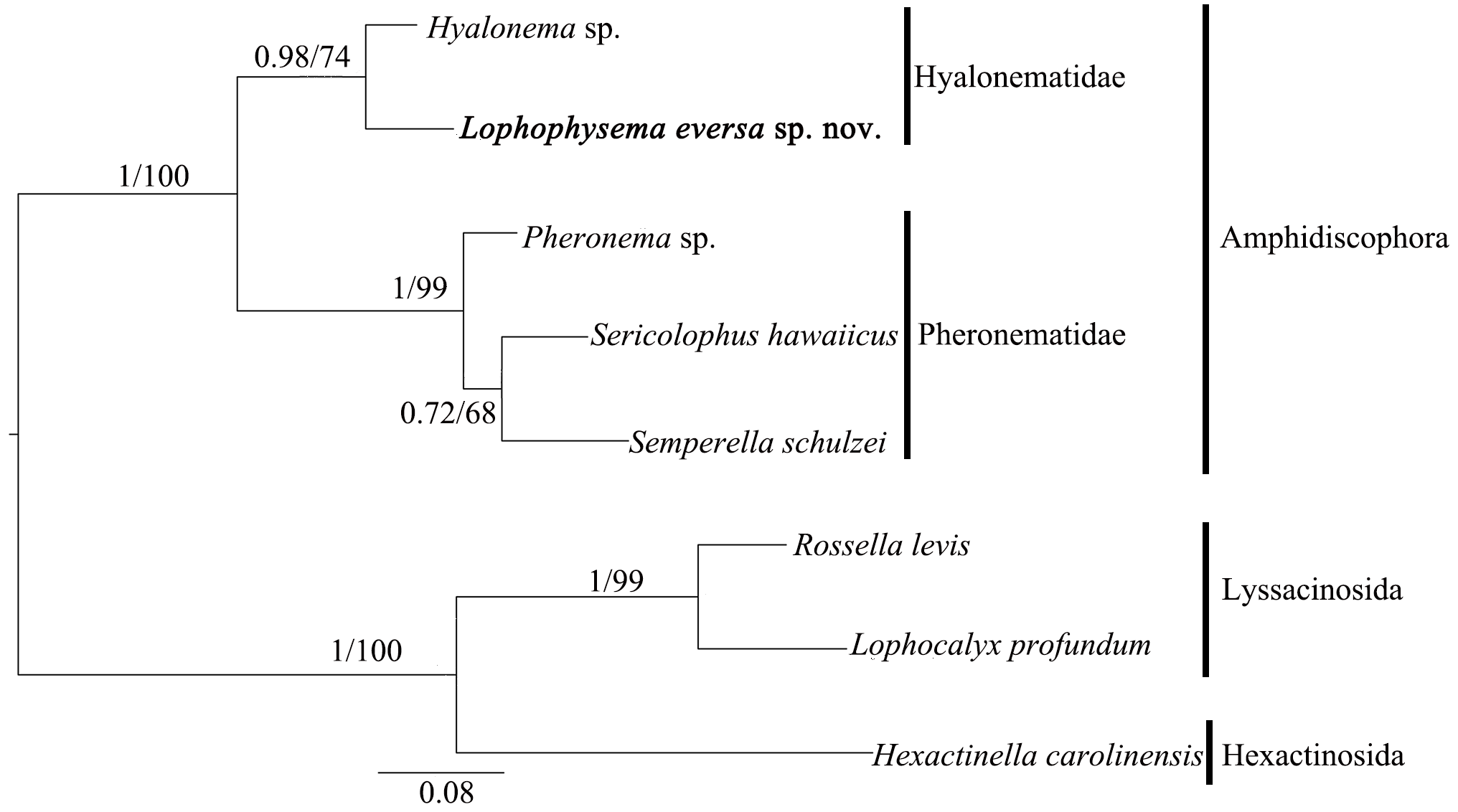
Molecular data. The phylogenies of ML and BI analyses were highly congruent. The phylogeny based on 16S rRNA ( Figure 4 View FIGURE 4 ) shows that L. eversa forms a clade with Hyalonema to the exclusion of the pheronematids, thus supporting the family assignment, with support values>70 for both the ML and BI analyses.
No known copyright restrictions apply. See Agosti, D., Egloff, W., 2009. Taxonomic information exchange and copyright: the Plazi approach. BMC Research Notes 2009, 2:53 for further explanation.
|
Kingdom |
|
|
Phylum |
|
|
Class |
|
|
Order |
|
|
Family |
|
|
Genus |
Lophophysema eversa
| Gong, Lin, Li, Xinzheng & Qiu, Jian-Wen 2014 |
Lophophysema eversa
| Zhang, Sun, Li & Qiu 2014 |