
Hoplichthys imamurai, Nagano, Yuki & Yabe, Mamoru, 2013
|
publication ID |
https://doi.org/ 10.11646/zootaxa.3681.3.3 |
|
publication LSID |
lsid:zoobank.org:pub:905443F4-A466-425D-BFED-8CBAF37938F1 |
|
DOI |
https://doi.org/10.5281/zenodo.6162217 |
|
persistent identifier |
https://treatment.plazi.org/id/7828879E-F118-332C-FF14-3109FCF6F89A |
|
treatment provided by |
Plazi |
|
scientific name |
Hoplichthys imamurai |
| status |
sp. nov. |
Hoplichthys imamurai View in CoL sp. nov.
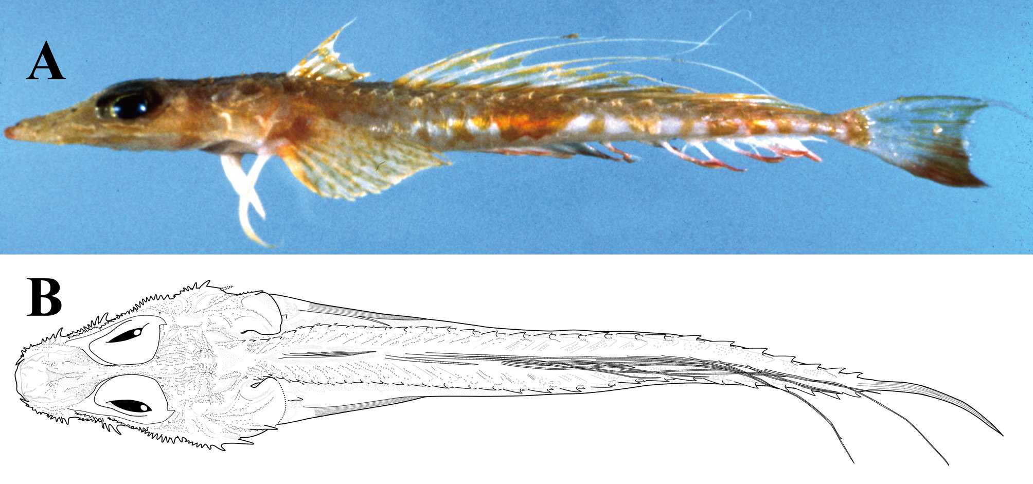
[New English name: Imamura's ghost flathead] ( Figures 1 View FIGURE 1 , 2 View FIGURE 2 , 3 View FIGURE 3 A, 4)
Hoplichthys regani View in CoL (not Jordan, 1908): Gloerfelt-Tarp & Kailola, 1984: 124 (southern Indonesia and northwestern Australia); Allen, 1997: 70, pl. 16 (southern Indonesia and northwestern Australia); Paxton et al., 2006: 950 (northwestern Australia). Hoplichthys View in CoL sp.: Sainsbury et al., 1984: 336 (northwestern Australia).
Holotype: AMS I.22807-040, 143.4 mm SL, male, 18°32’S, 118°17’E, 175 km north of Port Hedland, Northwest Shelf, Western Australia, 200–204 m depth, coll. J. Paxton and M. McGrouther on FRV Soela, 2 Apr. 1982.
Paratypes: 2 specimens. AMS I.22807-066, 143.6 mm SL, male, same data as holotype; AMS I.31156-016, 128.9 mm SL, male, 23°25.4’S, 113°03.9’E – 23°23.6’S, 113°05.1’E, off Point Cloates, Western Australia, 300–302 m depth, coll. J. Paxton, 26 Jan. 1991.
Non-type: 1 specimen. AMS I.22807-067, dissected, size unknown, same data as holotype.
Diagnosis. A species of Hoplichthys with the following combination of characters: dorsal-fin rays VI-15; analfin rays 17; pectoral-fin rays 13 + 3 = 16; short preoptic snout (31.2–32.7% HL); large eyes (23.8–24.7% HL); narrow interorbital width (2.6–4.1% HL); vomer without teeth; long, thin gill rakers; developed spines on ventral surface of lower jaw; single developed spine on each lateral scute; short free pectoral-fin rays (13.4–14.9% SL). In males, low first dorsal fin, its adpressed tips not reaching origin of second dorsal fin; some elongated second dorsal-fin rays filament-like; emarginate caudal fin, its upper edge elongated, filament-like (see Remarks below for sexual dimorphism of Hoplichthys ).
Description. Data for holotype are presented first, followed by that of paratypes in parentheses if different.
Dorsal-fin rays VI-15; anal-fin rays 17; pectoral-fin rays 13 + 3 = 16; pelvic-fin rays I, 5; gill rakers 2 + 12 = 14; branchiostegals 7; vertebrae 26; lateral scutes 27. Head spines: spines on lachrymal 9/8 (broken/9); inner row spines on first infraorbital 8/9 (8–10/9); outer row spines on first infraorbital 9/9 (9–10/9); inner row spines on second infraorbital 11/11 (8–12/9–12); outer row spines on second infraorbital 12/14 (12–14/11–14); preopercular spines 9/8 (7–8/8); spines on ventral surface of lower jaw 3/3 (3–4/3–4). Proportional measurements as % SL: HL 32.0 (31.9–32.1); body depth 7.0 (8.1–8.5); body width 8.7 (9.3–9.9); pre-first dorsal length 31.4 (30.7–31.0); length of first dorsal-fin base 6.7 (7.0–7.5); length of first dorsal-fin spine 9.4 (8.0–10.3); length of second dorsalfin spine 6.9 (7.1–8.2); length of third dorsal-fin spine 6.4 (6.3–7.2); length of sixth dorsal-fin spine 5.5 (4.7–5.4); pre-second dorsal length 43.2 (42.8–42.9); length of second dorsal-fin base 46.4 (46.4–47.4); length of first dorsalfin soft ray broken (11.1–11.2); length of second dorsal-fin soft ray broken (27.7); length of fifth dorsal-fin soft ray 42.1 (45.5); length of sixth dorsal-fin soft ray 17.0 (16.6–16.9); length of ninth dorsal-fin soft ray 18.3 (17.2–18.3); length of 12th dorsal-fin soft ray 21.8 (19.9–21.0); length of 14th dorsal-fin soft ray 27.2 (25.3); length of 15th dorsal-fin soft ray 13.9 (11.6–14.2); pre-anal length 41.0 (40.8–41.2); length of anal-fin base 51.8 (50.4–51.5); length of first anal-fin ray 5.2 (4.4–4.7); length of 17th anal-fin ray 7.3 (6.7–7.0); pectoral-fin length 20.2 (19.4– 19.5); length of first free pectoral-fin ray 17.6 (17.3–18.3); length of second free pectoral-fin ray 18.9 (18.2–18.8); length of third free pectoral-fin ray 16.5 (15.9–16.9); pelvic-fin length 13.5 (13.0–14.5); caudal peduncle depth 2.1 (2.2); length of upper caudal peduncle 11.0 (11.1–11.2); length of caudal peduncle 8.9 (8.5–8.7); caudal-fin length 20.2 (21.5–22.3). Proportional measurements as % HL: head depth 23.3 (24.5); head width at anterior margin of eye 34.1 (36.4); head width at inner base of posteriormost preopercular spine 48.3 (45.0–51.9); head width at outer base of posteriormost preopercular spine 59.0 (54.2–62.3); preoptic snout length 31.2 (32.0–32.7); preorbital snout length 23.2 (22.6–24.7); orbital diameter 35.6 (35.3–35.7); vertical eye diameter 24.7 (23.8–24.4); horizontal eye diameter 17.4 (17.6–18.0); interorbital width 2.6 (2.9–4.1); postorbital length 44.4 (43.6–45.5); upper jaw length 33.1 (31.2–32.6); lower jaw length 30.8 (29.8–30.4); length between upper edges of opercles on either side 19.0 (21.7).
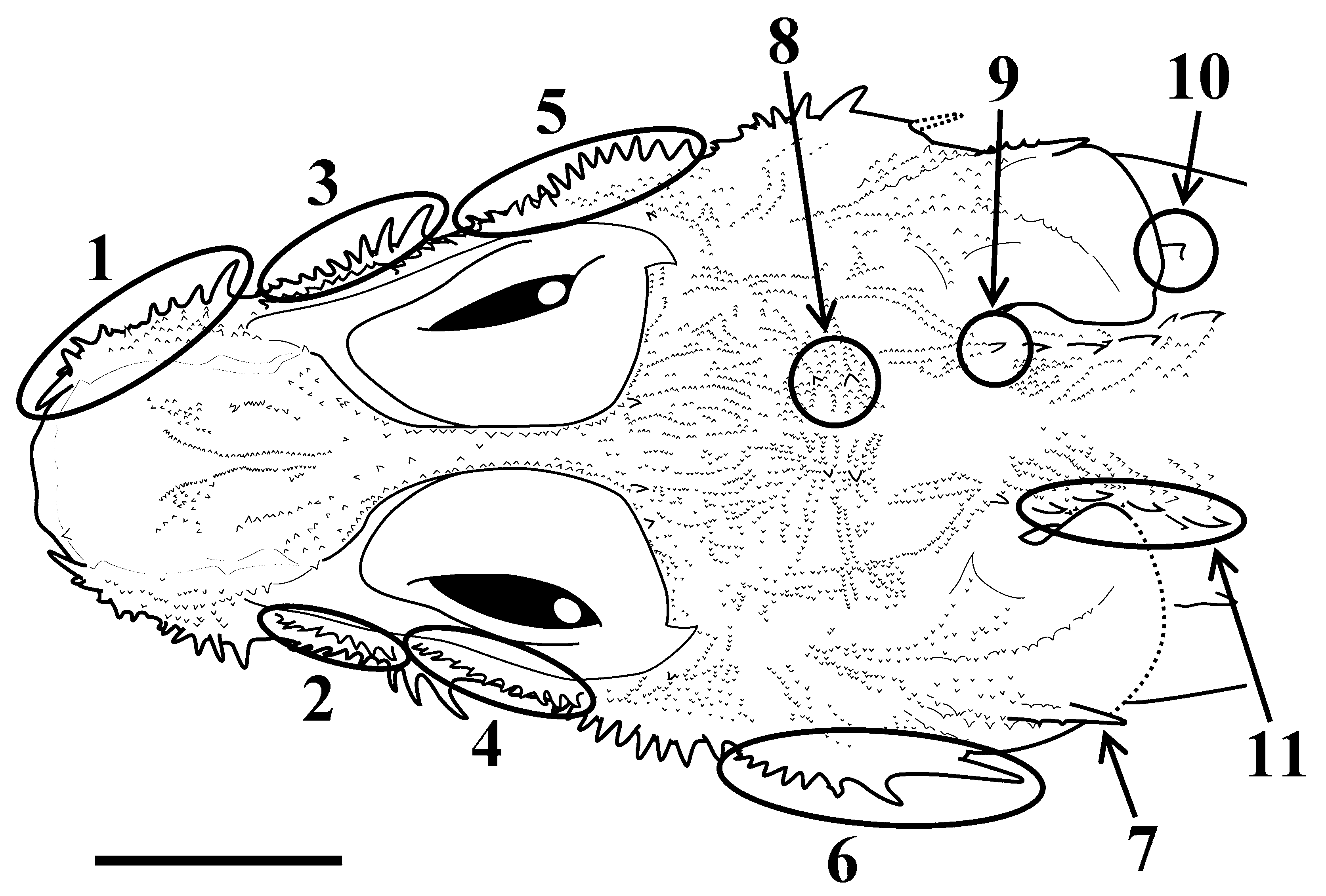
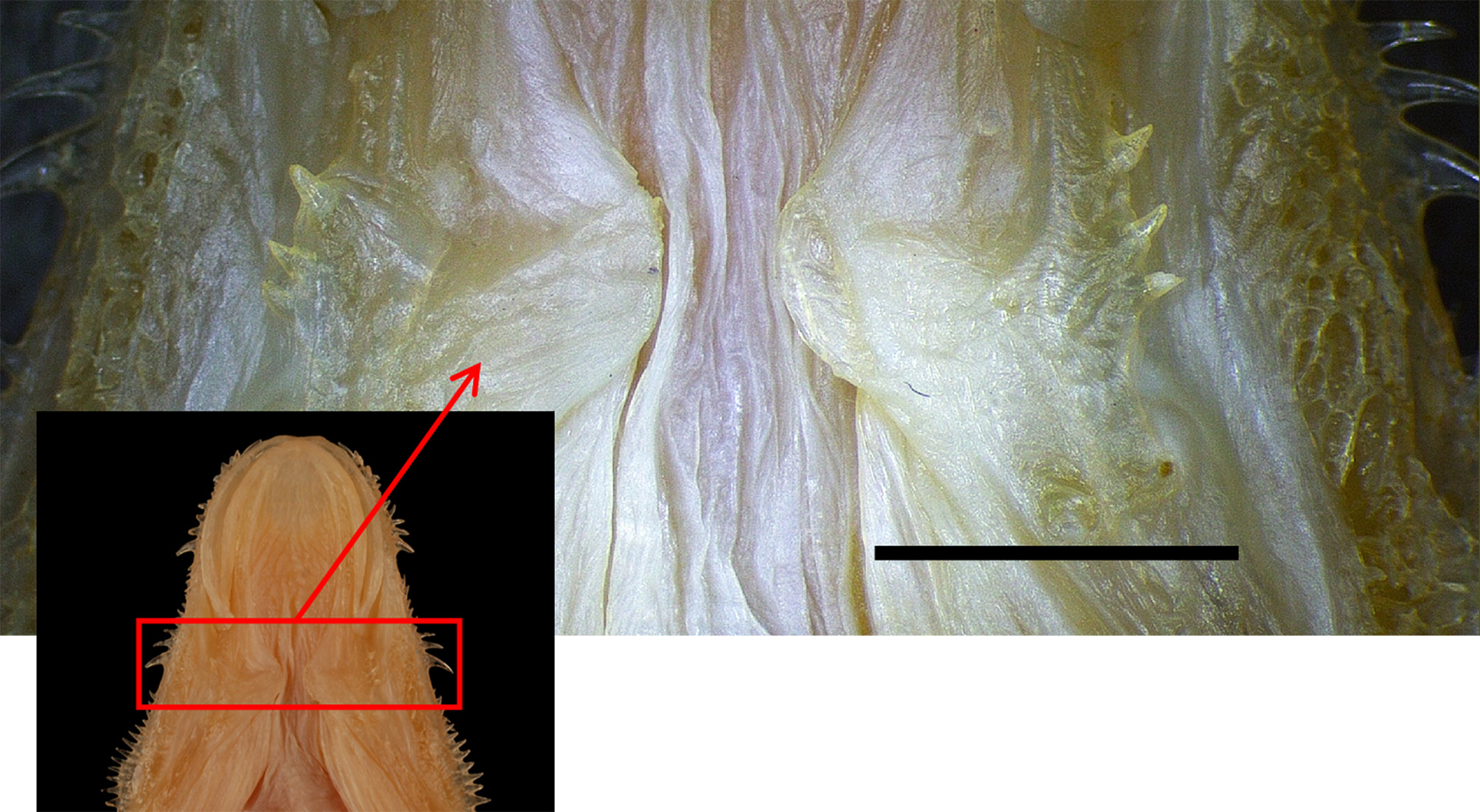
Head extremely depressed, depth 2.5 (2.2–2.5) in width at outer base of posteriormost preopercular spine. Body elongate, slightly depressed, naked, except for lateral scutes along body; depth at origin of first dorsal fin 1.3 (1.1–1.2) in body width at origin of second dorsal fin. Snout paddle-like; preoptic snout length longer than vertical eye diameter, 1.4 (1.3–1.4) in postorbital length. Anterior nostril located mesial to central part of lachrymal, possessing short tube and small cirrus on posterior margin; posterior nostril located mesial to posterior edge of lachrymal, possessing short tube, smaller than anterior nostril. Single cephalic sensory pore located anteromesial to both nostrils. Upper jaw length slightly longer than preoptic snout length; posterior edge of maxilla reaching anterior margin of eye, but not reaching base of posteriormost spine in outer row of spines on first infraorbital. Teeth villiform, forming tooth bands on jaws and palatines, but not on vomer. Anterior margin of basihyal slightly concave (flat). Eye relatively large; vertical eye diameter 1.3 (1.3–1.4) in preoptic snout length. Interorbit narrow and concave; interorbital width 9.5 (5.8–8.5) in vertical eye diameter. Posterior margin of opercle relatively rounded. Lower margin of branchiostegal membrane fused with isthmus. Gill rakers long and thin ( Fig. 3 View FIGURE 3 A); many minute spines on each raker. Many spines and finely serrated ridges on dorsal surface of head ( Fig. 2 View FIGURE 2 ). Single paired rows of spines on dorsal mid-line of snout. Single series of serrated ridges situated anterior to eye and interorbit, ending at posterior margin of orbit. Dorsal surface of lachrymal with sparsely scattered small spines. Single row of developed spines laterally on lachrymal; anteriormost spine largest, directed forward; second to fourth (broken) spines of similar size, directed upward; fifth to ninth (broken) spines on left side and fifth to eighth (broken) spines on right side directed laterally, becoming progressively larger posteriorly. Small spines on ventral side of lachrymal, of similar size, directed downward. Two rows of spines on first infraorbital, inner row on dorsal surface and outer row on lateral side; spines in inner row of similar size, directed upward; spines in outer row larger than those of inner row, becoming progressively larger posteriorly, anterior five spines on left side and six on right side (four spines on both sides) directed anterolaterally and posterior four spines on left side and three on right side (four or five spines on both sides) directed posterolaterally. A few small spines on ventral side of first infraorbital, directed downward. Two rows of spines on second infraorbital, inner row on dorsal surface and outer row laterally; inner row spines of similar size, directed upward, accompanied by sparse small spines posteriorly; outer row spines directed laterally, becoming progressively larger posteriorly. Outer rows of spines on first and second infraorbitals separated. Small spines scattered on dorsal surface of third and fourth infraorbitals. Single slightly developed spine present at center of third infraorbital. Single row of preopercular spines present laterally; penultimate spine large, directed posterolaterally; posteriormost spine largest, directed posteriorly, not reaching posterior margin of opercle; other anterior spines of similar size, directed laterally. Two rows of small spines on dorsal surface of preopercle directed laterally from inner edge of preopercle. Single opercular spine present and well developed, not reaching posterior edge of opercle. Two rows of small spines on dorsal surface of opercle arranged posteriorly and radially from anterodorsal corner of opercle; outer row accompanied by opercular spine; inner row directed toward posteromesial corner of opercle; small spines scattered on lateral, mesial and middle areas of these two rows. Two pairs of parietal spines present and developed on occiput; posterior spines larger than those anteriorly; small spines arranged radially from them. Small spines densely scattered in area between posterior margin of orbit and parietal spines. Single developed posttemporal spine present anterior to well developed spine on first lateral scute, accompanying small spines around posttemporal spine; serrated ridges radiating anteriorly from posttemporal spine. Large spines present on ventral surface of lower jaw posterior to maxilla, directed anterolaterally ( Fig. 4 View FIGURE 4 ). Single humeral spine posterior to opercle, without small spines on its anterior margin. Single row of spines on lateral scutes along body; single well developed spine directed upward and backward centrally on each scute, with single small spine directed posteriorly at its base; developed spines becoming progressively larger posteriorly, posterior three spines becoming smaller posteriorly; terminal scute extending to caudal fin base. One or two anteromesially arranged serrated ridges on upper part of each lateral scute, becoming progressively smaller posteriorly. First dorsal fin low, originating above third lateral scute (third or junction between second and third lateral scutes) and ending above seventh lateral scute (seventh or junction between sixth and seventh lateral scutes); length of first dorsal fin base 4.8 (4.3–4.6) in HL; tips of adpressed dorsal-fin spines not reaching origin of second dorsal fin; first dorsal-fin spine longest and second to sixth dorsal-fin spines becoming progressively shorter posteriorly. Second dorsal fin originating above ninth lateral scute (ninth or junction between ninth and 10th lateral scutes), and ending above junction between 23rd and 24th lateral scutes (23rd or 24th lateral scute); length of second dorsal-fin base 1.1 in length of anal-fin base; first dorsal-fin soft ray shortest; second to fifth and 12th to 15th (second to fifth and eighth to 14th or 15th) dorsal-fin soft rays elongated filament-like, and other rays except for first ray of almost equal length and not filamentosus . Pectoral fin originating at posterior edge of opercle, reaching junction between 11th and 12th lateral scutes (12th); fourth and fifth (fourth) rays longest, length 1.6 in HL. Lower three rays of pectoral fin thick and free, shorter than remaining joined pectoral-fin rays; second free pectoralfin ray, viz., 15th ray from uppermost pectoral-fin ray, longest, reaching beyond anus; its length 1.7 (1.7–1.8) in HL. Pelvic-fin base anterior to pectoral-fin base; distal edge of pelvic fin reaching below seventh lateral scute (seventh or junction between seventh and eighth lateral scutes); its length 2.4 (2.2–2.5) in HL. Anal fin lower than second dorsal fin, originating below eighth lateral scute (eighth or junction between eighth and ninth lateral scutes), ending below 24th lateral scute (24th or junction between 24th and 25th lateral scutes); first anal-fin ray short, its length 1.4 (1.4– 1.6) in length of last anal-fin ray. Caudal fin emarginate, with upper margin elongate, filament-like; depth of caudal peduncle 9.5 (10.0–10.2) in caudal-fin length. Anus slightly anterior to origin of anal fin.
Coloration. Color when fresh (based on photograph of holotype; Fig. 1 View FIGURE 1 A): head and body light brown dorsally, and white ventrally. Dorsal and lateral surface of body with yellow blotches. Dorsal fins semitransparent and yellowish basally and distally. Pectoral fin with scattered small yellowish spots. Pelvic fin white. Anal fin white, margined with red on lower edge. Caudal fin semitransparent; posterior part of lower lobe brown. Color in alcohol: mostly faded, head and body yellowish brown to light brown.
Distribution. Known only from the type series, off Port Hedland at 200–204 m depth, and off Point Cloates at 300–302 m depth, Western Australia.
Etymology. The new species is named for Dr. Hisashi Imamura, associate professor of Hokkaido University (HUMZ), in honor of his great contribution to the systematics of the Platycephaloidea, including the Hoplichthyidae .
Remarks. Hoplichthys imamurai is clearly distinguished from its congeners in having a vomer without teeth, and an emarginate caudal fin with an elongated filament-like upper margin in males. These two characters are unique to H. imamurai within the genus.
Hoplichthys imamurai is most similar to H. citrinus , H. gilberti and H. ogilbyi in having a narrow interorbit, large spines on the ventral surface of the lower jaw, a single spine on each lateral scute, short free pectoral-fin rays, and short remaining joined pectoral-fin rays. The new species is distinguished from H. citrinus and H. gilberti by its low first dorsal fin with the posterior tip of the adpressed fin not reaching the origin of the second dorsal fin in males (vs. high dorsal fin with the posterior tip reaching or over the origin of the second dorsal fin in the latter two), and long, thin gill rakers (vs. short and thick) ( Fig. 3 View FIGURE 3 ). Hoplichthys imamurai is distinguished from H. ogilbyi by its short preoptic snout (preoptic snout length 31.2–32.7 % HL in H. imamurai vs. more than 33.1 % HL in H. ogilbyi ; Fig. 5 View FIGURE 5 A), and several of the soft rays of the second dorsal fin filament-like in males (vs. all rays short, not filamentlike). Hoplichthys imamurai is also distinguished from the three species by the higher ratio of head depth to greatest head width, which is at the outer base of the posteriormost preopercular spine (39.3–45.2 % in H. imamurai vs. less than 37.6 % in the latter three; Fig. 5 View FIGURE 5 B).
Some species of Hoplichthys ( Hoplichthys langsdorfii Cuvier in Cuvier and Valenciennes, 1829, H. citrinus , H. gilberti , H. ogilbyi and Hoplichthys filamentosus Matsubara and Ochiai, 1950b ) have sexual dimorphism in the forms of the first and second dorsal fins and caudal fin (present observation). The first dorsal fin is longer in males of H. citrinus , H. gilberti and H. filamentosus than in females, some second dorsal-fin rays are longer and filamentosus in males but not in females of H. langsdorfii , H. citrinus , H. gilberti and H. filamentosus , and the caudal fin is lanceolate in males of H. langsdorfii and H. ogilbyi but shorter and rounded in females. Because all known specimens of H. imamurai are males, it is unclear if some diagnostic characters of this species, such as those of the dorsal and caudal fins, are sexually dimorphic.


No known copyright restrictions apply. See Agosti, D., Egloff, W., 2009. Taxonomic information exchange and copyright: the Plazi approach. BMC Research Notes 2009, 2:53 for further explanation.
|
Kingdom |
|
|
Phylum |
|
|
Class |
|
|
Order |
|
|
Family |
|
|
Genus |
Hoplichthys imamurai
| Nagano, Yuki & Yabe, Mamoru 2013 |
Hoplichthys regani
| Paxton 2006: 950 |
| Allen 1997: 70 |
| Gloerfelt-Tarp 1984: 124 |
| Sainsbury 1984: 336 |