
Stenosfemuraia González-Sponga, 1998
|
publication ID |
https://doi.org/ 10.11646/zootaxa.4341.4.3 |
|
publication LSID |
lsid:zoobank.org:pub:C476AF75-EF79-4C6E-9E16-9CC14540B25F |
|
DOI |
https://doi.org/10.5281/zenodo.6016016 |
|
persistent identifier |
https://treatment.plazi.org/id/7B0FFC7F-FFDC-FFED-56E5-FE5CFCC7884E |
|
treatment provided by |
Plazi |
|
scientific name |
Stenosfemuraia González-Sponga, 1998 |
| status |
|
Stenosfemuraia González-Sponga, 1998 View in CoL View at ENA
Stenosfemuraia González-Sponga, 1998: 21 View in CoL ; type species: S. parva González-Sponga, 1998 View in CoL .
Codazziella González-Sponga, 2005: 100 ; type species: C. pilosa González-Sponga, 2005 View in CoL . New synonymy. Chichiriviche González-Sponga, 2011: 41 ; type species: C. costanero González-Sponga, 2011 . New synonymy.
Justification of synonymies. The two type species of Codazziella and Chichiriviche are conspecific (see redescription of Stenosfemuraia pilosa View in CoL below), and the single species resulting from this synonymy is very similar to and shares all relevant characters with the type species of Stenosfemuraia View in CoL .
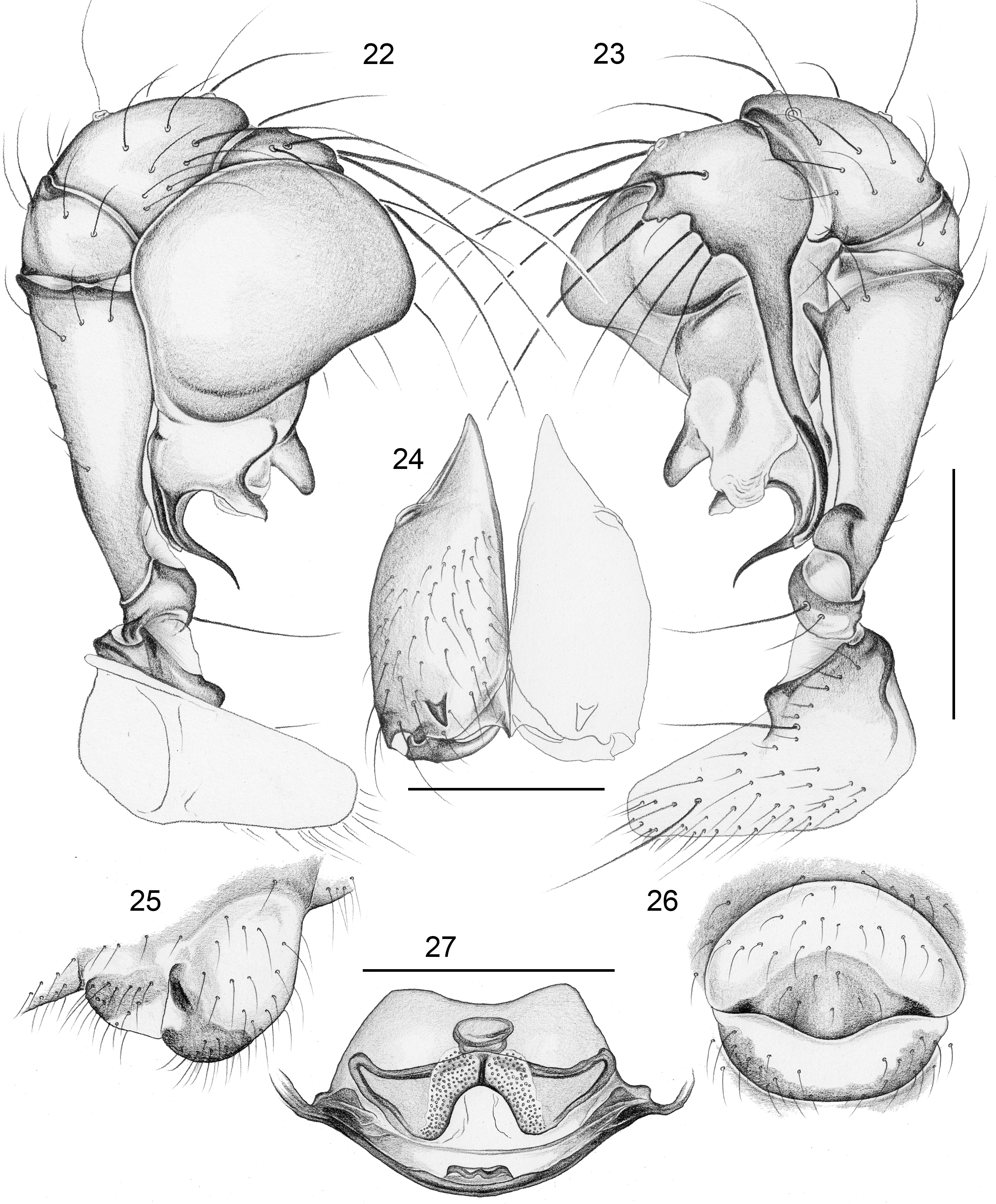
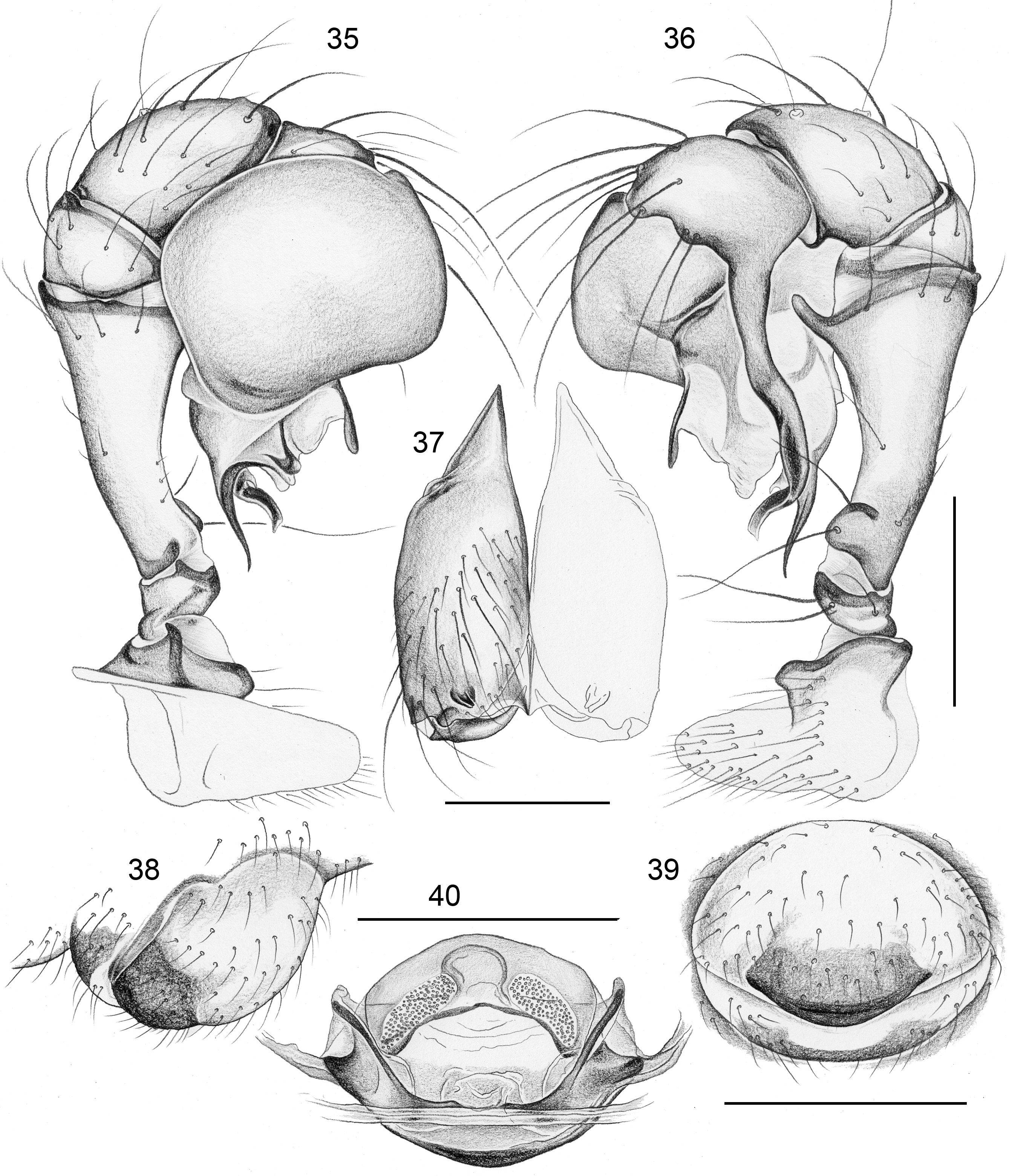
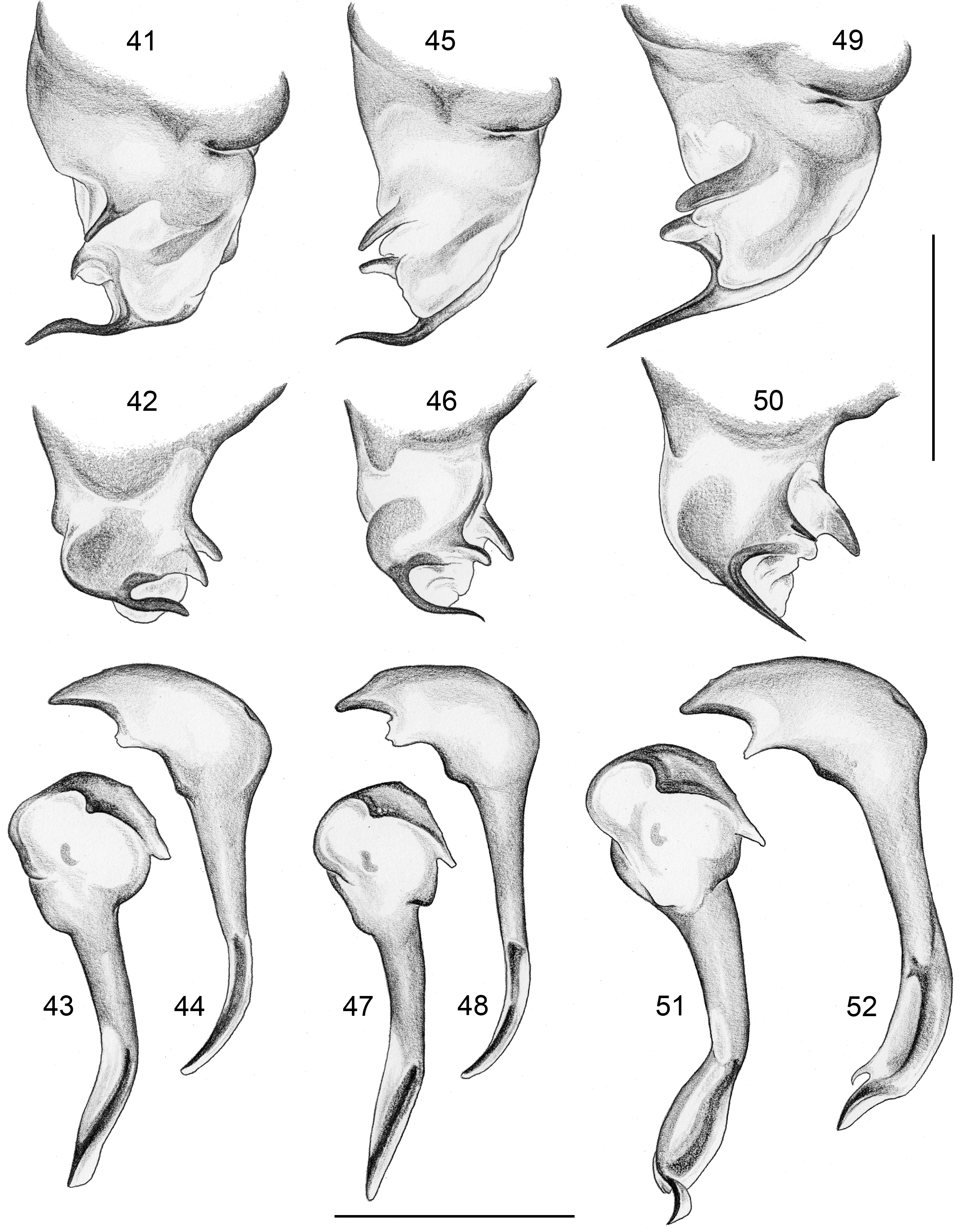
Diagnosis. Medium sized (~2.0– 3.5 mm total body length), eight-eyed spiders with round to oval abdomens and relatively short legs (tibia 1 L/d ~25–60). Distinguished from most species in related genera by shape of abdomen (elongated in Coryssocnemis Simon, 1893 , Systenita Simon, 1893 , and in most species of Mecolaesthus Simon, 1893 ). From similar species in Mecolaesthus by combination of: ventral apophysis distally on male palpal femur directed towards distal ( Figs 10 View FIGURES 9 – 14 , 23 View FIGURES 22 – 27 , 36 View FIGURES 35 – 40 ); genital bulb with slender distal apophysis and two dorsal processes ( Figs 41–42, 45–46, 49–50 View FIGURES 41 – 52 ); large membranous (whitish) area in front of small epigynal plate ( Figs 6 View FIGURES 2 – 8 , 19 View FIGURES 15 – 21 , 32 View FIGURES 28 – 34 ); internal female genitalia with anteriorly diverging sclerites/‘wings’ (arrows in Figs 8 View FIGURES 2 – 8 , 21 View FIGURES 15 – 21 , 34 View FIGURES 28 – 34 ); presence of curved hairs on male (rarely also female) legs (tibiae and metatarsi).
Description. Male. Total body length 2.5–3.5; carapace width 1.0–1.4. Carapace posteriorly elevated ( Figs 3 View FIGURES 2 – 8 , 16 View FIGURES 15 – 21 , 29 View FIGURES 28 – 34 ), with deep median furrow; ocular area weakly raised, eight eyes relatively close together, AME very small. Clypeus high, unmodified. Chelicerae never with stridulatory ridges, with one or two pairs of frontal apophyses ( Figs 11 View FIGURES 9 – 14 , 24 View FIGURES 22 – 27 , 37 View FIGURES 35 – 40 ), without modified hairs. Palpal coxa with retrolatero-ventral apophysis; trochanter barely modified; femur with retrolatero-ventral process proximally and ventral apophysis distally; procursus simple ( Figs 43–44, 47–48, 51–52 View FIGURES 41 – 52 ); bulb with slender distal apophysis and two dorsal processes ( Figs 41–42, 45–46, 49–50 View FIGURES 41 – 52 ).
Legs relatively short, leg 1 length 13–24, tibia 1 length 3.1–5.7, tibia 2/tibia 4 length 0.9–1.1. Tibia 1 L/d ~25– 60. Femora sometimes thickened ( S. parva , S. pilosa ). Legs without spines, with curved hairs on tibiae and metatarsi, retrolateral trichobothrium at 6–9%, prolateral trichobothrium always present (also on tibiae 1). Tarsal pseudosegments distinct.
Abdomen round to oval, higher than long, with dark internal marks dorsally and laterally ( Figs 2–3 View FIGURES 2 – 8 , 15–16 View FIGURES 15 – 21 , 28–29 View FIGURES 28 – 34 ).
Female. Very similar to male ( Figs 4–5 View FIGURES 2 – 8 , 17–18 View FIGURES 15 – 21 , 30–31 View FIGURES 28 – 34 ), carapace less elevated, legs usually without curved hairs, legs shorter than in male, leg femora never thickened. Epigynum with large protruding weakly sclerotized area in front of epigynal plate ( Figs 6–7 View FIGURES 2 – 8 , 19–20 View FIGURES 15 – 21 , 32–33 View FIGURES 28 – 34 ), the latter weakly curved, without processes or pockets; simple posterior plate. Internal female genitalia with anteriorly diverging sclerites/‘wings’, membranous median sac, and pair of pore plates ( Figs 14 View FIGURES 9 – 14 , 27 View FIGURES 22 – 27 , 40 View FIGURES 35 – 40 ).
Relationships. Previous molecular phylogenies have suggested a close relationship of Stenosfemuraia with the Venezuelan (or mostly Venezuelan) genera Mecolaesthus , Coryssocnemis , and Systenita ( Bruvo-Mađarić et al. 2005; Astrin et al. 2007). This is also supported in our most recent molecular phylogeny of the family (J. Eberle et al., unpublished data). Relationships among and within these genera remain poorly resolved. Even the monophylies of the genera need further study, i.e. some genera may actually be nested within a paraphyletic Mecolaesthus (as for example Coryssocnemis - but not Stenosfemuraia - in our most recent molecular phylogeny of the family; J. Eberle et al., unpublished data).
Natural history. All newly collected specimens below were found near the ground, mostly in forest litter, but also under larger objects like logs and rocks. Males and females were often found in close proximity. When disturbed, the spiders vibrated rapidly.
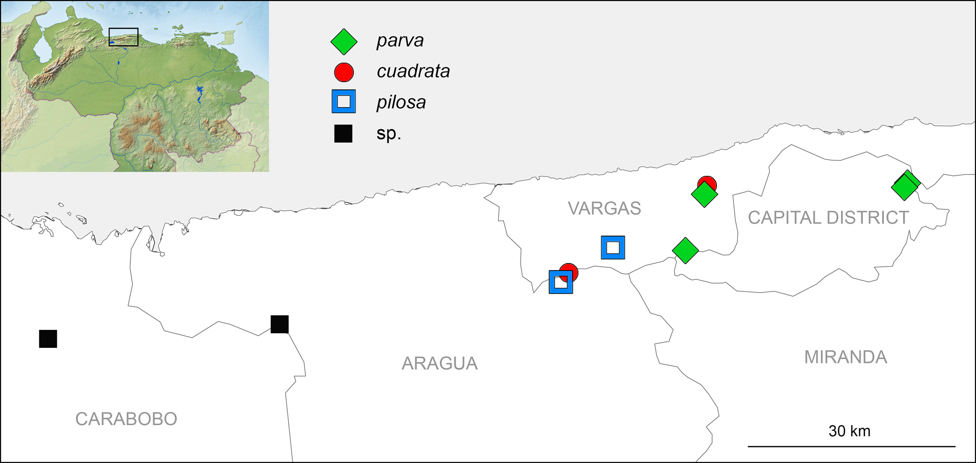
Distribution. The genus is apparently restricted to forests at medium to high elevation (~ 950–2200 m a.s.l.) in the Cordillera de la Costa region in Venezuela ( Fig. 1 View FIGURE 1 ). It has been found in the states Aragua, Vargas, Capital District, and Carabobo and likely ranges into the state Miranda.
Composition. The genus now includes the three named species treated below. A fourth (undescribed) species occurs in Henri Pittier National Park at Rancho Grande and at La Cumbre between San Estéban and Valencia (specimens in ZFMK, Ar 18256, AMNH, and MNHN, Ar 10556).


No known copyright restrictions apply. See Agosti, D., Egloff, W., 2009. Taxonomic information exchange and copyright: the Plazi approach. BMC Research Notes 2009, 2:53 for further explanation.
|
Kingdom |
|
|
Phylum |
|
|
Class |
|
|
Order |
|
|
Family |
Stenosfemuraia González-Sponga, 1998
| Huber, Bernhard A. & Arias, Quintin 2017 |
Codazziella González-Sponga, 2005 : 100
| Gonzalez-Sponga 2011: 41 |
| Gonzalez-Sponga 2005: 100 |
Stenosfemuraia González-Sponga, 1998 : 21
| Gonzalez-Sponga 1998: 21 |