
Pleurobranchus reticulatus Rang, 1832
|
publication ID |
https://doi.org/ 10.5281/zenodo.7225407 |
|
persistent identifier |
https://treatment.plazi.org/id/7B5BA125-7B41-0268-4E95-7FA8FB4C2C90 |
|
treatment provided by |
Valdenar |
|
scientific name |
Pleurobranchus reticulatus Rang, 1832 |
| status |
|
Pleurobranchus reticulatus Rang, 1832 View in CoL
( Figs. 1 View Fig A-B; 2-6)
Pleurobranchus reticulatus Rang, 1832 View in CoL : pl. 1; Pilsbry (1896: 216); Vayssière (1898: 354); Cervera et al. (1996: 154); Neves et al. (2007: 265, figs. 1-3); Goodheart et al. (2015: 338; figs. 7A-E).
Pleurobranchus areolatus View in CoL auct. non Mörch, 1863: Edmunds (1968: 85, figs. 2-3); Ev. Marcus (1976a: 16; 1977: 9; 1984: 60, figs. 1, 34-43); Rios (1994: 206, pl. 69; 2009: 417); García et al. (2002: 50, fig. 2H); García et al. (2008: 94); Troncoso et al. (2009: 409, figs. 17.2h; 17.6); Padula et al. (2012: 3, fig. 5D); Pereira et al. (2014: 1); Padula et al. (2014: 3, fig. 1B).
Type material: Type material of P. reticulatus is not known to exist, not found at MNHN ( Valdés and Héros, 1998; Goodheart et al. 2015).
Type locality: Gulf of Guinea.
Material examined: Brazil: Alagoas state: Recife da Jatiuca: MNRJ 13104, 22/xi/2003, M. Dorigo coll. [1; 1 dissected]; Saco de Pedra: MNRJ 12928, 12/i/2008, V. Padula coll. [3]; Bahia state: Guarapuá: Ilha de Tinharé: MNRJ 13290, xii/2008, P. M. S. Costa coll. [1]; Rio de Janeiro state: Cabo Frio: MZSP 75475, Marcus coll. [1]; MZSP 119938 [1 microscopic slide with radula and jaw platelets]; MZSP 119939 [1 microscopic slide with radula and jaw platelets]; Praia das Conchas: MZSP 50335, L. R. L. Simone coll. [1]; MZSP 97328, xii/2007, V. Padula coll. [2]; Enseada do norte: MNRJ 18223, 30/ii/2008, V. Padula coll. [1; 1 dissected]; Arraial do Cabo: Prainha: MNRJ 10844, ii/1984, G. Nunan & M. R. Sá coll. [4; 2 dissected]; MNRJ 14468, ii/1984, G. Nunan & M. R. Sá coll. [1, 1 dissected]; Praia do Forno: MNRJ 11710, 23/vi/2007, J. Alvim & V. Padula coll. [3]; MNRJ 12055, 20/x/2007, J. Alvim coll. [1; 1 dissected]; Angra dos Reis: MZSP 85984 [1]; Ilha Grande: MZSP 84228 ii/2007, L.
R. L. Simone coll. [1]; São Paulo state: Laje de Santos: MZSP 90723, ii/2003, C. M. Cunha coll. [2].
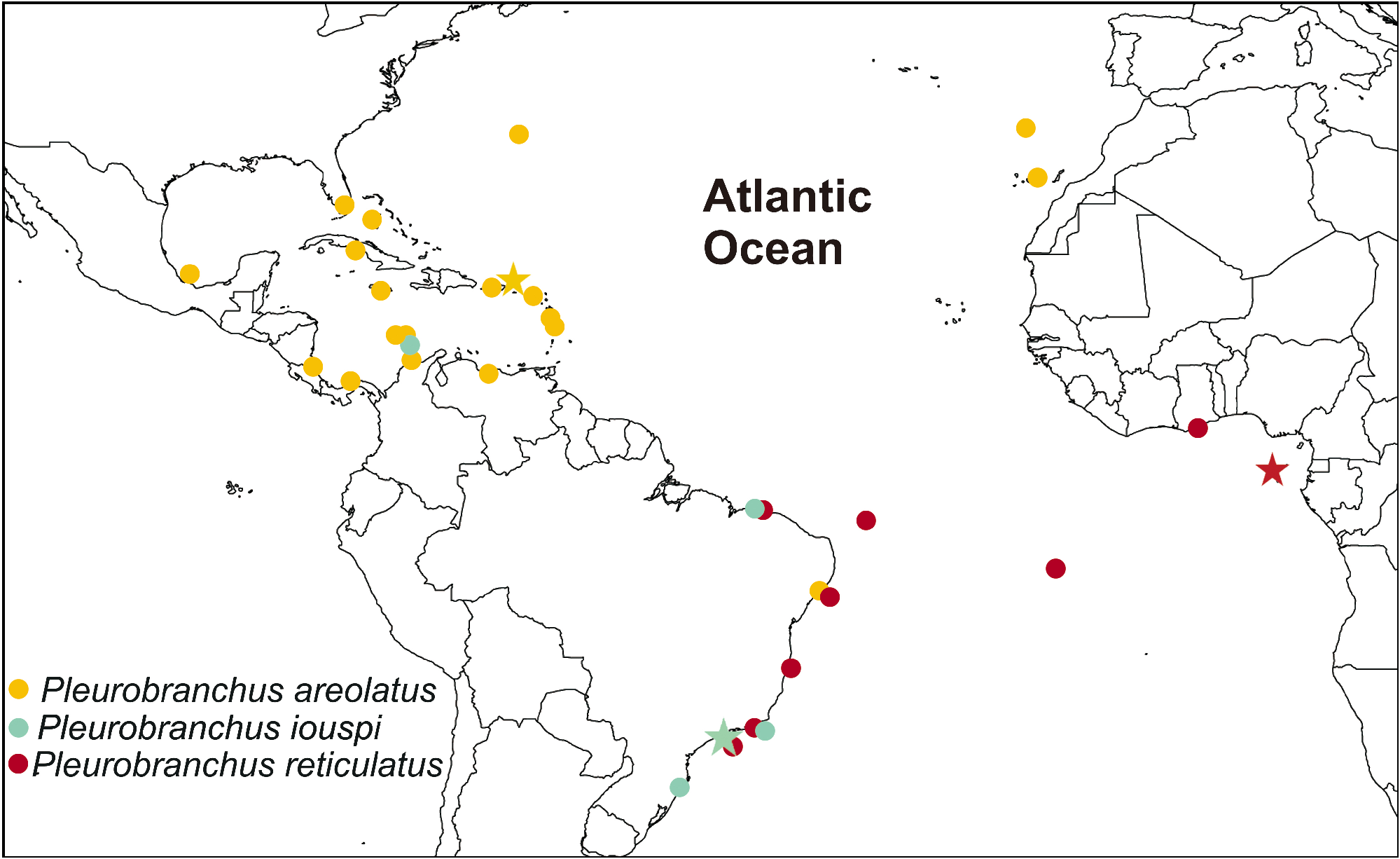
Specimen records ( Fig. 2 View Fig ): Ghana ( Edmunds 1968); Gulf of Guinea ( Rang 1832); English Bay and Soudan Bay, Ascension islands Padula et al. (2014); Brazil: Fernando de Noronha ( García et al. 2002); Maranhão state (Ev. Marcus 1984); Alagoas state ( Padula et al. 2012); Bahia state (present study); Rio de Janeiro state: Cabo Frio (Ev. Marcus 1977, 1984) and Arraial do Cabo (Ev. Marcus 1976a); São Paulo state: Laje de Santos (present study).
Description: External morphology ( Figs. 1 View Fig AB; 3A): Living specimens yellowish-orange with tubercles of many colors: dark red, red, bright orange and pale orange; space inter-tubercles white; mantle edge orange; red rhinophores with white ring-shaped ornamentation; oral veil white with orange marks and orange edge; foot dorsally white with orange blotches and edge. Living specimens up to 80 mm in length; length of preserved specimens 42-57 mm; width 26-34 mm; length of foot 20-56 mm; width of foot 32-39 mm. Body subquadrate, slightly depressed. Mantle covered foot partially. Mantle surface covered by many large rounded/oval/hexagons tubercles (1-2 mm in diameter) of different sizes and irregularly arranged; generally bigger and more concentrated in middle of mantle than at mantle edge. Oral veil broad connected with head region; laterally, oral tentacles with deep notch over almost its length. Rhinophores rolled joined at their bases, up to one third of the length. Gill exposed laterally ( Fig. 3A View Fig ); 2/3 length of body; main rachis with rounded tubercles at point of origin of pinnae, forming zig-zag line ( Fig. 3A View Fig ); small rounded tubercles at base of pinnae; bipinnate pinnae; 18- 30 pinnae; 12-16 pinnae free from the body wall, attached by branchial membrane. Anal opening lying over end of gill membrane ( Fig. 3A View Fig ). Pre-branchial pore opening approximately beside main rachis ( Fig. 3A View Fig ). Nephropore under second pinnae. Genital aperture surrounded by anterior flaps ( Fig. 3A View Fig ). Penis semi-internal, never completely internal when retracted. Foot round at posterior end; foot sometimes projecting beyond notum. Metapodial gland elongated, 1/7 times foot length; anteriorly bilabiated, upper lip notched, smaller than lower one.
Mantle ( Figs. 4 View Fig A-C): Mantle covered by calcareous spicules. Two types of spicules in mantle: linear, rod-like (length: 70.0-200.0 μm; thickness: 7.4-10.0 μm) ( Fig. 4A View Fig ); stellate (ray length: 17.3-27.2 μm; ray thickness: 4.2-5.5 μm) ( Fig. 4B View Fig ). Additionally, some elongated, but irregular (a not well definite shape) spicules grouped in some parts of mantle (length, difficult to evaluated because of shape, this measure refers to the distance, straight, between two ends: 60.0- 120.0 mm; thickness: 10.0 μm) ( Fig. 4C View Fig ).
Shell ( Figs. 5 View Fig A-B): Yellowish-cream, decalcified, flattened and fragile; two times longer than wide; brownish periostracum; not observed in some specimens; subquadrangular in outline. Length 4.6 mm, width 2.1 mm (in a preserved specimen with 52 mm in length); Length: 6.6 mm, width 3.3 mm (in a preserved specimen with 57 mm in length); and, length 3.9 mm, width 2.0 mm (in a preserved specimen with 42 mm in length). Spire has 1.5 whorls. Protoconch smooth. Lines of growth distinct; shell sculptured with longitudinal lines transverse to growth lines immediately after protoconch; anterior portion of last whorl only the lines of growth are recognizable. Shell above heart, on right side of blood gland.
Circulatory system: Pericardium well developed in anterior portion of body (near cerebro-pleural ganglia). Blood flowing into auricle from gills, kidney and venous sinuses. Efferent branchial vessel connecting the gill with auricle. Auricle on right side, ventricle on left; auricle with thin wall; ventricle muscular. Blood gland small, creamy covering left part of pericardium. Blood gland close or joined to aorta.
Digestive system ( Figs. 5 View Fig C-F; 6A-F): Transverse mouth in middle of snout tip. Oral canal muscular just posterior to mouth ( Fig. 6A View Fig ). Muscle surrounding jaws (mj) strong, pair of large jaws located in its inner surface; mj originating in lateral and dorsal surfaces of oral canal, inserting into lateral and dorsal regions of buccal mass ( Figs. 6 View Fig A-C). Jaws amber, lighter posteriorly; jaw of two plates surrounding radula inside buccal cavity ( Fig. 6D View Fig ); elongated, reaching level of radula. Each jaw plate showing alternate rows formed by elongated elements with slight cruciform lateral expansion (preserved specimen measuring 42 mm in length: 39 elements transversally, 59 elements longitudinally) ( Fig. 6C View Fig ); elements consist on a main cusp with 2-6 denticles of different sizes in each side, usually five ( Fig. 6C View Fig ); anterior elements worn, denticles sometimes unclear. Pair of jugal muscle m1v inserting into m5. Pair m4, main dorsal tensor muscle of radula, well developed, originating in lateral region of cartilages, surrounding them ventrally; inserting into subradular membrane ( Figs. 6 View Fig A-C). Pair m5, secondary dorsal tensor muscle of radula, covering median portions of cartilage, extending up to dorsal region; originating in posterior surface of cartilages; inserting laterally in mj ( Figs. 6 View Fig AC). Pair m7 absent. Pair m10d, originating in posterior portion of oral canal; inserting into m4 ( Figs. 6 View Fig B-C). Pair m10v, protractor muscle of odontophore, connected posterior portion of canal oral with ventral portion of m4 ( Fig. 6A View Fig ). Single auxiliary muscle m10a, ventral tensor muscle of radula, originates in anterior portion of oral canal, running in middle of buccal mass and inserting into radular sac ( Fig. 6A View Fig ). Pair of strong retractor muscles originates in most posterior portion of m5 ( Figs. 6A, C View Fig ); it is separated in 3/4 its total length and jointed in its posterior portion, lying above anterior portion of digestive gland. Odontophore cartilage subquadrate in outline ( Fig. 6F View Fig ). Radula cream; formula 69 × 165.0.165 (from preserved specimen 42 mm length). Innermost lateral tooth with enlarged base and hook-shaped cusp curved towards to base of tooth ( Fig. 5D View Fig ). Lateral teeth hook-shaped, larger and more developed in center of rows ( Fig. 5E View Fig ). Outermost lateral teeth less developed ( Fig. 5F View Fig ). Aperture of acid gland located between jaw plates ( Figs. 6 View Fig D-E). Duct of acid gland thin (same width as salivary duct) ( Figs. 6 View Fig B-C), passing within nerve ring. Acid gland slightly ramified, restrict to anterior portion of digestive organs. Esophagus tube-like passing into voluminous stomach ( Figs. 6 View Fig A-B, E). Salivary gland small and in front of digestive gland. Ducts of salivary glands entering pharynx musculature laterally to esophagus, opening into base of pharyngeal cavity between radula and jaw plates ( Fig. 6D View Fig ); not convoluted; without a visible ampullae. Stomach elongated and with thickened wall, it could be in different states of expansion ( Figs. 6 View Fig A-B, E); internally, could have some folds, wrinkled wall; posteriorly smooth, except by the ventral furrow that leads to intestine ( Fig. 6E View Fig ); stomach passes ventrally into digestive gland. Intestine short opening under gill membrane into anus; thin wall; internally, longitudinal folds near its apertures. Salivary, digestive and hermaphrodite glands forming a single aggregate ( Figs. 6 View Fig A-B).
Reproductive system ( Figs. 3 View Fig B-D): Triaulic. Ampulla elongated, three times wider than deferent duct; convoluted. Spermoviduct branching into two ducts, shorter oviduct and other duct leading to prostate. Prostate tubular and extremely convoluted; eight times wider than deferent duct. Deferent duct long, thin and convoluted ( Figs. 3 View Fig B-C), narrowing into a non cuticular and semi-contractile penis ( Fig. 3C View Fig ). Penis (in a preserved specimen with 42 mm length: length 8.5 mm; width 3.5 mm) ( Figs. 3 View Fig C-D); convex portion of penis with a wide leaflet ( Figs. 3 View Fig C-D); evident penial sheath, enlargement near gonopore ( Fig. 3C View Fig ). Vaginal duct slightly convoluted with two allosperm vesicles arranged semi-serially ( Fig. 3C View Fig ). Proximally, vagina divides into seminal receptacle and bursa copulatrix as a short duct, both allosperm receptacles lying close to each other, their stalks are crossed ( Fig. 3C View Fig ). Seminal receptacle shortstalked; elongated; highly convoluted ( Fig. 3C View Fig ). Bursa copulatrix rounded. Vaginal opening immediately ventral to penis. Vagina about three times wider than deferent duct; became wider near female aperture ( Fig. 3C View Fig ). Semimal receptacle approximately same wider than prostate. Genital aperture surrounded by anterior flaps, normally two ( Fig. 3B View Fig ).
Nervous system ( Fig. 3E View Fig ): Nerve ring above oral canal. Cerebral and pleural ganglia fused. Eyes located latero-centrally of cerebro-pleural complex; eyes borne upon very short optical nerves. Rhinophoral ganglia placed at bases of rhinophores, near cerebro-pleural ganglia; two main nerves leave from rhinophoral nerves, these runs until distal portion of rhinophores; rhinophoral nerves with many secondary nerves, perpendicular in relation of main nerves. Nerves leaving cerebro-pleural ganglia: cp1 inserting latero-ventrally, almost into foot; cp2 bifurcating near base, both cp2a and cp2b inserting dorsally into mantle; cp3 runs laterally until inserts into body wall, in right side nerve enters into mantle near the base of gonopore and, in left side nerve enters into mantle near anterior portion of digestive gland; cp4 runs until the most posterior portion of body; cp5 very short nerve, located in middle of ganglia, inserting dorsally into mantle; cp6 thin nerve, inserting into mantle dorsally; cp7 thin nerve, innervating latero-ventral side of body wall; cp8 innervates female gland mass. Connective between visceral and cerebro-pleural ganglia very short, almost imperceptible. Connective between buccal and cerebro-pleural ganglia leads from ventral view of cerebro-pleural ganglia, in the most posterior portion of ganglion. Nerves leaving buccal ganglia, in antero-posterior order: nb1 bifurcating near origin, both nb1a and nb1b inserts into esophagus; nb2 inserting into salivary ducts; connective cerebro-pleural-buccal shortly after nb2; nb3 bifurcating near origin, anterior branch (nb3a) inserting into m4; posterior branch (nb3b) leading to m5; nb4 inserting into radular sac. Connective cerebro-pleural-pedal extremely short and it can only be seen after carefully dissecting. Pedal commissure short and leaving from the most anterior posterior of pedal ganglion. Pedal ganglia smaller than cerebro-pleural complex, in antero-posterior order: np1 inserting ventrally into oral veil; np2 inserting anteriorly into foot; np3 innervates foot; np4 inserting ventrally into foot and runs until the most posterior portion of body.


| MNHN |
Museum National d'Histoire Naturelle |
| MNRJ |
Museu Nacional/Universidade Federal de Rio de Janeiro |
| V |
Royal British Columbia Museum - Herbarium |
| MZSP |
Sao Paulo, Museu de Zoologia da Universidade de Sao Paulo |
| R |
Departamento de Geologia, Universidad de Chile |
| AC |
Amherst College, Beneski Museum of Natural History |
No known copyright restrictions apply. See Agosti, D., Egloff, W., 2009. Taxonomic information exchange and copyright: the Plazi approach. BMC Research Notes 2009, 2:53 for further explanation.
|
Kingdom |
|
|
Phylum |
|
|
Class |
|
|
Order |
|
|
Family |
|
|
Genus |
Pleurobranchus reticulatus Rang, 1832
| Grzelak, Katarzyna & Sørensen, Martin V. 2022 |
Pleurobranchus areolatus
| Pereira FR & Santos MFC & Williams DE & Andersen RJ & Padula V & Ferreira AG & Berlinck RGS 2014: 1 |
| Padula V & Wirtz P & Schrodl M. 2014: 3 |
| Padula V & Bahia J & Correia MD & Sovierzoski HH 2012: 3 |
| Troncoso JS & Dominguez M & Garcia FJ 2009: 409 |
| Garcia FJ & Dominguez M & Troncoso JS & Feito, S. L. 2008: 94 |
| Garcia FJ & Troncoso JS & Dominguez M. 2002: 50 |
| Rios E. 1994: 206 |
| Marcus Ev. 1984: 60 |
| Marcus Ev. 1977: 9 |
| Marcus Ev. 1976: 16 |
| Edmunds M. 1968: 85 |
Pleurobranchus reticulatus
| Goodheart J & Camacho-Garcia Y & Padula V & Schrodl M & Cervera JL & Gosliner TM & Valdes A. 2015: 338 |
| Neves R & Cervera JL & Calado G. 2007: 265 |
| Cervera JL & Cattaneo-Vietti R & Edmunds M. 1996: 154 |
| Vayssiere A. 1898: 354 |
| Pilsbry HA 1896: 216 |