
Pleurobranchus areolatus Mörch, 1863
|
publication ID |
https://doi.org/ 10.5281/zenodo.7225407 |
|
persistent identifier |
https://treatment.plazi.org/id/7B5BA125-7B4A-026F-4E79-7F88FECB29B0 |
|
treatment provided by |
Valdenar |
|
scientific name |
Pleurobranchus areolatus Mörch, 1863 |
| status |
|
Pleurobranchus areolatus Mörch, 1863 View in CoL
( Figs. 1 View Fig C-D; 2; 7-10)
Pleurobranchus areolatus Mörch, 1863: 28 View in CoL ; Pilsbry (1896: 199); Vayssière (1898: 338); Ev. Marcus and Er. Marcus (1962: 466, figs. 14-15; 1963: 25; 1967: 44; 1967: 163, fig. 19); Er. Marcus and Ev. Marcus (1970: 55); Keen (1971: 811, fig. 16, pl. 19); Abbott (1974: 347); Thompson (1977: 108, figs. 12c-d; 13a-b); Cervera et al. (2004: 32); Ardila and Rachello (2005: 60, fig. 2); Espinosa et al. (2005: 63); Valdés et al. (2006: 113); Redfern (2013: 295, figs. 812AB); Goodheart et al. (2015: 343, figs. 18A-G, 19C-F, 20- 22).
Oscaniella areolata : Bergh (1897: 111, figs. 31-41, pls. 9).
Pleurobranchus crossei Vayssière, 1897: 353 View in CoL , fig. 1; Vayssière (1898: 332, figs. 148-154); Valdés et al. (2006: 114); Redfern (2013: 295, figs. 813B-E); Ortea et al. (2014: 120, figs. 1B-C, 3A-B). Synonymized by Thompson (1977).
Pleurobranchus (Susania) atlanticus Abbott, 1949: 73 View in CoL , figs. 1-10, pl. 5. Synonymized by Thompson (1977).
Pleurobranchus atlanticus View in CoL : Padula et al. (2012: 3, fig. 5E).
Susania gardineri White, 1952: 106 View in CoL , figs. 2-5, pl. 6, fig. 1. Synonymized by Ev. Marcus (1984).
Pleurobranchus reesi White, 1952: 107 View in CoL , figs. 3, 6, pl. 6, fig. 2. Synonymized by Goodheart et al. (2015).
Pleuroranchus evelinae Thompson, 1977: 108 , figs. 12E-F, 13C-E; Ev. Marcus (1984: 63, figs. 44-50, in part); Redfern (2001: 167, pl. 72, fig. 691E-F); Espinosa et al. (2005: 63); Valdés et al. (2006: 112); Ortea et al. (2014: 122, figs. 1D, 2). Synonymized by Goodheart et al. (2015).
Type material: Not located in Natural History Museum of Denmark and Natural History Museum.
Type locality: St. Thomas, United States Virgin Islands.
Material examined: Syntypes of P. reesi : NHMUK 1934.9.11.102-104, Bird Key reef, Dry Tortugas, G. Tandy and J.S. Colman leg. [3]. Holotype of P. evelinae : NHMUK 19773 View Materials W, Discovery Bay , Jamaica, T. E. Thompson coll. [1] . Holotype of P. crossei : MNHN-IM-2000-30102, Caribbean Sea [1]. Syntypes of P. gardineri : NHMUK 1934.9.11.99-101, Dry Tortugas, G. Tandy and J.S. Colman leg. [3]. Colombia: MZSP 119937, K. Bandel leg. [1 microscopic slide with radula and jaw platelets]; Brazil: Alagoas state: Galés de Maragogi: MNRJ 18760, 29/xii/2007, V. Padula coll. [1; 1 dissected].
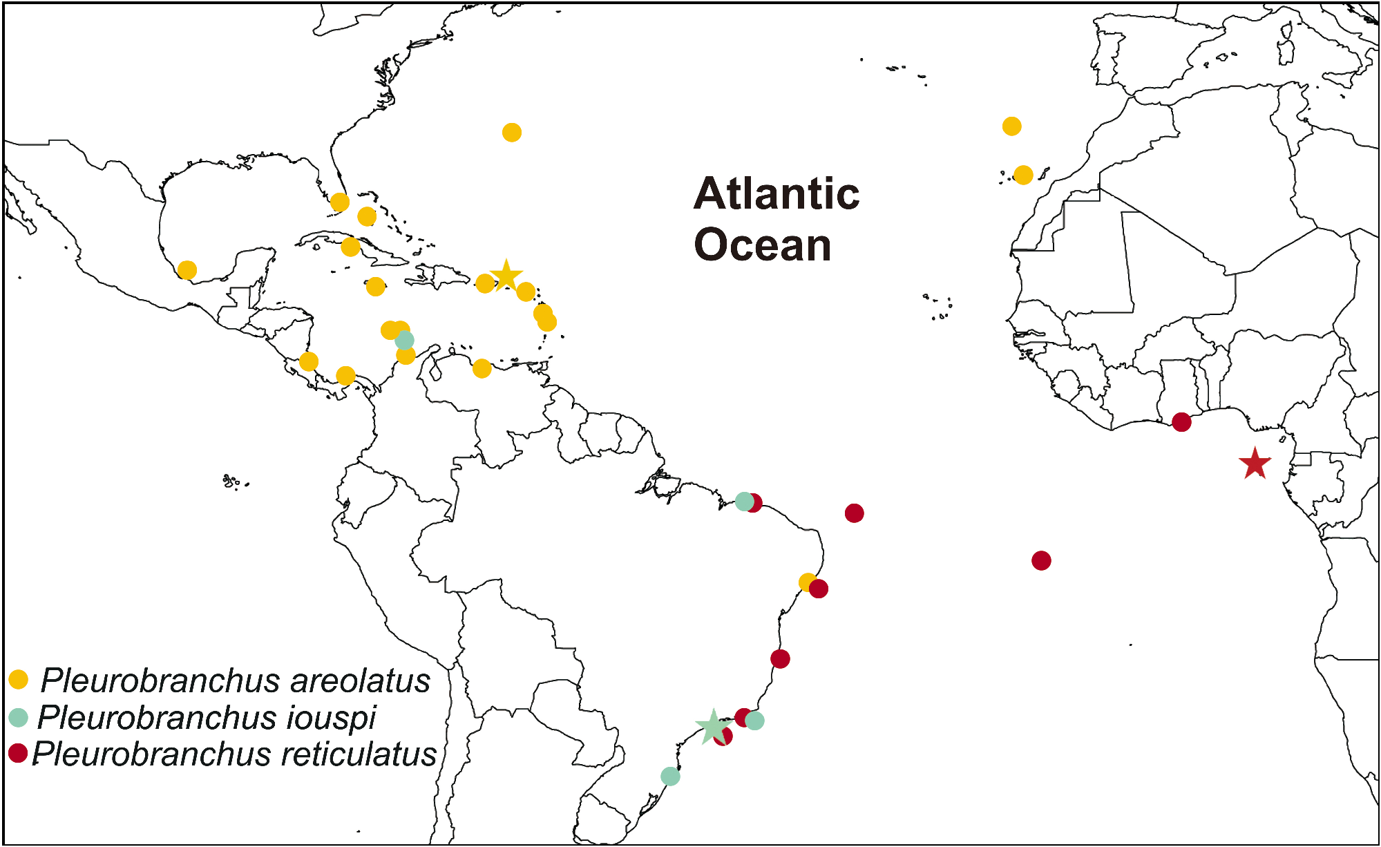
Specimen records ( Fig. 2 View Fig ): Florida ( Abbott 1949; White 1952; Ev. Marcus and Er. Marcus 1962; 1967); Puerto Rico (Er. Marcus and Ev. Marcus 1970; Valdés et al. 2006); Virgin Islands: St. Thomas ( Mörch 1863; Ev. Marcus and Er. Marcus 1962; Valdés et al. 2006); Mexico (Ev. Marcus and Er. Marcus 1967; Valdés et al. 2006); Bahamas ( Redfern 2001, 2013; Valdés et al. 2006; Goodheart et al. 2015); Cuba ( Espinosa et al. 2005, 2007), Guadeloupe and Martinique ( Ortea et al. 2014); Jamaica ( Thompson 1977; Valdés et al. 2006); Curaçao (Ev. Marcus and Er. Marcus 1963; Er. Marcus and Ev. Marcus 1970); Aruba, Costa Rica, Bermuda, St. Marteen/ St. Martin, Venezuela ( Valdés et al. 2006); Panama ( Valdés et al. 2006; Goodheart et al. 2015); Colombia ( Ardila and Rachello 2005); Brazil: Alagoas ( Padula et al. 2012); Canary Islands ( Ortea et al. 2014); Madeira ( Cervera et al. 2004). Specimens recorded from Pacific Ocean (Ev. Marcus and Er. Marcus 1967; Bertsch and Smith 1973; Bertsch 1979) under the name P. areolatus may in fact be this species, but judgment must be reserved at present.
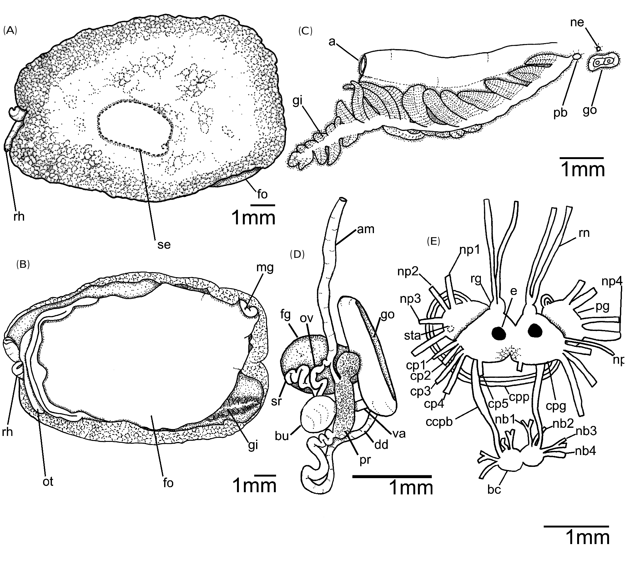
Description: External morphology ( Figs. 1 View Fig C- D; 7A-C): External morphology of P. areolatus is very similar to P. reticulatus as described above, with the following exceptions: Living specimens white with brownish-red blotches on dorsum; some tubercles with pale-orange tips; mantle edge orange; rhinophores with dark red, orange and white blotches; rhinophoral apex white; oral veil same color pattern of mantle; ventrally, foot translucent white with its edge with orange and white alternate pigment ( Fig. 1D View Fig ). Living specimen 27 mm in length. Preserved specimen: length 12 mm; width 8 mm; length of foot 11 mm; width of foot 6 mm. Mantle covered foot entirely. Mantle surface covered by pointed low tubercles (125- 375 μm in diameter); tubercles at mantle edge smaller in diameter, higher and more concentrated than middle of mantle; irregularly arranged. Gill 2/3 length of body; main rachis with tiny rounded tubercles at point of origin of pinnae, forming zig-zag line; 19 pinnae; 9 pinnae free from body wall, attached by branchial membrane. Pre-branchial pore opening approximately beside main rachis ( Fig. 7C View Fig ). Nephropore lying above female aperture. Genital aperture surrounded by a thin fold. Penis not protruded ( Fig. 7C View Fig ). Foot posteriorly with small white semi-circled metapodial gland ( Fig. 7B View Fig ); metapodial gland 1/ 20 foot length.
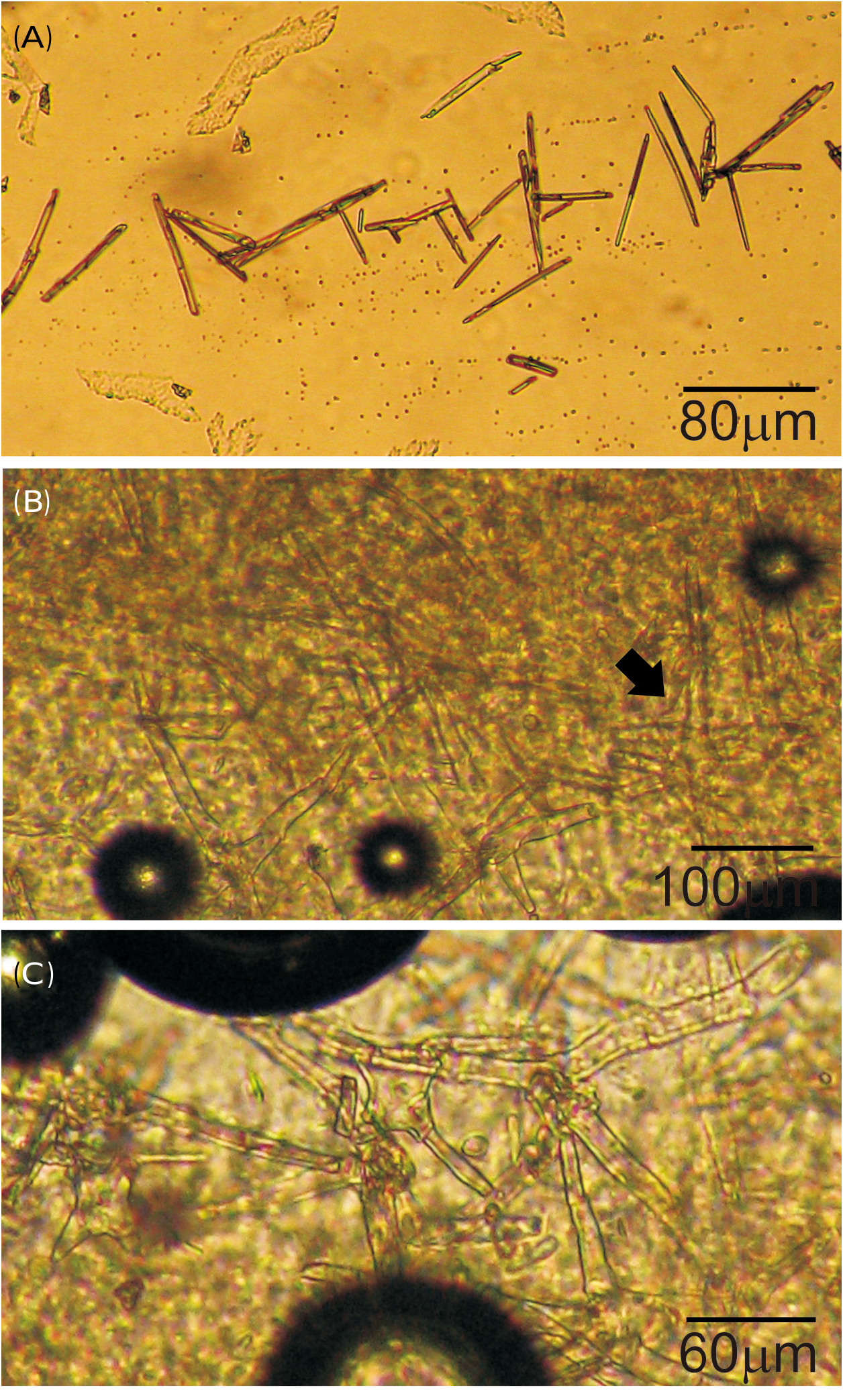
Mantle ( Figs. 8 View Fig A-C): Mantle covered by sparse spicules, only in some parts of mantle it was found. Two types of spicules in mantle: linear, rod-like (length: 70.0 μm -160.5 μm, mainly between 70.0 μm 90.0 μm; thickness: 5.0 μm) ( Fig. 8A View Fig ) and stellate with four or five irregular rays (ray length: 70.0 μm- 112.5 μm; ray thickness: 10.0 μm) ( Figs. 8 View Fig B-C). Rod-like spicules calcareous; stellate spicules composed of an unidentified organic matrix.
Shell ( Figs. 7A View Fig ; 9 View Fig A-B): White to brownish with light golden tones in some parts of shell, visible according to incidence of light; subquadrangular in outline. Length 3.4 mm, width 2.3 mm (in a specimen with 27 mm long alive); approximately 1.5 times longer than wide. Spire has 1.5 whorls. Protoconch smooth. Lines of growth distinct ( Fig. 9B View Fig ). Shell sculptured with longitudinal lines transverse to growth lines immediately after protoconch. Shell situated in middle portion of body, on the left side ( Fig. 7A View Fig ); partially above pericardium, blood gland and digestive gland.
Circulatory system: Circulatory system of P. areolatus identical to the P. reticulatus as described above.
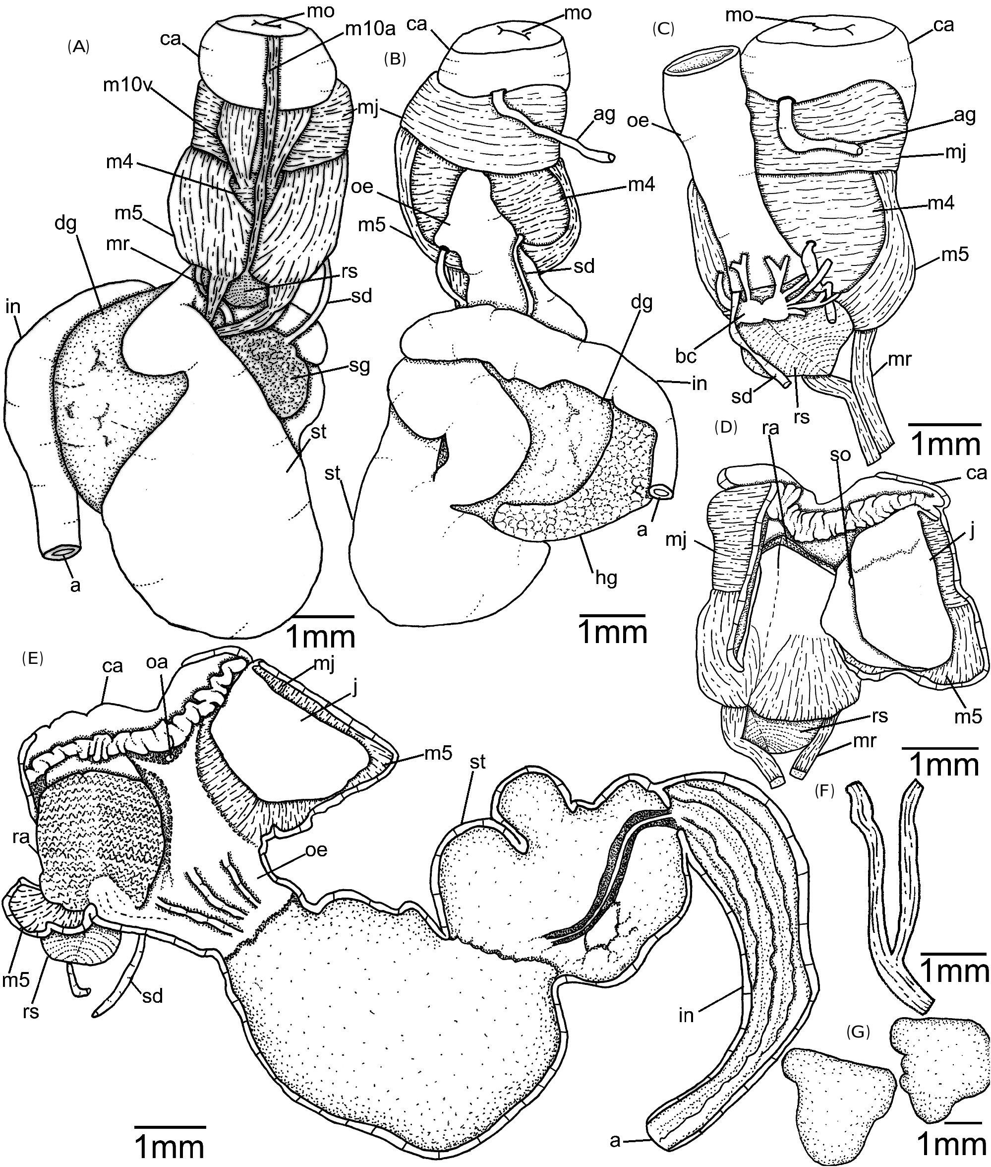
D i g e sti v e s y s te m ( Figs. 9 View Fig C-F; 10A-G): Digestive system of P. areolatus very similar to P. reticulatus as described above, with the following exceptions: Each jaw plate showing alternate rows formed by elongated elements with cruciform lateral expansion ( Fig. 9C View Fig ); elements consist on a main cusp with 2-3 denticles in each side, sometimes asymmetrical ( Fig. 9C View Fig ). Pair of jugal muscle m1v absent. Pair m5, secondary dorsal tensor muscle of radula, large and broad, covering median portions of cartilage, extending up to dorsal region; originating in posterior surface of cartilages; inserting laterally in mj ( Figs. 10 View Fig A-C). Pair m10d (dorsal) absent. Pair of strong retractor muscles originates in most posterior portion of m5 ( Figs. 10A, C, F View Fig ); it is separated in almost its total length. Odontophore cartilage wider anteriorly than posteriorly ( Fig. 10G View Fig ). Radula translucent-white; formula 66 × 90.0.90 (in a specimen of 27 mm long alive). Outermost lateral teeth elongated, less developed ( Fig. 10F View Fig ). Aperture of acid gland located between jaw plates ( Fig. 10E View Fig ). Duct of acid gland thin (twice diameter of salivary duct) and short ( Figs. 10 View Fig B-C); aperture located between jaw plates. Stomach elongated ( Figs. 10 View Fig A-B, E); internally, stomach wall thin without longitudinal folds; posteriorly smooth, except by a ventral furrow that leads to intestine ( Fig. 10E View Fig ); stomach passes ventrally into digestive gland. Intestine long opening under gill membrane into anus; thin wall; internally, longitudinal folds well-developed. Salivary, digestive and hermaphrodite glands forming a single aggregate ( Figs. 10 View Fig A-B).
Reproductive system ( Fig. 7D View Fig ): Triaulic. Ampulla elongated with same width as vas deferent and vagina; not convoluted. Spermoviduct branching into two ducts, oviduct other duct leading to prostate. Prostate tubular, sausage-like; proximal portion rounded; not convoluted; three times wider than deferent duct. Near gonopore without trace of any penial sheath. Penis not observed. Vaginal duct not convoluted with two allosperm vesicles arranged semiserially. Vaginal duct slightly convoluted with two allosperm vesicles arranged semiserially, both allosperm receptacles lying close to each other, their stalks are crossed. Seminal receptacle elongate; same wider than deferent duct. Bursa copulatrix bulky, rounded; connected with vagina approximately in its first 1/3. Vaginal opening immediately ventral to penis. Vagina about same diameter of deferent duct.
Nervous system ( Fig. 7E View Fig ): Nervous system of P. areolatus very similar to P. reticulatus as described above, except by the absence of cp6, cp7 and cp8.


No known copyright restrictions apply. See Agosti, D., Egloff, W., 2009. Taxonomic information exchange and copyright: the Plazi approach. BMC Research Notes 2009, 2:53 for further explanation.
|
Kingdom |
|
|
Phylum |
|
|
Class |
|
|
Order |
|
|
Family |
|
|
Genus |
Pleurobranchus areolatus Mörch, 1863
| Grzelak, Katarzyna & Sørensen, Martin V. 2022 |
Pleurobranchus atlanticus
| Padula V & Bahia J & Correia MD & Sovierzoski HH 2012: 3 |
Pleuroranchus evelinae
| Ortea J & Leopoldo M & Caballer M. 2014: 122 |
| Valdes A & Hamann J & Behrens DW & DuPont A. 2006: 112 |
| Espinosa J & Ortea J & Caballer M & Moro L. 2005: 63 |
| Redfern C. 2001: 167 |
| Marcus Ev. 1984: 63 |
| Thompson TE 1977: 108 |
Susania gardineri
| White KM 1952: 106 |
Pleurobranchus reesi
| White KM 1952: 107 |
Pleurobranchus (Susania) atlanticus
| Abbott RT 1949: 73 |
Oscaniella areolata
| Bergh R. 1897: 111 |
Pleurobranchus crossei Vayssière, 1897: 353
| Ortea J & Leopoldo M & Caballer M. 2014: 120 |
| Redfern C. 2013: 295 |
| Valdes A & Hamann J & Behrens DW & DuPont A. 2006: 114 |
| Vayssiere A. 1898: 332 |
| Vayssiere A. 1897: 353 |
Pleurobranchus areolatus Mörch, 1863: 28
| Goodheart J & Camacho-Garcia Y & Padula V & Schrodl M & Cervera JL & Gosliner TM & Valdes A. 2015: 343 |
| Redfern C. 2013: 295 |
| Valdes A & Hamann J & Behrens DW & DuPont A. 2006: 113 |
| Ardila NE & Rachello P. 2005: 60 |
| Espinosa J & Ortea J & Caballer M & Moro L. 2005: 63 |
| Cervera JL & Calado G & Gavaia C & Malaquias MAE & Castano JT & Ballesteros M & Megina Martinez C & Garcia-Gomez JC 2004: 32 |
| Thompson TE 1977: 108 |
| Abbott RT 1974: 347 |
| Keen M. 1971: 811 |
| Marcus Er & Marcus Ev. 1970: 55 |
| Marcus Ev & Marcus Er. 1962: 466 |
| Vayssiere A. 1898: 338 |
| Pilsbry HA 1896: 199 |
| Morch OAL 1863: 28 |