
Infernotropis Taszakowski et al., 2022
|
publication ID |
https://doi.org/ 10.11646/zootaxa.5209.1.5 |
|
publication LSID |
lsid:zoobank.org:pub:D65B575C-5C0F-45CA-B56F-EE6EB4386931 |
|
DOI |
https://doi.org/10.5281/zenodo.7331585 |
|
persistent identifier |
https://treatment.plazi.org/id/50F5C989-3039-462E-880A-CE0C75A9D7B5 |
|
taxon LSID |
lsid:zoobank.org:act:50F5C989-3039-462E-880A-CE0C75A9D7B5 |
|
treatment provided by |
Plazi |
|
scientific name |
Infernotropis Taszakowski et al. |
| status |
gen. nov. |
Genus: Infernotropis Taszakowski et al. gen. nov.
Type species: Infernotropis madagascariensis Taszakowski et al. sp. nov., here designated.
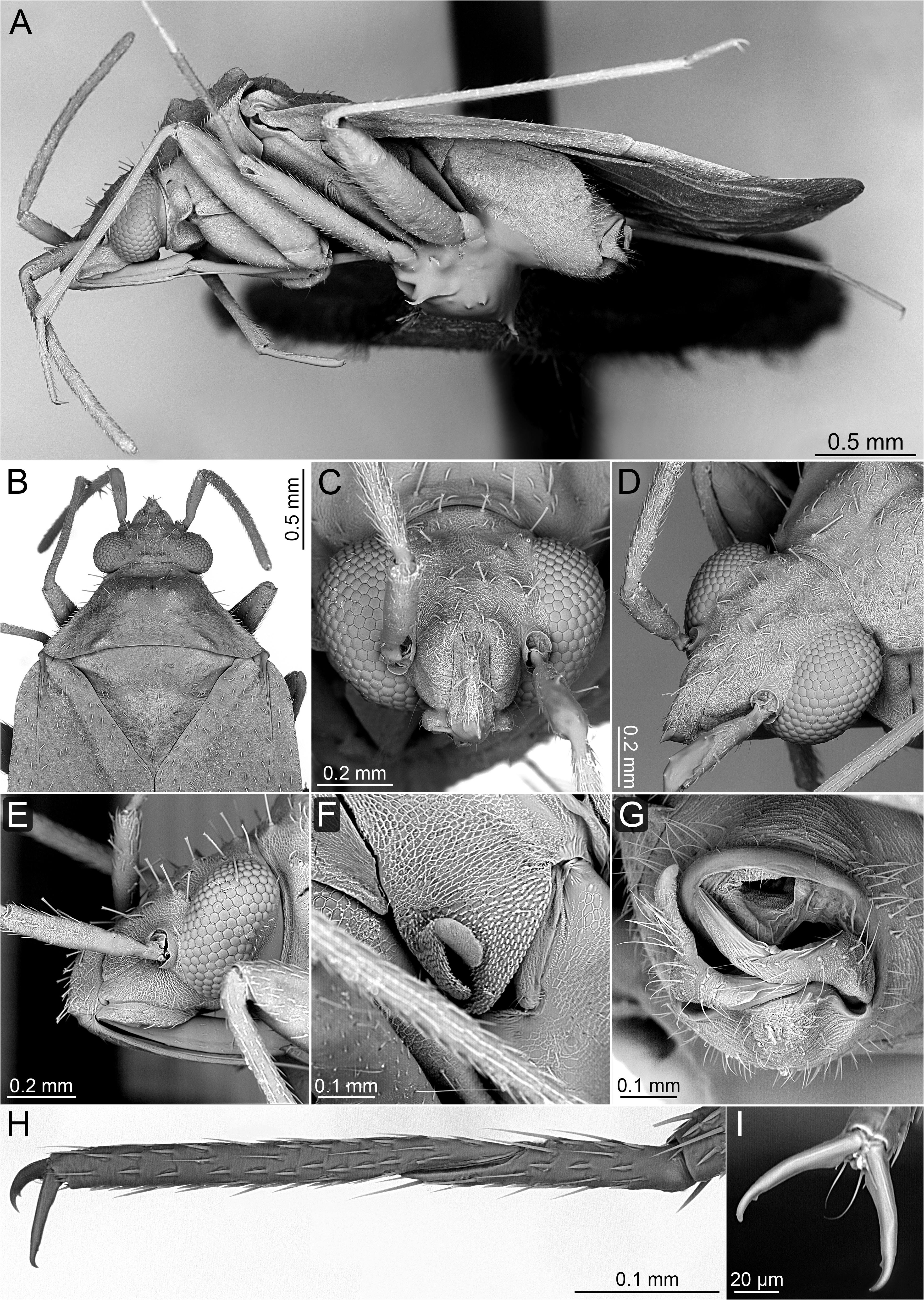
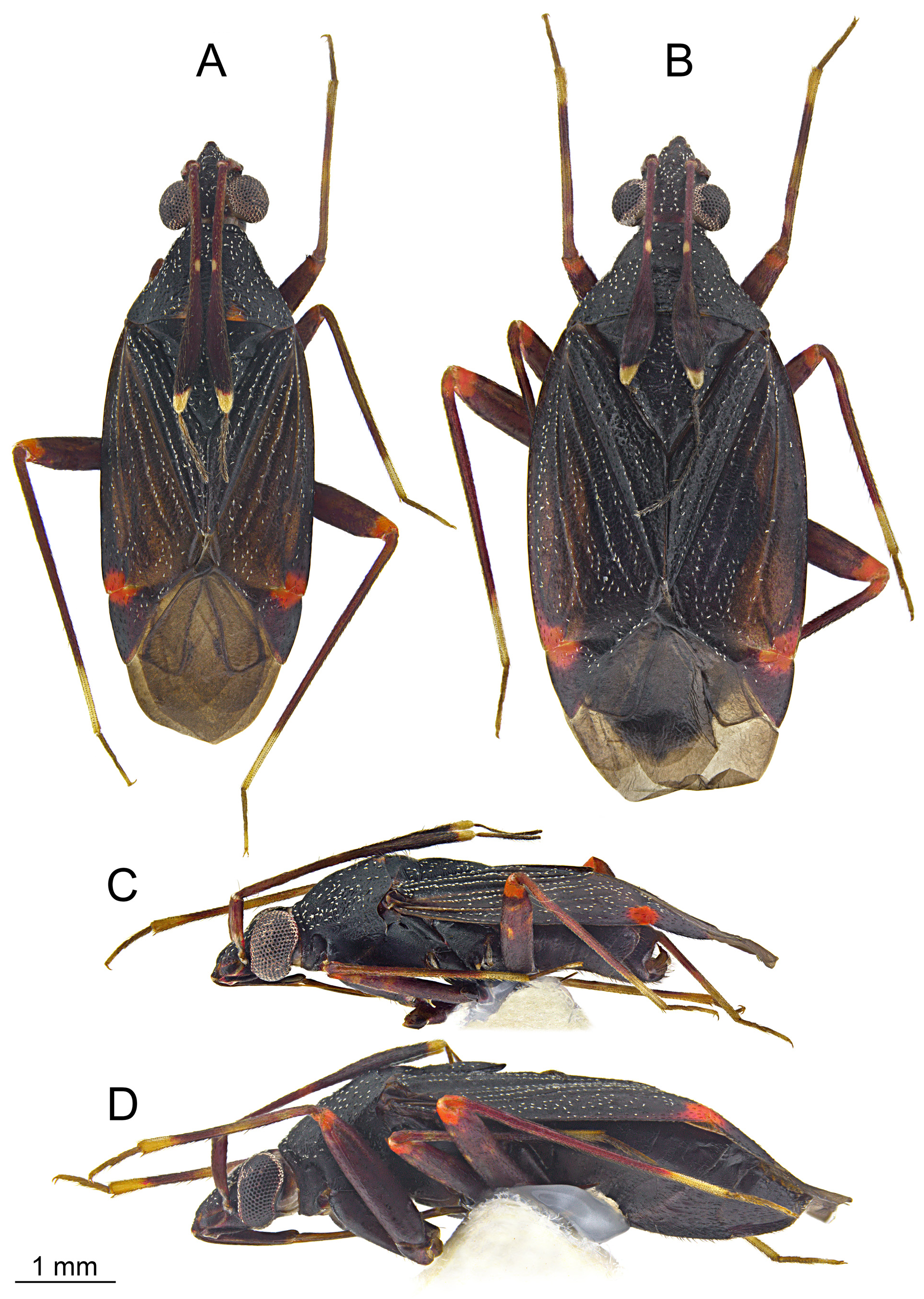
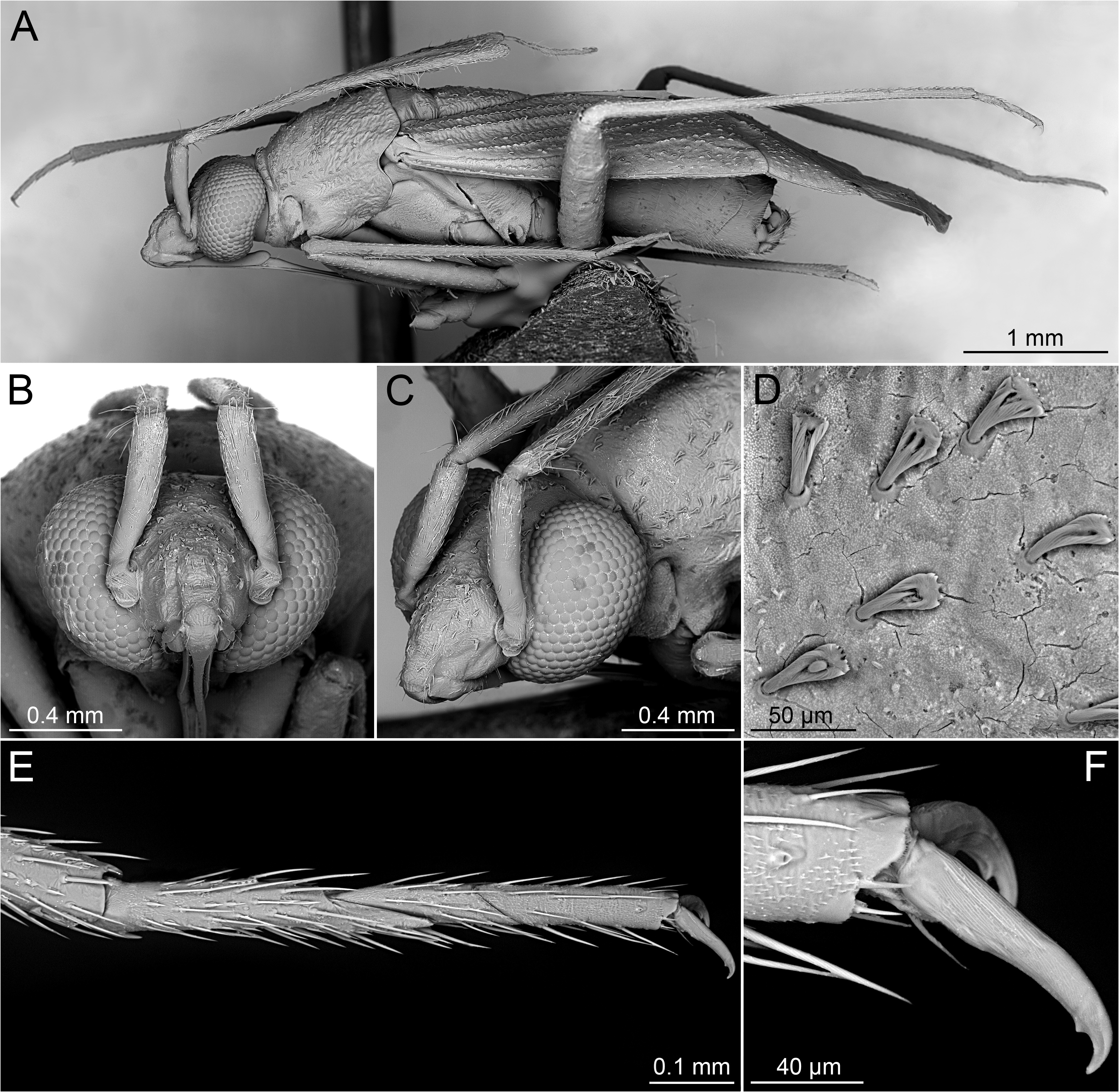
Diagnosis. Recognized by the following combination of characters: body large (> 6.20 mm); dorsum clothed with fine, adpressed, regularly distributed, moderately dense scale-like, silvery setae that are distinctly broadened toward apex ( Figs 5 View FIGURE 5 , 6A, D View FIGURE 6 ); antennal segment II stout, with basal half cylindrical and apical half strongly flattened dorsoventrally ( Figs 5 View FIGURE 5 , 6A View FIGURE 6 ), its outer surface in males with dense, long, erect and semirecumbent setae ( Fig. 6A View FIGURE 6 ); pronotum distinctly narrowing toward anterior part, bell-like ( Figs 5A, B View FIGURE 5 ); tarsus three-segmented; tarsomere I about as long as tarsomeres II and III combined ( Fig. 6E View FIGURE 6 ); sclerotized portion of ductus seminis inside endosoma with basal twothirds thin and cylindrical, apical one-third broadened, irregularly shaped with apico-lateral portions well expanded beyond middle of endosoma; apical part of endosoma with large, inflated, serrated lobe embraced basally by apicolateral parts of distal portion of ductus seminis; endosoma also with long, flattened sclerotized lobe originated on its base and terminated subapically ( Figs 7A–C View FIGURE 7 ); dorsal labiate plate (dlp) with large sclerite occupying most of it, laterally possessing rib-like projections directed dorsad ( Figs 8B, D, E View FIGURE 8 ); ventral labiate plate (vlp) with elongate sclerotization laterally (sv) ( Figs 8B–E View FIGURE 8 ).
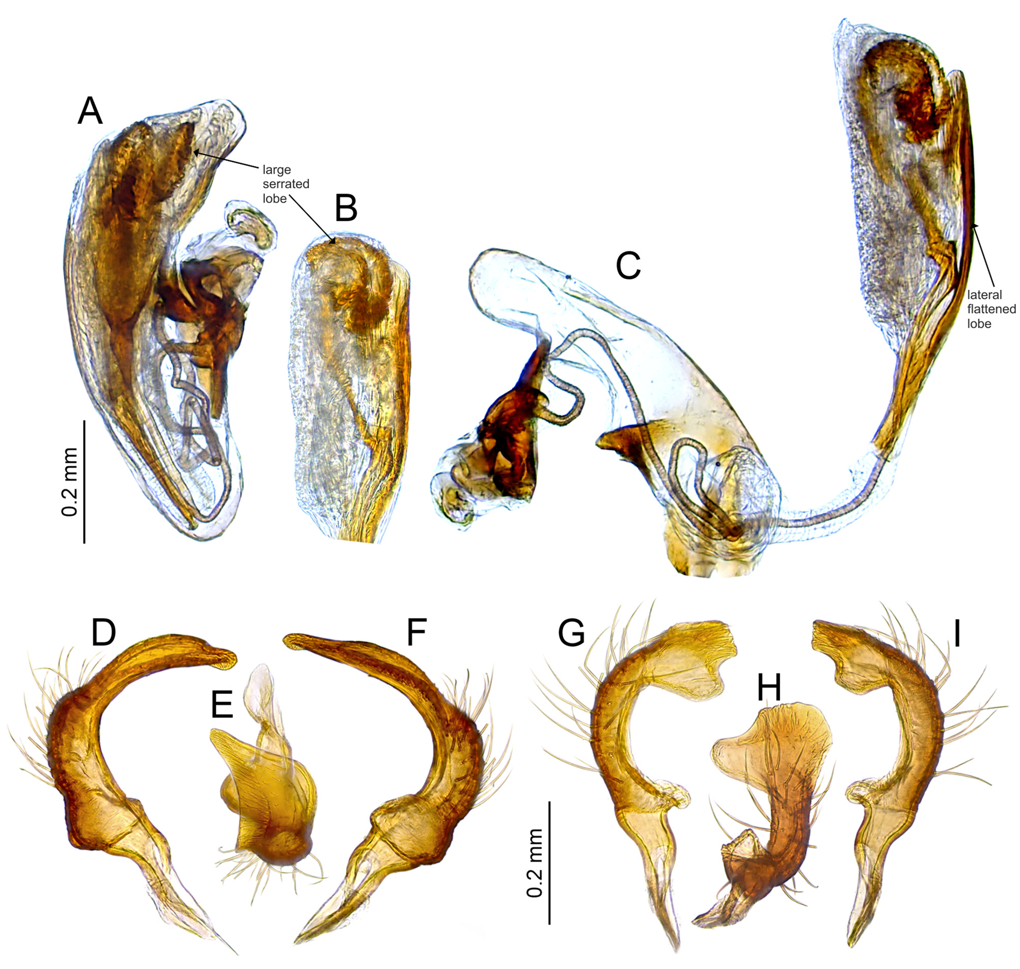
Description. Male. Macropterous. TEXTURE AND VESTITURE. Dorsum weakly rugose, clothed with fine, adpressed, regularly distributed, moderately dense scale-like, silvery setae that are distinctly broadened toward apex ( Figs 5 View FIGURE 5 , 6A, D View FIGURE 6 ). Head. Weakly rugose ( Figs 6B, C View FIGURE 6 ); antennal segment I covered with short and moderately dense, adpressed simple setae; segment II with inner surface clothed with setae similar to those present on segment I, outer surface with dense, long, erect and semirecumbent setae; segment III covered with relatively dense and long, erect and semirecumbent fine setae ( Figs 5A, C View FIGURE 5 , 6A View FIGURE 6 ) (segment IV missing in male specimen). Thorax. Thoracic pleura. Weakly rugose, covered with sparse, fine, recumbent setae ( Fig. 6A View FIGURE 6 ). Hemelytron. Clavus and endocorium with setae organized in regular rows along their veins; exocorium, embolium, and clavus with more irregularly distributed vestiture ( Figs 5A, C View FIGURE 5 , 6A View FIGURE 6 ). Legs. Coxae and femora covered with fine, recumbent, rather short setae; tibiae and tarsi with relatively dense, stiff, semirecumbent setae, tibiae also with small black spinules organized in several straight and regular rows ( Figs 5A, C View FIGURE 5 , 6A View FIGURE 6 ). Abdomen. Covered with relatively long and dense, recumbent and semirecumbent setae ( Fig. 6A View FIGURE 6 ). STRUCTURE. Body stout, elongate oval ( Fig. 5A View FIGURE 5 ). Head. Porrect, in anterior view about twice wider than high, frons forming obtuse angle with vertex; vertex with weakly developed, longitudinal sulcus medially; base of clypeus situated above antennal insertions and ventral margin of eye; antennal insertion contiguous with sulcus between maxillary and mandibular plates; eye relatively large, reniform, its ventral margin reaching gula; mandibular plate without sulcus posteriorly; antenna short, equal to median of body; segment I mostly cylindrical, somewhat narrowed basally; segment II stout, its basal half cylindrical, apical half strongly flattened dorso-ventrally; segment III thin, cylindrical; labium thin, reaching well beyond hind coxae; segment I equal to length of head, subdivided near apex ( Figs 5A, C View FIGURE 5 , 6A–C View FIGURE 6 ). Thorax. Pronotum. Distinctly narrowing toward anterior part, bell-like; collar present, convex, well separated from remainder of pronotum by deep sulcus; lateral margin weakly sinuate, almost straight, not carinate; posterior margin somewhat concave; humeral angle relatively broad. Mesoscutum and scutellum. Mesoscutum well exposed; scutellum weakly convex ( Figs 5A, C View FIGURE 5 , 6A View FIGURE 6 ). Thoracic pleura. Mesepimeral spiracle slit-like; scent gland evaporative area triangular, occupying entire ventral portion of metepisternum; peritreme moderately upraised ( Fig. 6A View FIGURE 6 ). Hemelytron. Claval commissure as long as scutellum; embolium narrow; membrane with both cells well developed; major cell relatively short, its apex weakly reaching beyond apex of cuneus ( Figs 5A, C View FIGURE 5 ). Legs. Tarsus three-segmented; tarsomere I about as long as tarsomeres II and III combined ( Fig. 6E View FIGURE 6 ); pretarsal claw with subapical tooth ( Fig. 6F View FIGURE 6 ). Abdomen. Genitalia. Pygophore with dorsal wall as long as ventral wall, its opening terminated posteriorly; aedeagus with theca moderately sclerotized; ductus seminis outside endosoma long and thin, sclerotized portion of ductus seminis inside endosoma with basal two-thirds thin and cylindrical, apical one-third broadened, irregularly shaped with apico-lateral portions well expanded beyond middle of endosoma; secondary gonopore undifferentiated; endosoma divided into vesica and conjunctiva; apical part of endosoma with large, inflated, serrated lobe embraced basally by apico-lateral parts of distal portion of ductus seminis; endosoma laterally with long, flattened sclerotized lobe originated on its base and terminated subapically ( Figs 7A–C View FIGURE 7 ); left paramere C-shaped; apical process relatively long; paramere body with long, stiff setae ( Figs 7D–F View FIGURE 7 ); right paramere with apical process short, broadened and irregularly shaped in lateral view; paramere body covered with long, stiff setae; sensory lobe somewhat protruding, round ( Figs 7G–I View FIGURE 7 ).
Female. Similar to male in coloration, texture, vestiture, and structure. Body more oval than in male ( Fig. 5B, D View FIGURE 5 ). Head. Antennal segment II entirely covered with short, moderately dense, adpressed setae; segment IV longer than III, thin, cylindrical. Abdomen. Genitalia. Bursa copulatrix relatively broad, weakly extending laterally beyond rami of the first valvulae (ra1) ( Fig. 8A View FIGURE 8 );dorsad labiate plate (dlp) with large sclerite occupying most of it, laterally possessing rib-like projections directed dorsal ( Figs 8A–E View FIGURE 8 ); sclerotized ring (sr) situated laterally, thickrimmed, relatively large ( Figs 8A–C View FIGURE 8 ); lateral oviducts (odl) placed centrally on genital chamber, relatively thick and short; spermathecal gland (sgl) opens centrally in the genital chamber, between the lateral oviducts ( Fig. 8B View FIGURE 8 ); ventral labiate plate (vlp) with elongate sclerotization laterally (sv) ( Figs 8B–E View FIGURE 8 ); posterior wall of genital chamber with interramal lobe strongly sclerotized, triangular ( Fig. 8F View FIGURE 8 ); vestibulum without any sclerite around vulva ( Fig 8C View FIGURE 8 ); first and second valvulae (gonapophyses 8 and 9) (va1) with apex sharpened and not serrate ( Figs 8G, H View FIGURE 8 ); first valvulae (gonapophyses 8) connected by membrane ( Fig. 8A View FIGURE 8 ).
Etymology. This name is derived from the Latin adjective infernus meaning of the lower origin, infernal, of hell and is used to denote distinct, flame-colored patches of the hemelytron and apical portions of each femur found in the type species. The gender is feminine.
Remarks. Infernotropis is a usual genus distinguished from other cylapines by the combination of diagnostic characters presented above. It is superficially similar to the Afrotropical genus Linnavuorifulvius Gorczyca & Wolski, 2016 in both sharing the stout, elongate-oval body and the dorsal surface weakly rugose. The new genus can be easily distinguished among others by the dorsum sprinkled with fine, adpressed, regularly distributed silvery setae, the antennal segment II flattened dorsoventrally on the apical half, and the shape of the male genitalia ( Fig. 7 View FIGURE 7 ; Gorczyca et al. 2016: Figs 1 View FIGURE 1 , 2A–C View FIGURE 2 ).


No known copyright restrictions apply. See Agosti, D., Egloff, W., 2009. Taxonomic information exchange and copyright: the Plazi approach. BMC Research Notes 2009, 2:53 for further explanation.
|
Kingdom |
|
|
Phylum |
|
|
Class |
|
|
Order |
|
|
Family |
|
|
SubFamily |
Cylapinae |
|
Tribe |
Fulviini |