
Coniophora yunnanensis Y. Yang & C.L. Zhao, 2023
|
publication ID |
https://doi.org/10.11646/phytotaxa.591.1.1 |
|
DOI |
https://doi.org/10.5281/zenodo.7785757 |
|
persistent identifier |
https://treatment.plazi.org/id/7D752C75-393D-9005-FF50-F8EDFDC2FE17 |
|
treatment provided by |
Plazi |
|
scientific name |
Coniophora yunnanensis Y. Yang & C.L. Zhao |
| status |
sp. nov. |
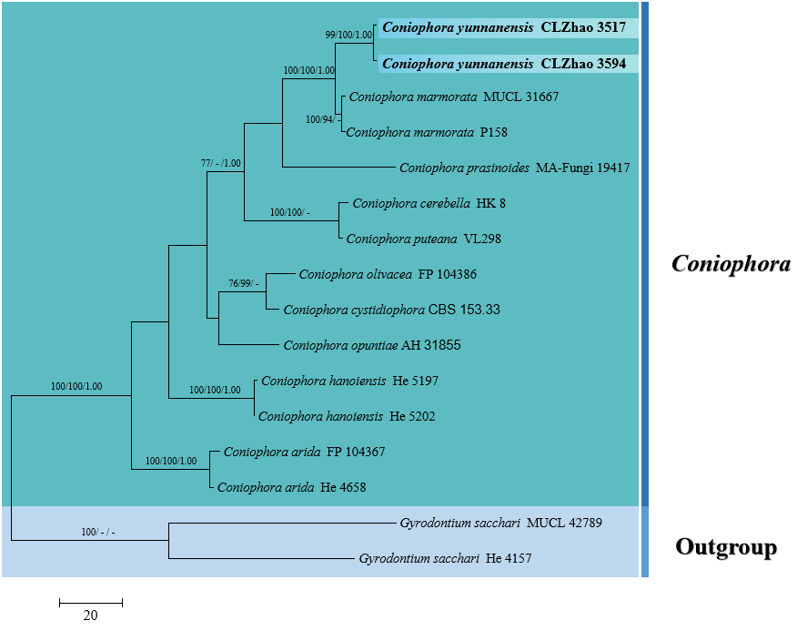
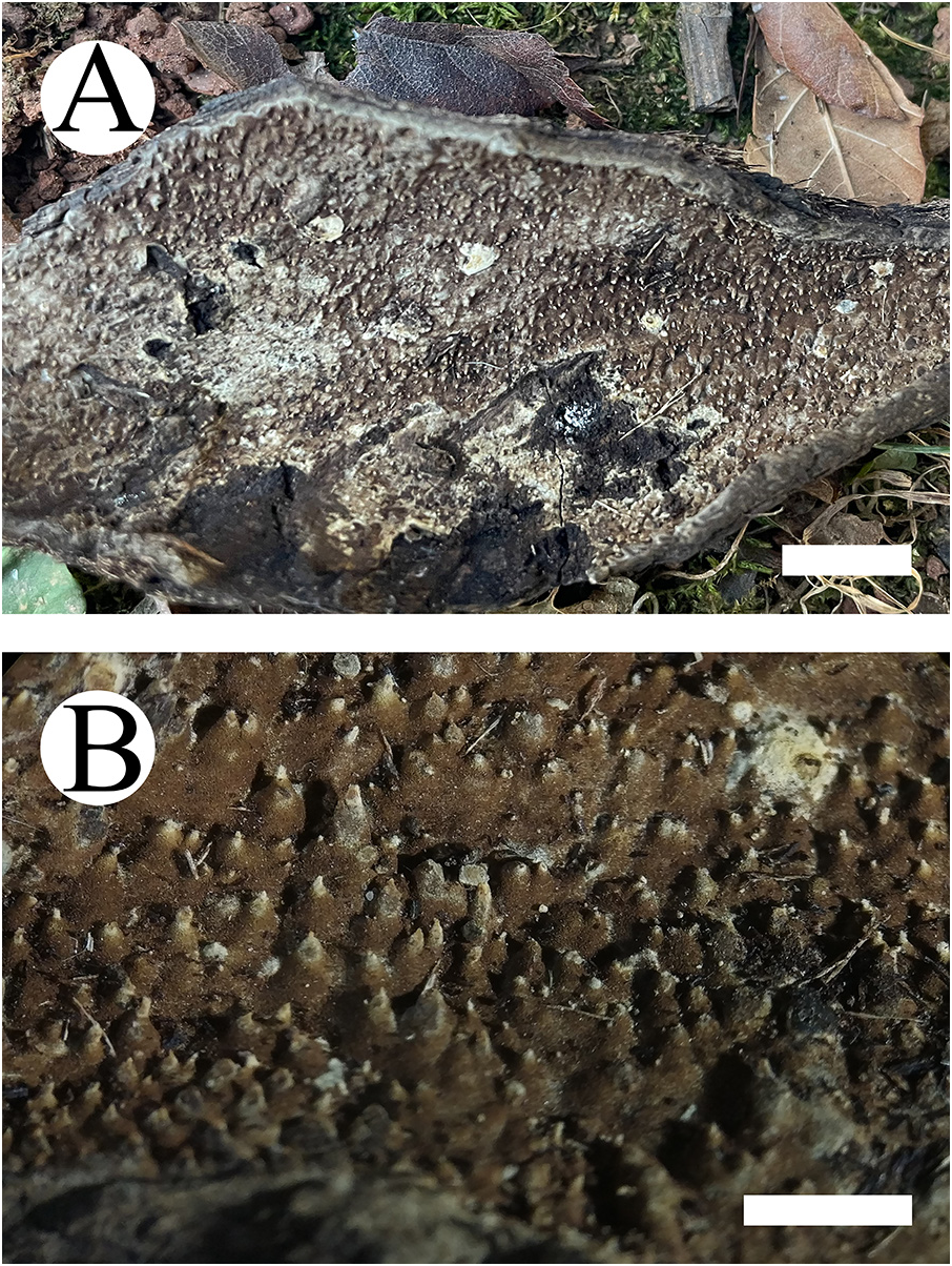
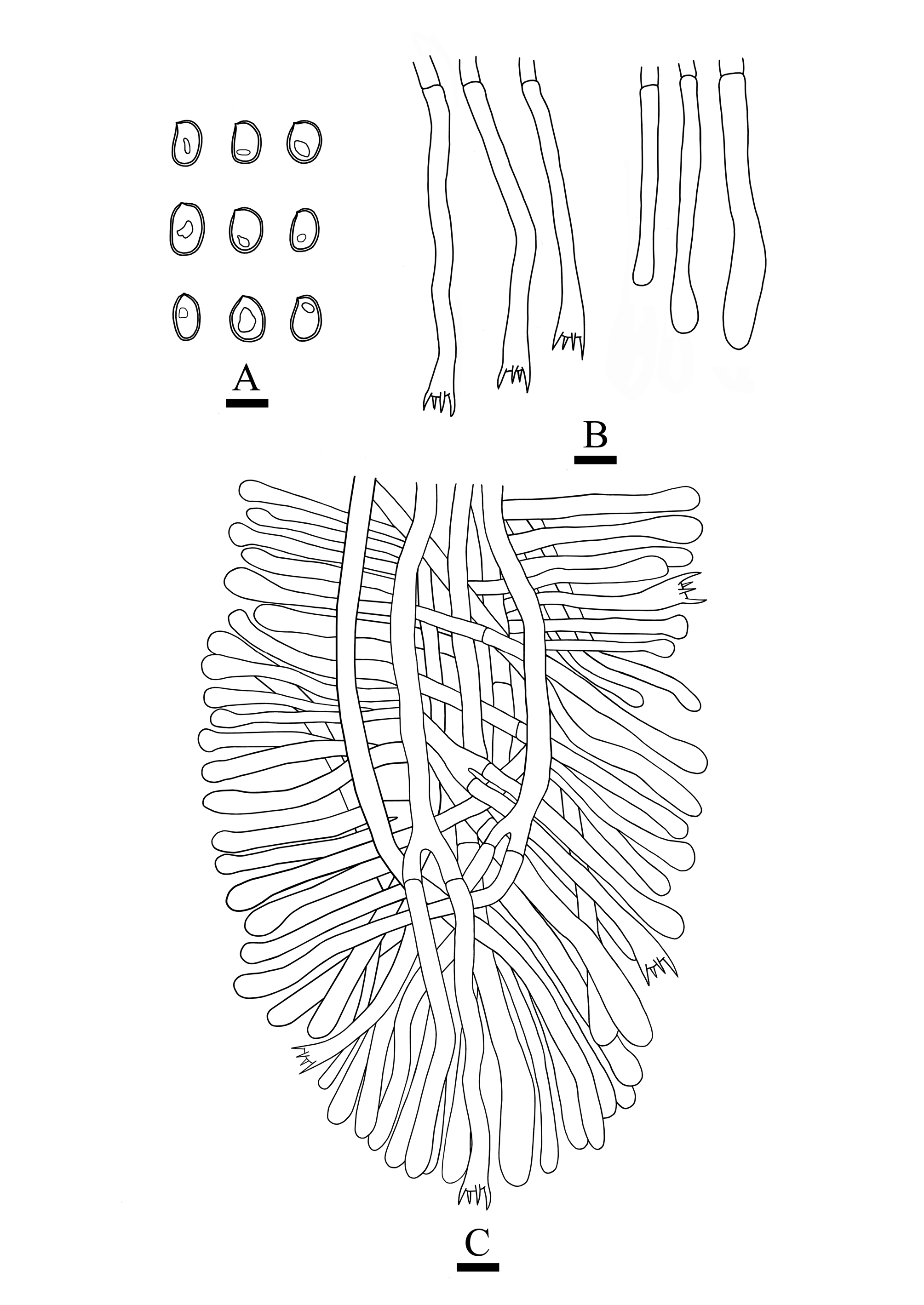
Coniophora yunnanensis Y. Yang & C.L. Zhao , sp. nov. Figs. 1 View FIGURE 1 , 2 View FIGURE 2 , 4 View FIGURE 4 , 5 View FIGURE 5
MycoBank no.: MB 847019
Holotype:— CHINA. Yunnan Province, Pu’er, Jingdong County, Wuliangshan National Nature Reserve , E 100°31′10′′, N 24°25′07′′, elev. 1500 m, on the dead bamboo, 2 October 2017, CLZhao 3594 ( SWFC!), GenBank No. (ITS OP901837; nLSU OP904196). GoogleMaps
Etymology: — yunnanensis (Lat.) : refers to the locality ( Yunnan Province) of the type specimen.
Basidiomata: —Annual, resupinate to effused, separable, soft ceraceous, without odor or taste when fresh, becoming hard corky upon drying, up to 10 cm long, 4 cm wide, 0.5–1 mm thick; hymenial surface odontoid, 100–500 µm long, olivaceous buff (4C4) when fresh, olivaceous buff (4C4) to greyish brown (5/6E4) drying; sterile margin indistinct and olivaceous buff (4C4).
Hyphal structure: — Hyphal system monomitic; generative hyphae with simple septa, colorless, thin-walled, rarely branched, interwoven, 2.5–6 μm in diam; IKI–, CB–, tissues unchanged in KOH.
Hymenium : — Cystidia and cystidioles absent; basidia cylindrical, constricted, 40–90 × 5–7 µm, with 4-sterigmata and a basal simple septum, basidioles dominant, in shape similar to basidia, but slightly smaller.
Basidiospores: — Ovoid to broadly ellipsoid, smooth, thick-walled, pale yellowish, with oil drops, IKI–, CB–, (8–)8.5–12.5 × (5.5–)6.5–8.5 µm, L = 10.7 µm, W = 7.27 µm, Q = 1.46–1.53 (n = 60/2), Qm = 1.47.
Additional specimen examined: — CHINA. Yunnan Province, Pu’er, Jingdong County, Wuliangshan National Nature Reserve , E 100°31′10′′, N 24°25′07′′, elev. 1500 m, on the dead bamboo, 2 October 2017, CLZhao 3517 ( SWFC!), GenBank No. (ITS OP901836; nLSU OP904195) GoogleMaps .
Notes: Morphologically, Coniophora arida (Fr.) P. Karst. (1868: 370) , C. hanoiensis Pat. (1907: 76) , C. olivacea (Fr.) P. Karst. (1879: 162) , and C. puteana (Schumach.) P. Karst. (1803: 397) are similar to C. yunnanensis by having ovoid to ellipsoid basidiospores. However, C. arida differs from C. yunnanensis by its wider basidia (40–70 × 7–10 µm), and presence of the hyphal strands ( Bernicchia & Gorjón 2010); C. hanoiensis is separated from C. yunnanensis by its white to yellowish-brown hymenial surface and dimitic hyphal system ( Ginns 1982); C. olivacea is distinguished from C. yunnanensis by smooth hymenial surface, presence of the thick-walled septate cystidia and dextrinoid basidiospores ( Bernicchia & Gorjón 2010); C. puteana is separated from C. yunnanensis by having the tabular, yellowish-brown, smooth basidiomata and brownish basidiospores ( Bernicchia & Gorjón 2010).
Coniophora yunnanensis resembles C. arachnoidea , C. fusispora (Cooke & Ellis) Cooke (1889: 650) and C. ladoi Tellería (1991: 236) in having a odontoid hymenial surface. However, Coniophora arachnoidea is different from C. yunnanensis by brownish hymenophore and smaller basidiospores (6–8 × 4–5 µm) ( Blanco et al. 2009); C. fusispora can be delimited from C. yunnanensis by its orange-yellow to brown hymenophore and longer basidiospores (14–20 × 5–8 µm) ( Bernicchia & Gorjón 2010); C. ladoi is distinguished from C. yunnanensis by small basidiospores (5.5–7.5 × 3.5–4.5 µm) ( Bernicchia & Gorjón 2010).

| SWFC |
Southwest Forestry College |
No known copyright restrictions apply. See Agosti, D., Egloff, W., 2009. Taxonomic information exchange and copyright: the Plazi approach. BMC Research Notes 2009, 2:53 for further explanation.
|
Kingdom |
|
|
Phylum |
|
|
Class |
|
|
Order |
|
|
Family |
|
|
Genus |