
Tychius subsulcatus Tournier, 1847
|
publication ID |
https://doi.org/ 10.11646/zootaxa.4568.1.10 |
|
publication LSID |
lsid:zoobank.org:pub:3EF72F1D-21B8-4729-8141-7B490D820496 |
|
DOI |
https://doi.org/10.5281/zenodo.5925471 |
|
persistent identifier |
https://treatment.plazi.org/id/7E0F8795-A206-FFB3-1FA5-AA35FD987DFD |
|
treatment provided by |
Plazi |
|
scientific name |
Tychius subsulcatus Tournier, 1847 |
| status |
|
Tychius subsulcatus Tournier, 1847 View in CoL
Material examined. 20 L 3 larvae and 16 pupae: 2.vii.2015 and 14.vii.2015, Cerová vrchovina Mts., Gemerský Jablonec env., Slovakia, N 48°11.9´E 19°59.6´, 250 m a. s. l., lgt. and det. M. Košťál. GoogleMaps
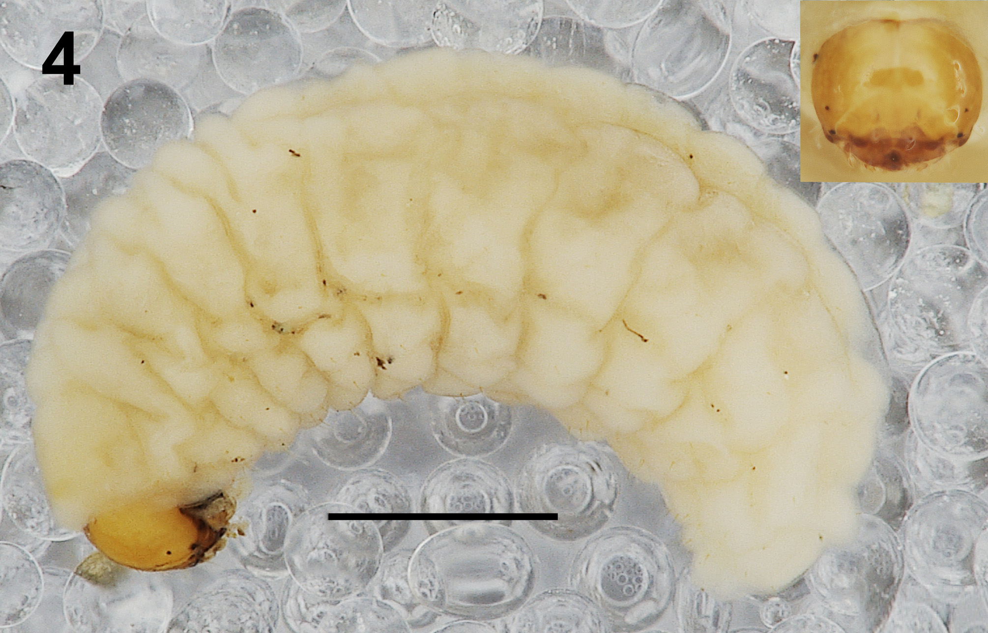
Descriptions. Mature larva. Measurements (in mm). Body length: 4.52–6.15 (mean 5.30). Body width (abdominal segments II–V) up to 1.80. Head width: 0.60–0.71 (mean 0.66).
General. Body elongate, slender, curved, rounded in cross section ( Fig. 4 View FIGURE 4 ).
Coloration. Head light brown ( Fig. 4 View FIGURE 4 ). All thoracic and abdominal segments whitish to yellowish ( Fig. 4 View FIGURE 4 ).
Vestiture. Setae on body thin, pale orange to brown, distinctly unequal in length (minute to very short or long).
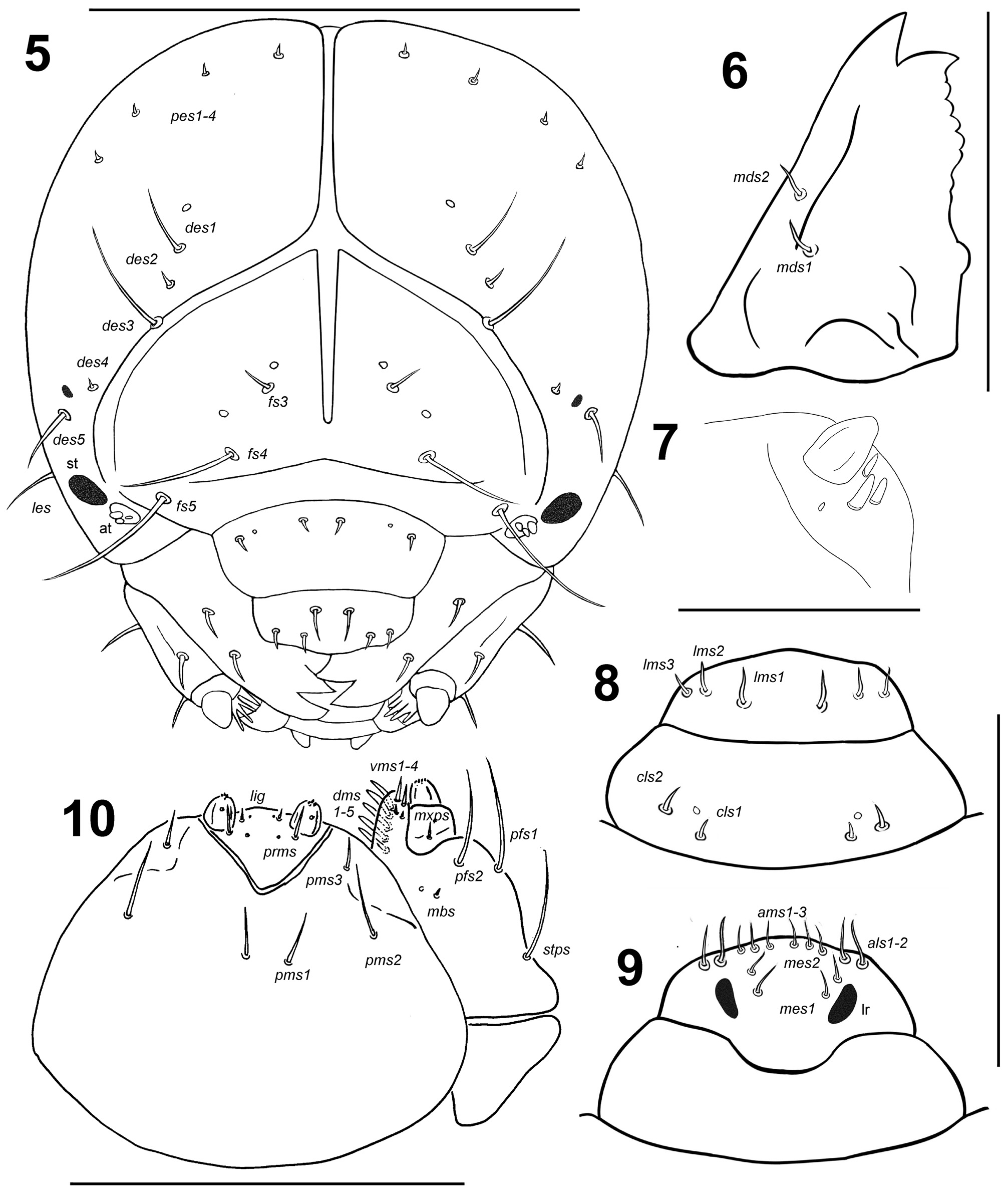
Head capsule. Head oval, slightly or more flattened laterally. Frontal sutures distinct, extended to stemmata, forming a “Y” with the epicranial suture. Endocarinal line present, long and wide, reaching 2/3 of frons length. Two small stemmata (st), one larger, in the form of a pigmented spot with convex cornea behind antenna, second rudimentary, close to des 5. Des 1 located almost in central part of epicarnium, des 2 very close to frontal suture, des 3 at lateral margin of frontal suture; des 4 close to des 5, des 5 located anterolaterally; des 1, des 3 and des 5 long to very long, des 2 short to very short, des 4 very short to minute ( Fig. 5 View FIGURES 5–10 ). Fs 1 and fs 2 absent, fs 3 located medially, fs 4 more laterally, close to epistome, fs 5 anterolaterally almost at epistome; fs 3 short, fs 4 and fs 5 very long ( Fig. 5 View FIGURES 5–10 ). Les 1 absent, les 2 as long as half of des 3 length. Both ves very short to short. Epicranial area with 1 sensillum (close to des 1) and 4 minute pes in line.
Antennae bearing one relatively elongate, conical sensorium; and basal membranous article with three coneshaped sensilla equal in length, and 1 minute sensillum ( Fig. 7 View FIGURES 5–10 ).
Clypeus ( Fig. 8 View FIGURES 5–10 ) approximately 2.9 times as wide as long; with 2 cls of short size, almost equal in length, cls 1 located postero-medially, and cls 2 more laterally, and 1 sensillum between both cls; anterior margin slightly, posterior margin clearly arched.
Mouthparts. Labrum ( Fig. 8 View FIGURES 5–10 ) approximately 2.8 times as wide as long; with three piliform lms, lms 1 slightly longer than lms 2, lms 3 moderately shorter than lms 2, lms 3 approximately as long as half of length lms 1; anterior margin medially slightly bulged, paramedially indistinctly sinuose. Epipharynx ( Fig. 9 View FIGURES 5–10 ) with 2 medium sized piliform als, als 1 slightly longer than als 2; with 3 equally long ams as long as half of als 1 length; with 2 mes moderately longer than ams; labral rods relatively short, slightly unevenly shaped, elongate, divergent anteriad. Mandible ( Fig. 6 View FIGURES 5–10 ) bifid; cutting edge with blunt tooth; bearing 2 piliform mds of medium size, aligned longitudinally. Maxilla ( Fig. 10 View FIGURES 5–10 ) stipes with 1 long stps and 2 long pfs, equal in length, minute mbs, and 1 sensillum close to mbs; mala with 5 medium sized finger-like dms; 4 vms, unequal in length, two very short to short. Maxillary palpi: basal palpomere with 1 short mxps and 2 sensilla; distal palpomeres with a medium, cuticular apical processes; length ratio of basal and distal palpomeres 1:1.2. Prelabium ( Fig. 10 View FIGURES 5–10 ) with 1 medium prms; ligula with 1 minute ligs; premental sclerite narrow, ring-shaped. Labial palpi with only 1 palpomere; with 1 sensillum, and with a medium, cuticular apical processes. Postlabium ( Fig. 10 View FIGURES 5–10 ) with long pms 1 located basally, very long pms 2 located medially, and long pms 3 located apically; membranous area basolaterally sparsely and finely asperate.
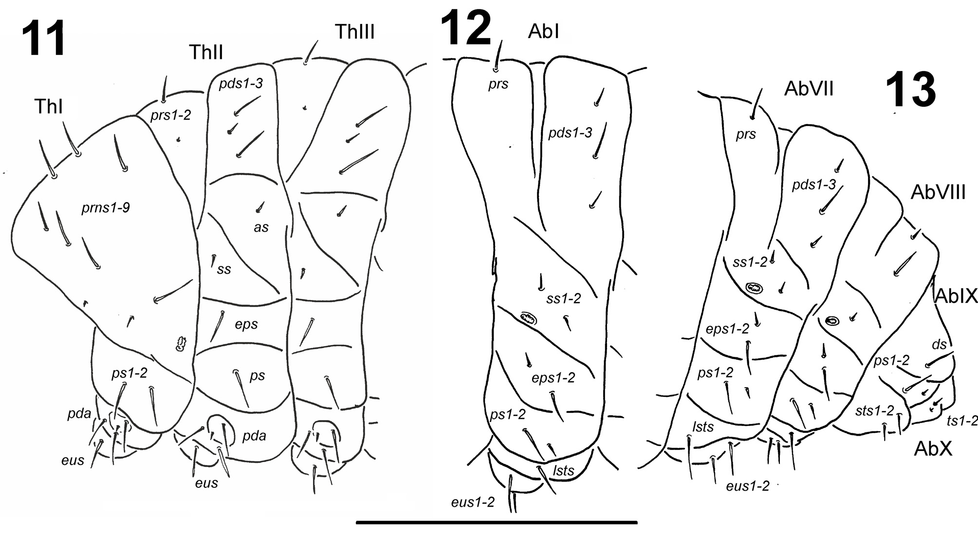
Thorax. Prothorax smaller than meso- and metathorax. Spiracle bicameral, placed between the pro- and mesothorax (see e.g., Skuhrovec et al. 2015). Prothorax ( Fig. 11 View FIGURES 11–13 ) with nine prns (seven very long located on weakly pigmented dorsal sclerite, and 2 very short (one sometimes absent) located below); 2 long ps; and 1 long eus. Meso- and metathorax ( Fig. 11 View FIGURES 11–13 ) with 1 long and 1 very short prs, sometimes absent; 2 long and 1 short pds (order: long, short, long); 1 short as; 1 short ss; 1 long eps; 1 long ps; and 1 long eus. Each pedal area of thoracic segments with 3 long and 1 very short pda.
Abdomen. Abdominal segments I–II slightly smaller than others, abdominal segments III–VI almost of equal length; following abdominal segments decreasing gradually to terminal part of body. Abdominal segment X reduced to four anal lobes of unequal size, lateral lobes being distinctly largest, dorsal and the ventral lobes being very small. Anus located terminally. Eight spiracles, unicameral, all spiracles functional, close to anterior margin. Abdominal segments I–VII ( Figs 12–13 View FIGURES 11–13 ) with 1 long prs; 1 long and 2 medium sized pds (order: medium, long, medium); 2 medium sized ss; 1 long and 1 medium eps; 1 long and 1 medium ps; 1 long lsts; and 2 long eus. Abdominal segment VIII ( Fig. 13 View FIGURES 11–13 ) without prs; 1 short and 1 long pds (order: short, long); 1 short ss; 1 long and 1 medium eps; 1 long and 1 medium ps; 1 long lsts; and 2 long eus. Abdominal segment IX ( Fig. 13 View FIGURES 11–13 ) with 1 long ds; 1 long and 1 short ps; and 2 medium sts. Abdominal segment X ( Fig. 13 View FIGURES 11–13 ) on lateral lobes with 1 short and 1 minute ts.
Pupa. Measurements (in mm). Body length: 3.00–4.05 (mean 3.46). Body width: 1.30–1.84 (mean 1.59). Head width: 0.45–0.68 (mean 0.55).
Coloration. Body bright yellow.
Morphology. Body moderately stout ( Figs 14–16 View FIGURES 14–16 ). Rostrum medium long, approximately 4 times as long as wide, reaching mesocoxae. Antennae elongate. Pronotum 1.6 times as wide as long. Mesonotum distinctly shorter than metanotum. Abdominal segments I–V of equal length; segments VI and VII diminish gradually; segment VIII almost semicircular; segment IX distinctly reduced. Spiracles on abdominal segments placed dorsolaterally: on segments I–V functional, and on segment VI atrophic, on next ones invisible. Urogomphi (ur) slender and short, conical ( Figs 14–16 View FIGURES 14–16 ).
Identification of the sex of pupae was very difficult in the studied species. The differences in size of body between males and females were very similar. The shape of the gonothecae was typical of the pupae of weevils (bilobed in females and undivided in males; see Stejskal et al. 2014) but was weakly visible in some specimens.
Chaetotaxy. Setae distinct, medium sized to short, very thin, piliform, unequal in length, reddish-brown, on head and pronotum anchored on top of small protuberances. Head capsule including 1 vs, 1 sos, 1 os, and 1 pas; pas relatively close to anterior margin of eye ( Figs 14–16 View FIGURES 14–16 ). Rostrum with 1 paramedially located rs. Vs almost twice longer than sos, os, pas, and rs, as long as setae on pronotum. Pronotum with 2 as, 2 ls, 2 ds, and 1 pls; all pronotal setae almost equal in length ( Figs 14–16 View FIGURES 14–16 ). Each of dorsal part of meso- and metathorax with 1 seta. All femora with 1 fes on apex ( Figs 14–16 View FIGURES 14–16 ). Setae on abdominal segments I–VIII arranged in one dorsal and one lateral row; setae on abdominal segments I–IV anchored on tiny, indistinct protuberances, setae on segments V–VII on small, conical protuberances ( Figs 14–16 View FIGURES 14–16 ). Ventral parts of segments I–IX without setae. Urogomphi small, slightly medially curved.
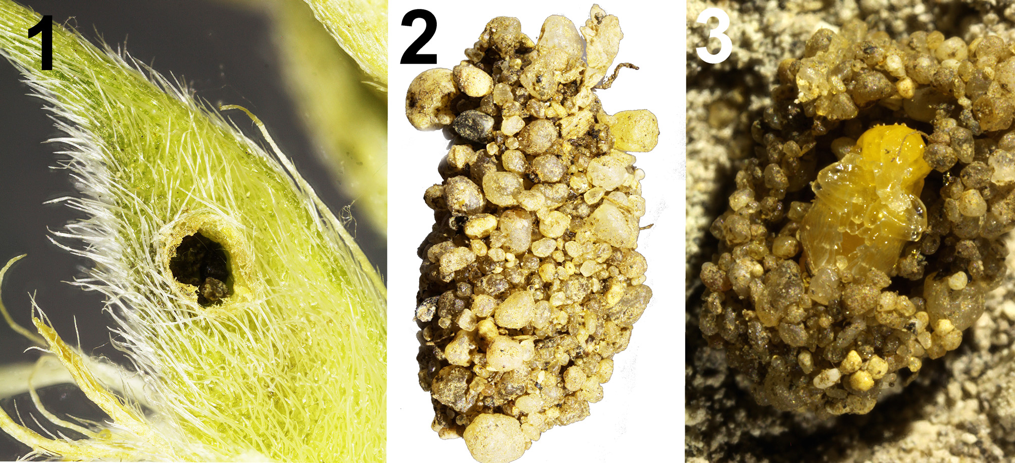
Biological observations. Large plants of Astragalus onobrychis at the visited locality had almost half of their pods infested by larvae of T. subsulcatus , especially large pods. Dozens of larvae emerged through relatively small holes ( Fig. 1 View FIGURES 1–3 ) after four days of incubation and for several days later. Coarse pupal cells were considerably strengthened by sand grains ( Figs 2, 3 View FIGURES 1–3 ). Pupation followed in 7–10 days after the larvae burrowed into the soil. Duration of the pupal stage at a stable temperature was on average 14 days and could be shortened to 10 days by wetting the soil in some containers.
Remarks. Caldara (1990) in the revision of Palaearctic species of the genus Tychius , based exclusively on adult morphological characters, included T. subsulcatus in the speciose T. intrusus Faust, 1889 group. Imagoes of this group are characterized by broad scales on elytral interstriae “often arranged in double rows with single median rows of seta-like scales”.

No known copyright restrictions apply. See Agosti, D., Egloff, W., 2009. Taxonomic information exchange and copyright: the Plazi approach. BMC Research Notes 2009, 2:53 for further explanation.
|
Kingdom |
|
|
Phylum |
|
|
Class |
|
|
Order |
|
|
Family |
|
|
Genus |