
Polycystis ali Schockaert, 1982
|
publication ID |
https://doi.org/ 10.11646/zootaxa.4446.1.3 |
|
publication LSID |
lsid:zoobank.org:pub:AC2EA154-7E31-4E91-AF4A-9C95FE504123 |
|
DOI |
https://doi.org/10.5281/zenodo.5958670 |
|
persistent identifier |
https://treatment.plazi.org/id/7E20083B-FFD0-0A4E-FF74-C2E1CDA0FEBC |
|
treatment provided by |
Plazi |
|
scientific name |
Polycystis ali Schockaert, 1982 |
| status |
|
Polycystis ali Schockaert, 1982 View in CoL
( Figs. 4–6 View FIGURE 4 View FIGURE 5 View FIGURE 6 )
Material examined. Holotype: PLA-Po031, whole-mounted specimen. Paratypes: PLA-Po032~034, wholemounted specimen. Other specimens: PLA-Po036, mounted slides of stylet; PLA-Po035-1~035-5, seriallysectioned specimens. Specimens were collected at the same location as that of Paulodora sinensis n. sp. All specimens were preserved in IZCAS .
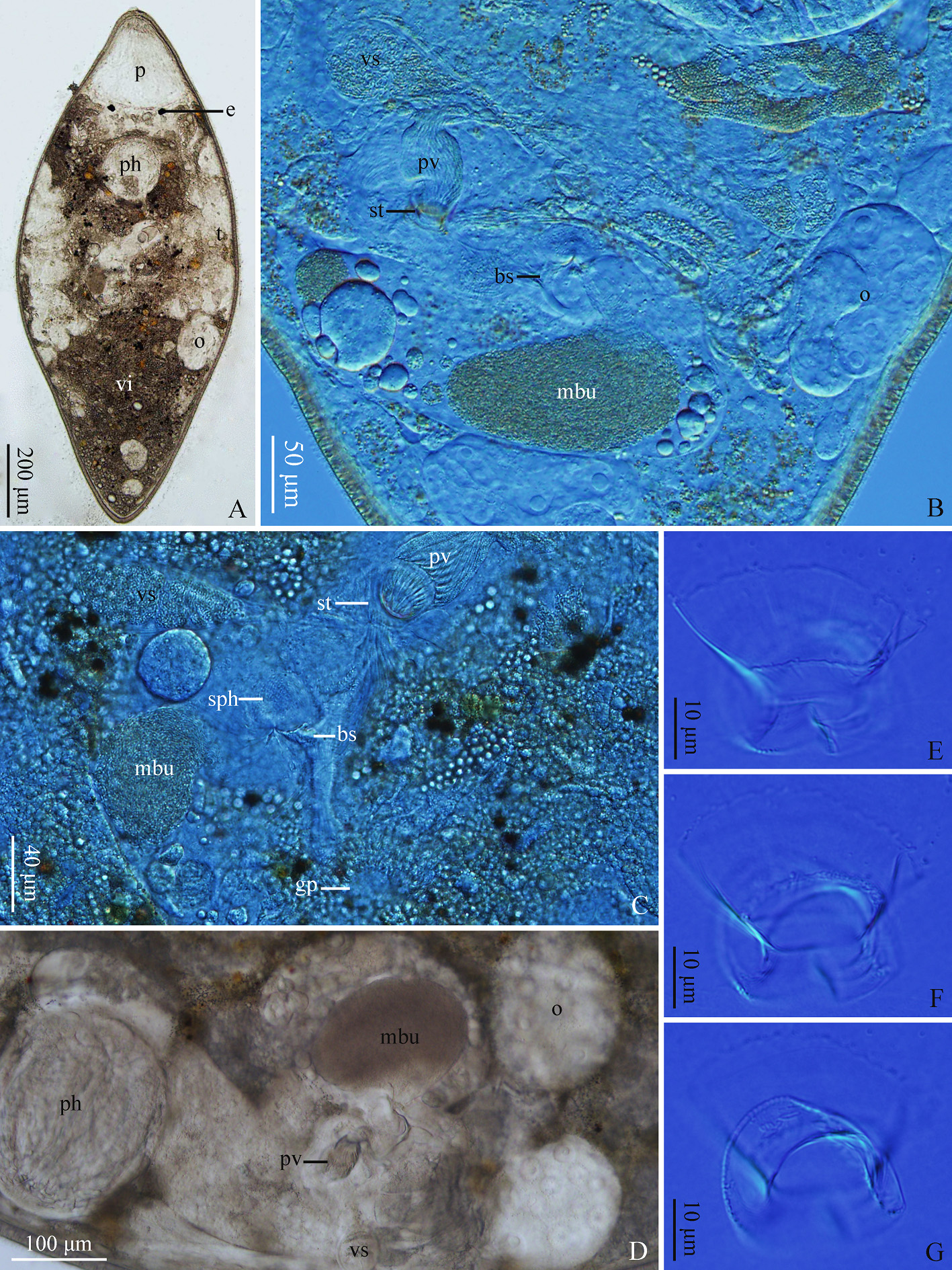
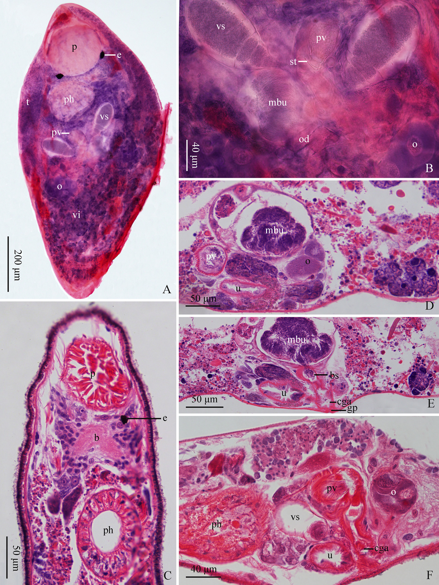
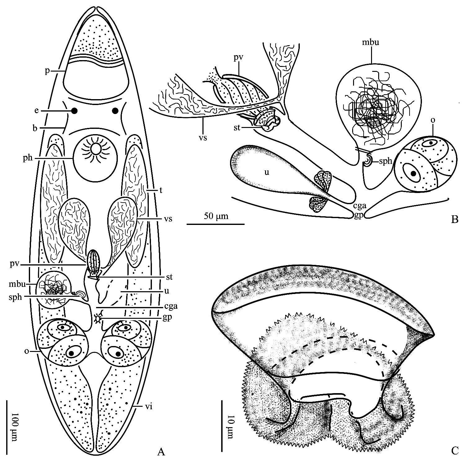
Description. Mature individual is 1,400–1,700 µm in length, 590–600 µm in width, and has no pigment. The bullet-shaped proboscis is 250–320 µm in length (n=3) ( Fig. 4A View FIGURE 4 , 5A & C View FIGURE 5 , 6A View FIGURE 6 ). A pair of pigmented circular eyes (16–22 µm in diameter, n=3) is located posterior to proboscis and the distance between two eyes is 120–200 µm (n=3) ( Fig. 4A View FIGURE 4 , 5A & C View FIGURE 5 , 6A View FIGURE 6 ). The brain is situated posterior to eyes at the ventral side ( Fig. 5C View FIGURE 5 , 6A View FIGURE 6 ). The pharynx (170–190 µm in diameter, n=3) is located at 33% of the body ( Fig. 4A & D View FIGURE 4 , 5A & C & F View FIGURE 5 , 6A View FIGURE 6 ).
The paired elongated rod-like testes (300–400 µm in length, 70–90 µm in width, n=3) are located at middle of body ( Fig. 4A View FIGURE 4 , 5A View FIGURE 5 , 6A View FIGURE 6 ) and are connected to the seminal vesicle via the vas deferens. Two seminal vesicles (120– 130 µm in length, n=3) are located at ventral side of the middle body ( Fig. 4B–D View FIGURE 4 , 5A–B & F View FIGURE 5 , 6A–B View FIGURE 6 ). The ejaculatory duct is formed posterior to the fusion of seminal vesicles and enters the male atrium proximally. The prostatic vesicle is spindle-shaped (32–36 µm in length, n=3), with its distal end embedded in the base of the stylet ( Fig. 4B–D View FIGURE 4 , 5A–B, D & F View FIGURE 5 , 6A–B View FIGURE 6 ). The doubled-walled stylet is 32–36 µm in length (n=3) ( Fig. 4B–C & E–G View FIGURE 4 , 5B View FIGURE 5 , 6A–C View FIGURE 6 ). The proximal part of the stylet is funnel-shaped and is 35–38 µm (n=3) in basal diameter. The distal part of the stylet is folded outwards to form a collar (25–27 µm in width), while it is partially concave and finally forms a jagged edge. The stylet enters the male atrium anteriorly. The total length of inner stylet is about half of the outer stylet, while its basal diameter is relatively small (18–22 µm, n=3). The narrow bursa stalk connects the male atrium to the male bursa, with strong asymmetric sphincter at the junction ( Fig. 4B–C View FIGURE 4 , 5E View FIGURE 5 ). The globular male bursa (80–100 µm in diameter, n=3) is located right anterior to the ovary and is filled with sperm ( Fig. 4B–D View FIGURE 4 , 5B & D–E View FIGURE 5 , 6A–B View FIGURE 6 ).
* Measurement based on references.
The two vitellaria are located dorsally at both sides of the body, extending from the anterior end of pharynx to the cauda of the body ( Fig. 4A View FIGURE 4 , 5A View FIGURE 5 , 6A View FIGURE 6 ). A pair of ovaries (150–158 µm in diameter, n=3) are located at 65% of the body ( Fig. 4A–B & D View FIGURE 4 , 5A–B, D & F View FIGURE 5 , 6A–B View FIGURE 6 ). The two oviducts merge and form the female duct, which subsequently connects the common genital atrium. The uterus is located anterior to the ovary on the left side and connects to the common genital atrium at the left anterior end of the female duct ( Fig. 5D–F View FIGURE 5 , 6A–B View FIGURE 6 ).
Remarks. Previously, Karling categoriZed P. ali into three forms ( Karling 1986), namely ‘Somali’, ‘Galapagos’, and ‘California’. Based on more detailed comparison, Artois & Tessens (2008) suggested that f. ‘California’ is a separate species: P. californica . Among them, P. ali f. ‘Galapagos’ and P. californica have apparent slit in their collar, which cannot be found in P. ali and the newly recorded specimens from China. The stylet of the newly recorded specimen is highly similar to that of P. ali . However, minor differences could still be found between them. For example, stylets of the new P. ali specimens are 32–36 µm in length, which is much longer than that of P. ali ( Tab. 3). Besides, the distal end of the new specimens is partially concave and forms an inverted Vshaped structure, which is not described in P. ali . Taking the above comparisons into consideration, we described the new specimens as P. ali , a newly recorded species within the genus Polycystis in China.
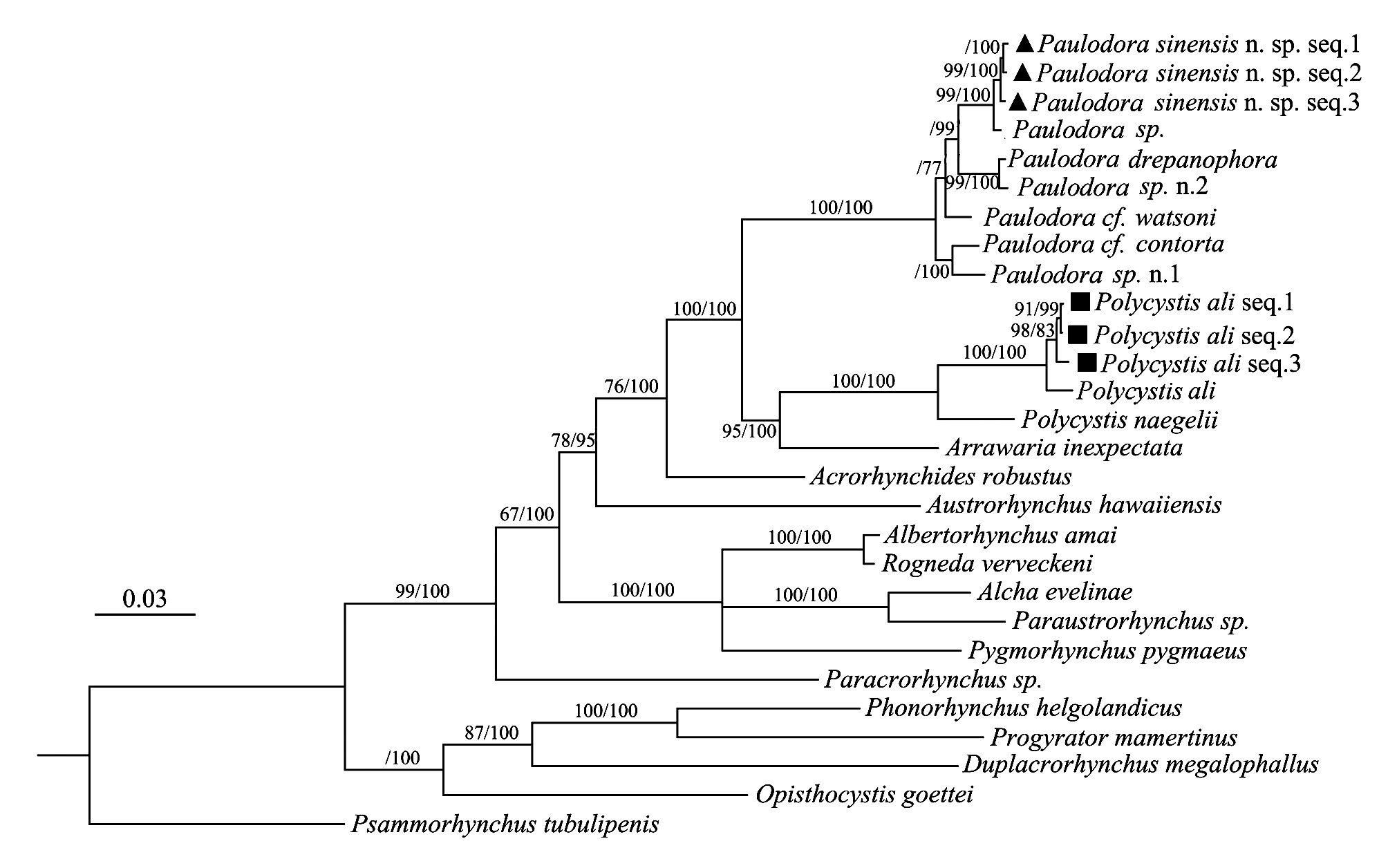
Phylogenetic analysis. The concatenated sequences of 18S and 28S rDNA genes phylogenetic analysis showed that three specimens of the new species clustered together and form a clade independently, which are distinct from other species ( Fig. 7 View FIGURE 7 ). Paulodora sinensis n. sp. is closely related to Paulodra sp., Paulodra drepanophora, Paulodra sp. n. 2, which belong to the genus Paulodora , while the newly recorded species of Polycystis is closely related to Polycystis ali , which belongs to the genus Polycystis .
Discussion. There are 16 species within the genus Paulodora ( Paulodora sinensis n. sp. included), of which the stylets are all double-walled and consist of the funnel-shaped proximal part and the curved distal part. In the current study, according to the morphological characteristics of their stylet, we have categoriZed the genus Paulodora into three groups, namely spiral stylet group (eight species), unciform stylet group (five species) and lamellar stylet group (three species) ( Tab. 2). In spiral stylet group, the stylet is helix-like and bends at distal part with an angle larger than 180°. In unciform stylet group, the stylet is hook-like and bends at distal part with an angle between 90°–180°. As for lamellar stylet group, the stylet is plate-like and bends at distal part with an angle approximately 90°. In addition, the stylet has two plate-like structures attached to the outer stylet.
There are eight species within the genus Polycystis . We have also categoriZed the genus Polycystis into three groups based on the morphological characteristics of their stylet ( Tab. 4). In Group I, the stylet consists of a funnelshaped proximal part and a collar distal part. As for Group II, the overall morphology of stylet is similar to that of the Group I, but the collar has extension structure at distal end. In Group III, the stylet only has funnel-shaped proximal part, but has no distal collar part.
In flatworm taxonomy, although the morphological analysis of the stylet is an efficient tool to differentiate species, the siZe of the stylet can vary due to the differences of their body siZe, making the identification of new species particular difficult in some cases. As such, more thorough molecular phylogenetic analyses are of great significance to the study of diversity of flatworm. In this study, we have supplemented 18S rDNA and 28S rDNA sequences of Paulodora sinensis n. sp. and new specimens of Polycystis ali , which can be served as useful information for more comprehensive phylogenetic mapping of reproductive character (stylet morphology) in future study.
| IZCAS |
Institute of Zoology, Chinese Academy of Sciences |
No known copyright restrictions apply. See Agosti, D., Egloff, W., 2009. Taxonomic information exchange and copyright: the Plazi approach. BMC Research Notes 2009, 2:53 for further explanation.